
Search Count: 39,366
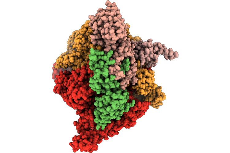 |
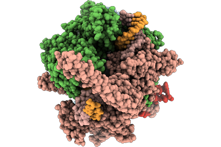
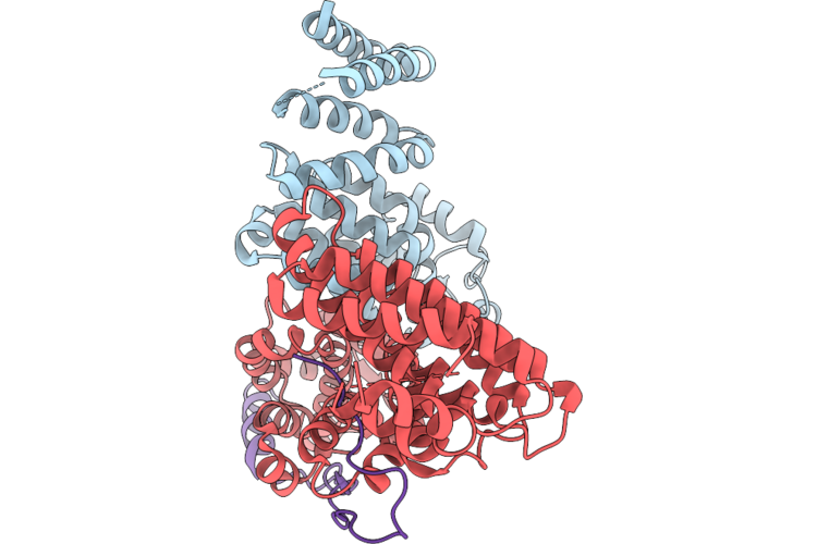
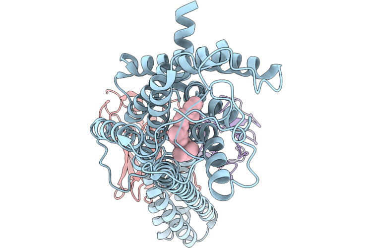
Semiclosed Eco-Epec: Cryo-Em Structure Of Eco Rnap His-Elemental Paused Elongation Complex With A Semi-Closed Active Site (Closed Tl And Si3, Open Rh-Fl)
Organism: Escherichia coli
Method: ELECTRON MICROSCOPY Resolution:4.00 Å Release Date: 2026-06-24 Classification: Transcription/DNA/RNA Ligands: 1N7, ZN, MG |
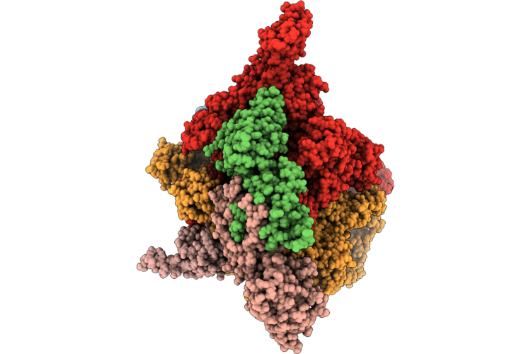 |
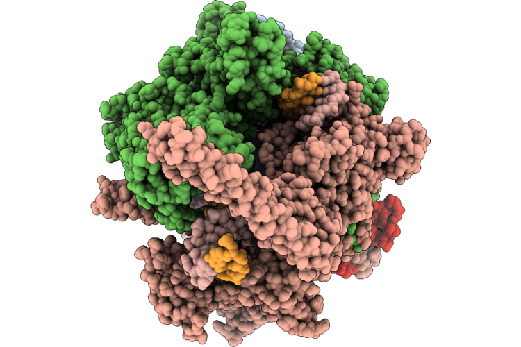
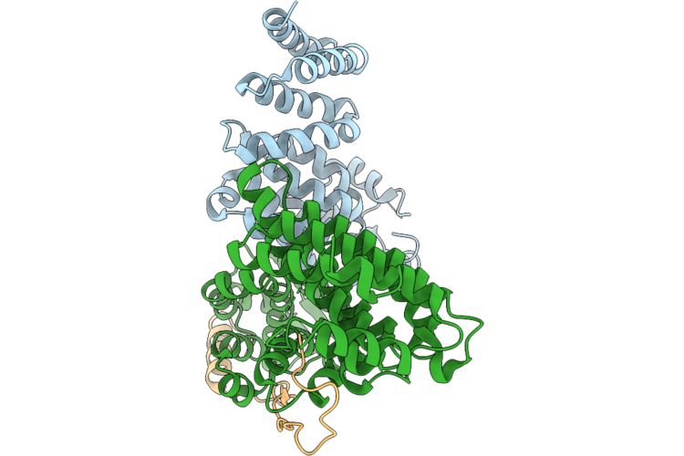
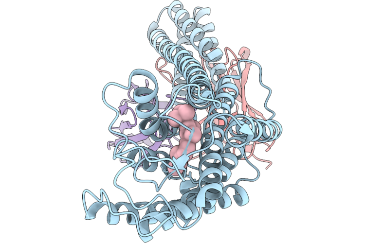
Cbr9379 Bound Open1 Eco-Epec: Cryo-Em Structure Of Eco Rnap His-Elemental Paused Elongation Complex With An Open Active Site (Open Tl, Si3 And Rh-Fl)
Organism: Escherichia coli
Method: ELECTRON MICROSCOPY Resolution:2.80 Å Release Date: 2026-06-24 Classification: TRANSCRIPTION Ligands: 1N7, MG, ZN, 42T |
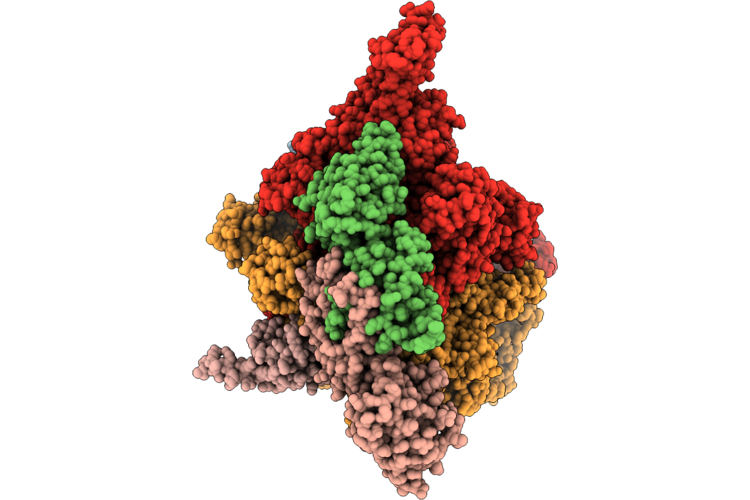 |
Cbr9379 Bound Open2 Eco-Epec: Cryo-Em Structure Of Eco Rnap His-Elemental Paused Elongation Complex With An Open Active Site (Open Tl, Si3 And Rh-Fl)
Organism: Escherichia coli
Method: ELECTRON MICROSCOPY Resolution:2.90 Å Release Date: 2026-06-24 Classification: TRANSCRIPTION Ligands: 1N7, MG, ZN, 42T |
 |
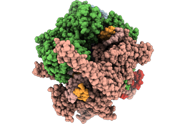
Closed Mtb-Ec: Cryo-Em Structure Of Mtb Rnap Elongation Complex (Substrate Loading Mimic) With A Closed Active Site (Closed Tl And Rh-Fl)
Organism: Mycobacterium tuberculosis, Escherichia coli
Method: ELECTRON MICROSCOPY Resolution:3.00 Å Release Date: 2026-06-24 Classification: TRANSCRIPTION Ligands: ZN, MG, GTP |
 |
Open Mtb-Ec: Cryo-Em Structure Of Mtb Rnap Elongation Complex (Substrate Loading Mimic) With An Open Active Site (Open Tl And Rh-Fl)
Organism: Mycobacterium tuberculosis, Escherichia coli
Method: ELECTRON MICROSCOPY Resolution:3.30 Å Release Date: 2026-06-24 Classification: TRANSCRIPTION Ligands: ZN, MG |
 |
Semiclosed Mtb-Ec: Cryo-Em Structure Of Mtb Rnap Elongation Complex (Substrate Loading Mimic) With A Semiclosed Active Site (Closed Tl, Open Rh-Fl)
Organism: Mycobacterium tuberculosis, Escherichia coli
Method: ELECTRON MICROSCOPY Release Date: 2026-06-24 Classification: TRANSCRIPTION Ligands: GTP, ZN, MG |
 |
Aap-So2 Bound Open Mtb-Ec: Cryo-Em Structure Of Mtb Rnap Elongation Complex (Substrate Loading Mimic) With An Open Active Site (Open Tl And Rh-Fl)
Organism: Mycobacterium tuberculosis, Escherichia coli
Method: ELECTRON MICROSCOPY Resolution:3.00 Å Release Date: 2026-06-24 Classification: TRANSCRIPTION Ligands: GTP, A1BNV, MG, ZN |
 |
Organism: Escherichia coli k-12
Method: ELECTRON MICROSCOPY Resolution:3.00 Å Release Date: 2026-06-24 Classification: PROTEIN TRANSPORT |
 |
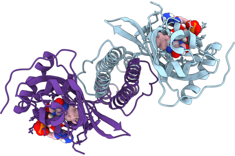
Organism: Escherichia coli, Homo sapiens
Method: ELECTRON MICROSCOPY Release Date: 2026-06-24 Classification: GENE REGULATION |
 |
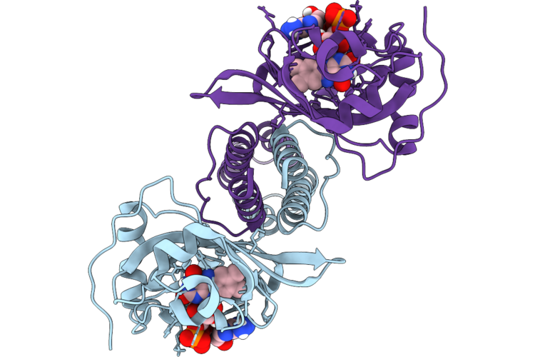
Organism: Escherichia coli, Homo sapiens
Method: ELECTRON MICROSCOPY Release Date: 2026-06-24 Classification: GENE REGULATION |
 |
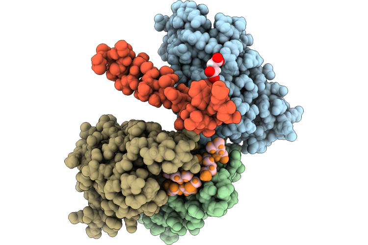
Organism: Escherichia coli, Homo sapiens
Method: ELECTRON MICROSCOPY Release Date: 2026-06-24 Classification: GENE REGULATION |
 |
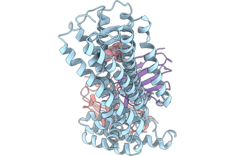
Organism: Escherichia coli, Homo sapiens
Method: ELECTRON MICROSCOPY Release Date: 2026-06-24 Classification: GENE REGULATION |
 |
Seryl-Trna Synthetase In Complex With A Fragment-Sized Inhibitor (3-Cyclopropyl Benzoic Acid)
Organism: Escherichia coli
Method: X-RAY DIFFRACTION Resolution:1.47 Å Release Date: 2026-06-24 Classification: LIGASE Ligands: A1J8R, GOL |
 |
Organism: Escherichia coli k-12
Method: ELECTRON MICROSCOPY Release Date: 2026-06-24 Classification: FLAVOPROTEIN Ligands: FAD |
 |
Organism: Escherichia coli k-12
Method: ELECTRON MICROSCOPY Resolution:3.50 Å Release Date: 2026-06-24 Classification: FLAVOPROTEIN Ligands: FAD |
 |
Organism: Escherichia coli
Method: X-RAY DIFFRACTION Resolution:2.49 Å Release Date: 2026-06-24 Classification: DE NOVO PROTEIN |
 |
Organism: Homo sapiens, Escherichia coli, Synthetic construct
Method: ELECTRON MICROSCOPY Resolution:3.70 Å Release Date: 2026-06-24 Classification: MEMBRANE PROTEIN Ligands: RET |
 |
Organism: Homo sapiens, Escherichia coli, Synthetic construct
Method: ELECTRON MICROSCOPY Resolution:3.22 Å Release Date: 2026-06-24 Classification: MEMBRANE PROTEIN Ligands: RET |
 |
Pre-Active Dark-State Structure Of Human Short-Wave-Sensitive Opsin (Opn1Sw)
Organism: Homo sapiens, Escherichia coli, Synthetic construct
Method: ELECTRON MICROSCOPY Resolution:3.06 Å Release Date: 2026-06-24 Classification: MEMBRANE PROTEIN Ligands: RET |
 |
Organism: Escherichia coli, Podisus maculiventris
Method: SOLUTION NMR Release Date: 2026-06-24 Classification: PEPTIDE BINDING PROTEIN |