

Search Count: 17
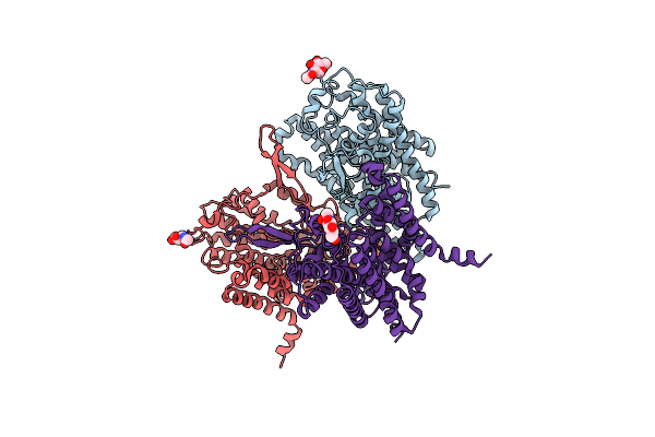 |
Cryo-Em Structure Of The Human Neutral Amino Acid Transporter Asct2
Organism: Homo sapiens
Method: ELECTRON MICROSCOPY Release Date: 2019-11-06 Classification: TRANSPORT PROTEIN Ligands: NAG |
Organism: Homo sapiens
Method: ELECTRON MICROSCOPY
Release Date: 2019-11-06
Ligands: NAG
 |
Cryo-Em Structure Of The Human Neutral Amino Acid Transporter Asct2
Organism: Homo sapiens
Method: ELECTRON MICROSCOPY Release Date: 2019-11-06 Classification: TRANSPORT PROTEIN Ligands: GLN, NAG |
Organism: Homo sapiens
Method: ELECTRON MICROSCOPY
Release Date: 2019-11-06
Ligands: GLN, NAG
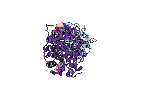 |
Structure Of Human Ketohexokinase Complexed With Hits From Fragment Screening
Organism: Homo sapiens
Method: X-RAY DIFFRACTION Resolution:2.16 Å Release Date: 2017-09-13 Classification: TRANSFERASE Ligands: A4G, SO4 |
Organism: Homo sapiens
Method: X-RAY DIFFRACTION
Release Date: 2017-09-13
Ligands: A4G, SO4
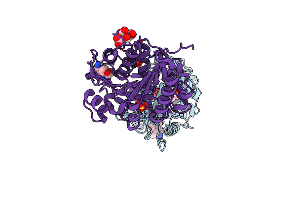 |
Structure Of Human Ketohexokinase Complexed With Hits From Fragment Screening
Organism: Homo sapiens
Method: X-RAY DIFFRACTION Resolution:2.25 Å Release Date: 2017-09-13 Classification: TRANSFERASE Ligands: A1Y, SO4, CIT, GOL |
Organism: Homo sapiens
Method: X-RAY DIFFRACTION
Release Date: 2017-09-13
Ligands: A1Y, SO4, CIT, GOL
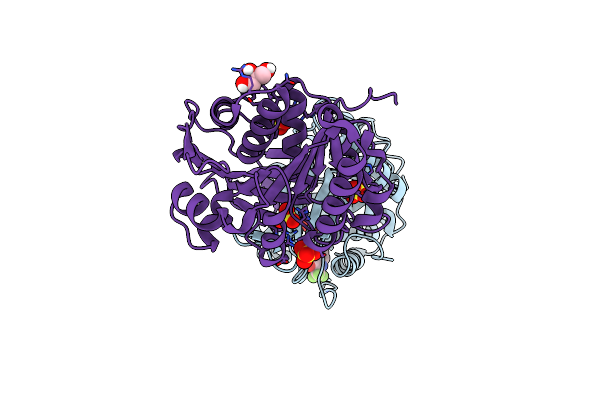 |
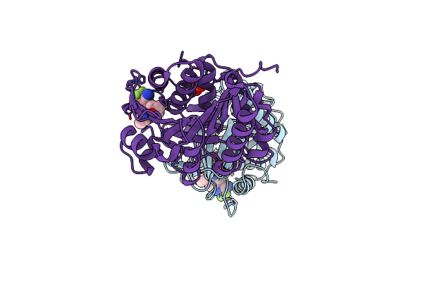
Structure Of Human Ketohexokinase Complexed With Hits From Fragment Screening
Organism: Homo sapiens
Method: X-RAY DIFFRACTION Resolution:2.74 Å Release Date: 2017-09-13 Classification: TRANSFERASE Ligands: A2J, SO4, GOL |
Organism: Homo sapiens
Method: X-RAY DIFFRACTION
Release Date: 2017-09-13
Ligands: A2J, SO4, GOL
 |
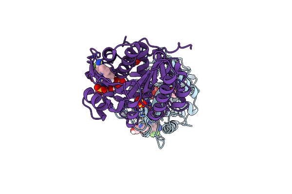
Structure Of Human Ketohexokinase Complexed With Hits From Fragment Screening
Organism: Homo sapiens
Method: X-RAY DIFFRACTION Resolution:2.40 Å Release Date: 2017-09-13 Classification: TRANSFERASE Ligands: A3J, SO4, CL |
Organism: Homo sapiens
Method: X-RAY DIFFRACTION
Release Date: 2017-09-13
Ligands: A3J, SO4, CL
 |
Structure Of Human Ketohexokinase Complexed With Hits From Fragment Screening
Organism: Homo sapiens
Method: X-RAY DIFFRACTION Resolution:2.58 Å Release Date: 2017-09-13 Classification: TRANSFERASE Ligands: A3Y, SO4, CIT, GOL |
Organism: Homo sapiens
Method: X-RAY DIFFRACTION
Release Date: 2017-09-13
Ligands: A3Y, SO4, CIT, GOL
 |
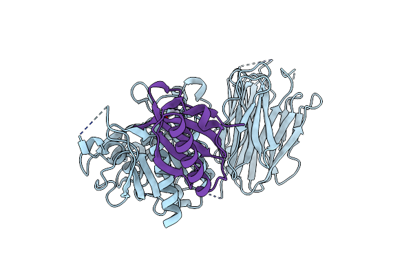
Structure Of Human Ketohexokinase Complexed With Hits From Fragment Screening
Organism: Homo sapiens
Method: X-RAY DIFFRACTION Resolution:2.40 Å Release Date: 2017-09-13 Classification: TRANSFERASE Ligands: A4J, SO4, CIT |
Organism: Homo sapiens
Method: X-RAY DIFFRACTION
Release Date: 2017-09-13
Ligands: A4J, SO4, CIT
 |
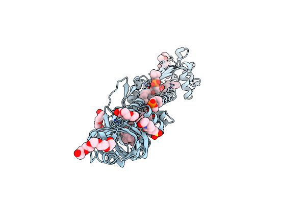
Crystal Structure Of Pcsk9
Organism: Homo sapiens
Method: X-RAY DIFFRACTION Resolution:1.98 Å Release Date: 2007-04-10 Classification: HYDROLASE Ligands: HG |
Organism: Homo sapiens
Method: X-RAY DIFFRACTION
Release Date: 2007-04-10
Ligands: HG
 |
Crystal Structure Of Cholesteryl Ester Transfer Protein
Organism: Homo sapiens
Method: X-RAY DIFFRACTION Resolution:2.10 Å Release Date: 2007-01-23 Classification: LIPID TRANSPORT Ligands: CL, 2OB, PCW, EPE, 1PE, PG4 |
Organism: Homo sapiens
Method: X-RAY DIFFRACTION
Release Date: 2007-01-23
Ligands: CL, 2OB, PCW, EPE, 1PE, PG4
 |
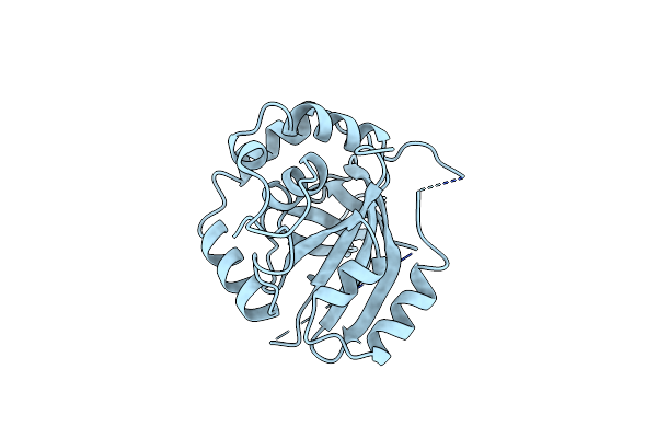
Structure Of Varicella-Zoster Virus Protease
Organism: Human herpesvirus 3
Method: X-RAY DIFFRACTION Resolution:3.00 Å Release Date: 1998-09-16 Classification: SERINE PROTEASE |
Organism: Human herpesvirus 3
Method: X-RAY DIFFRACTION
Release Date: 1998-09-16
 |
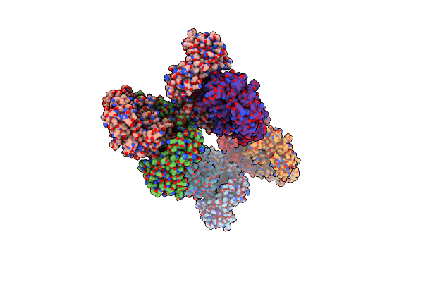
Crystal Structure Of Cytomegalovirus Protease
Organism: Cytomegalovirus
Method: X-RAY DIFFRACTION Resolution:2.50 Å Release Date: 1997-09-26 Classification: SERINE PROTEASE |
Organism: Cytomegalovirus
Method: X-RAY DIFFRACTION
Release Date: 1997-09-26
 |
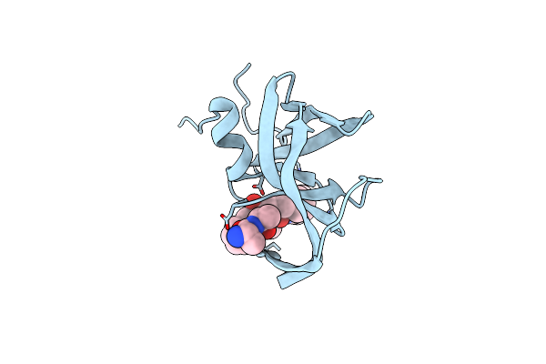
The Structure Of Unliganded Reverse Transcriptase From The Human Immunodeficiency Virus Type 1
Organism: Human immunodeficiency virus 1
Method: X-RAY DIFFRACTION Resolution:3.20 Å Release Date: 1995-03-31 Classification: NUCLEOTIDYLTRANSFERASE Ligands: MG |
Organism: Human immunodeficiency virus 1
Method: X-RAY DIFFRACTION
Release Date: 1995-03-31
Ligands: MG
 |
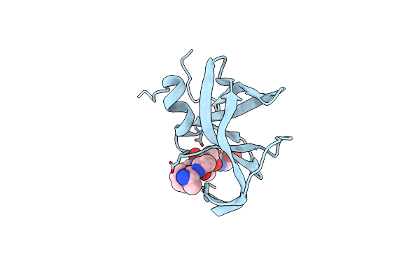
The Crystal Structures At 2.2 Angstroms Resolution Of Hydroxyethylene-Based Inhibitors Bound To Human Immunodeficiency Virus Type 1 Protease Show That The Inhibitors Are Present In Two Distinct Orientations
Organism: Human immunodeficiency virus 1
Method: X-RAY DIFFRACTION Resolution:2.20 Å Release Date: 1994-05-31 Classification: HYDROLASE/HYDROLASE INHIBITOR |
Organism: Human immunodeficiency virus 1
Method: X-RAY DIFFRACTION
Release Date: 1994-05-31
 |
The Crystal Structures At 2.2 Angstroms Resolution Of Hydroxyethylene-Based Inhibitors Bound To Human Immunodeficiency Virus Type 1 Protease Show That The Inhibitors Are Present In Two Distinct Orientations
Organism: Human immunodeficiency virus 1
Method: X-RAY DIFFRACTION Resolution:2.20 Å Release Date: 1994-05-31 Classification: HYDROLASE/HYDROLASE INHIBITOR Ligands: PSI |
Organism: Human immunodeficiency virus 1
Method: X-RAY DIFFRACTION
Release Date: 1994-05-31
Ligands: PSI
 |
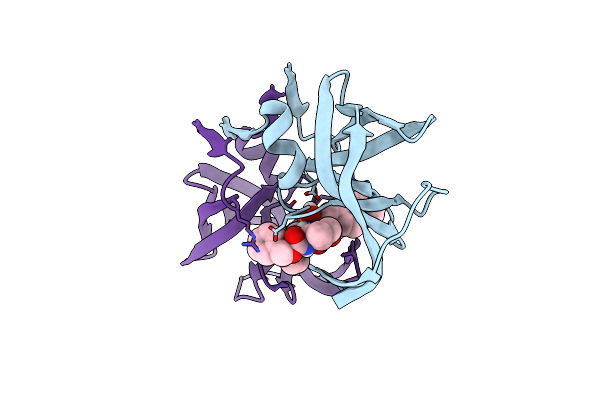
Three-Dimensional Structure Of A Siv Protease(Slash)Inhibitor Complex. Implications For The Design Of Hiv-1 And Hiv-2 Protease Inhibitors
Organism: Simian immunodeficiency virus
Method: X-RAY DIFFRACTION Resolution:2.50 Å Release Date: 1994-01-31 Classification: HYDROLASE/HYDROLASE INHIBITOR Ligands: PSI |
Organism: Simian immunodeficiency virus
Method: X-RAY DIFFRACTION
Release Date: 1994-01-31
Ligands: PSI
 |
Inhibition Of Human Immunodeficiency Virus-1 Protease By A C2-Symmetric Phosphinate Synthesis And Crystallographic Analysis
Organism: Human immunodeficiency virus 1
Method: X-RAY DIFFRACTION Resolution:2.30 Å Release Date: 1993-10-31 Classification: HYDROLASE(ACID PROTEINASE) Ligands: PHP |
Organism: Human immunodeficiency virus 1
Method: X-RAY DIFFRACTION
Release Date: 1993-10-31
Ligands: PHP