
Search Count: 135
 |
Organism: Streptococcus pneumoniae, Homo sapiens
Method: ELECTRON MICROSCOPY Release Date: 2026-06-17 Classification: ANTIMICROBIAL PROTEIN Ligands: ZN |
 |
Organism: Homo sapiens
Method: ELECTRON MICROSCOPY Resolution:3.47 Å Release Date: 2026-05-06 Classification: MEMBRANE PROTEIN Ligands: PT5, CA |
 |
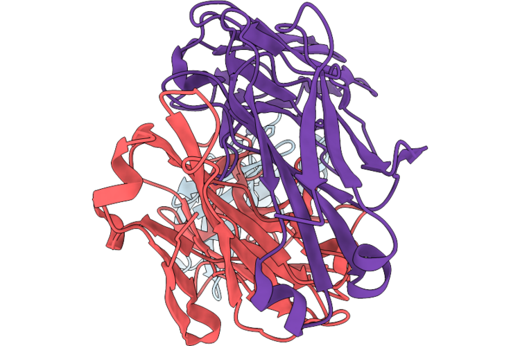
Organism: Homo sapiens, Human immunodeficiency virus 1
Method: ELECTRON MICROSCOPY Release Date: 2026-04-22 Classification: VIRAL PROTEIN/IMMUNE SYSTEM Ligands: NAG |
 |
Organism: Homo sapiens
Method: X-RAY DIFFRACTION Resolution:2.50 Å Release Date: 2026-04-15 Classification: DNA BINDING PROTEIN/DNA Ligands: GOL, PO4 |
 |
Organism: Homo sapiens
Method: X-RAY DIFFRACTION Resolution:2.70 Å Release Date: 2026-04-08 Classification: LIGASE Ligands: A1CTS |
 |
Organism: Homo sapiens
Method: X-RAY DIFFRACTION Resolution:1.27 Å Release Date: 2026-04-08 Classification: LIGASE Ligands: A1CTR |
 |
Organism: Homo sapiens
Method: X-RAY DIFFRACTION Resolution:1.27 Å Release Date: 2026-04-08 Classification: LIGASE Ligands: A1CTQ |
 |
Organism: Homo sapiens
Method: X-RAY DIFFRACTION Resolution:1.33 Å Release Date: 2026-04-08 Classification: LIGASE Ligands: A1CTE |
 |
Organism: Homo sapiens
Method: X-RAY DIFFRACTION Resolution:1.31 Å Release Date: 2026-04-08 Classification: LIGASE Ligands: A1CTL |
 |
Organism: Homo sapiens
Method: X-RAY DIFFRACTION Resolution:1.29 Å Release Date: 2026-04-08 Classification: LIGASE Ligands: A1CTM |
 |
Organism: Homo sapiens
Method: X-RAY DIFFRACTION Resolution:1.20 Å Release Date: 2026-04-08 Classification: LIGASE Ligands: A1CTO, GOL |
 |
Organism: Homo sapiens
Method: X-RAY DIFFRACTION Resolution:1.33 Å Release Date: 2026-04-08 Classification: LIGASE Ligands: A1CTP |
 |
Organism: Homo sapiens
Method: X-RAY DIFFRACTION Resolution:1.29 Å Release Date: 2026-04-08 Classification: LIGASE Ligands: A1CTN |
 |
Organism: Homo sapiens
Method: X-RAY DIFFRACTION Resolution:1.40 Å Release Date: 2026-04-08 Classification: LIGASE Ligands: A1CTJ |
 |
Organism: Homo sapiens
Method: X-RAY DIFFRACTION Resolution:1.01 Å Release Date: 2026-04-08 Classification: LIGASE Ligands: A1CTK |
 |
Organism: Homo sapiens
Method: X-RAY DIFFRACTION Resolution:1.27 Å Release Date: 2026-04-08 Classification: LIGASE Ligands: A1CTF |
 |
The Crystal Structure Of Human Dead-Box Rna Helicase Ddx28 Reca1 Domain In Complex With Adp
Organism: Homo sapiens
Method: X-RAY DIFFRACTION Resolution:2.60 Å Release Date: 2026-03-04 Classification: RIBOSOMAL PROTEIN Ligands: PO4, ADP |
 |
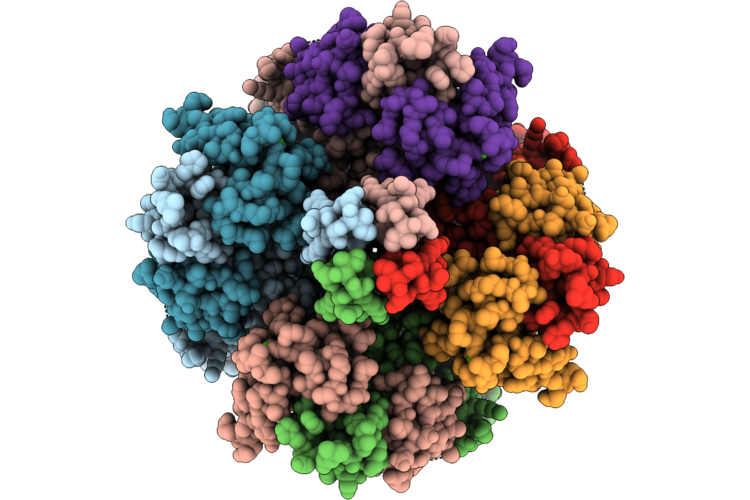
Sub-Particle Structure Of The Iterative Acetyltransferase From Actinomycetes In Complex With Accoa And Monoacetylated Lasso Peptides
Organism: Actinosynnema mirum dsm 43827
Method: ELECTRON MICROSCOPY Resolution:3.43 Å Release Date: 2026-02-25 Classification: TRANSFERASE Ligands: ACO |
 |
Organism: Macaca mulatta, Human immunodeficiency virus 1
Method: ELECTRON MICROSCOPY Release Date: 2026-02-11 Classification: VIRAL PROTEIN Ligands: NAG |
 |
Organism: Homo sapiens, Human immunodeficiency virus 1
Method: ELECTRON MICROSCOPY Release Date: 2026-02-04 Classification: VIRAL PROTEIN Ligands: NAG |