

Search Count: 171
 |
Bacterial Protein - Dp_3-4
Organism: Escherichia coli
Method: X-RAY DIFFRACTION Resolution:2.00 Å Release Date: 2026-08-19 Classification: TRANSFERASE Ligands: PO4 |
Organism: Escherichia coli
Method: X-RAY DIFFRACTION
Release Date: 2026-08-19
Ligands: PO4
 |
Cjman26C Bound To Covalent Beta-Mannanase Inhibitor
Organism: Cellvibrio japonicus ueda107
Method: X-RAY DIFFRACTION Resolution:1.20 Å Release Date: 2026-07-22 Classification: HYDROLASE Ligands: BMA, YLL, NA |
Organism: Cellvibrio japonicus ueda107
Method: X-RAY DIFFRACTION
Release Date: 2026-07-22
Ligands: BMA, YLL, NA
 |
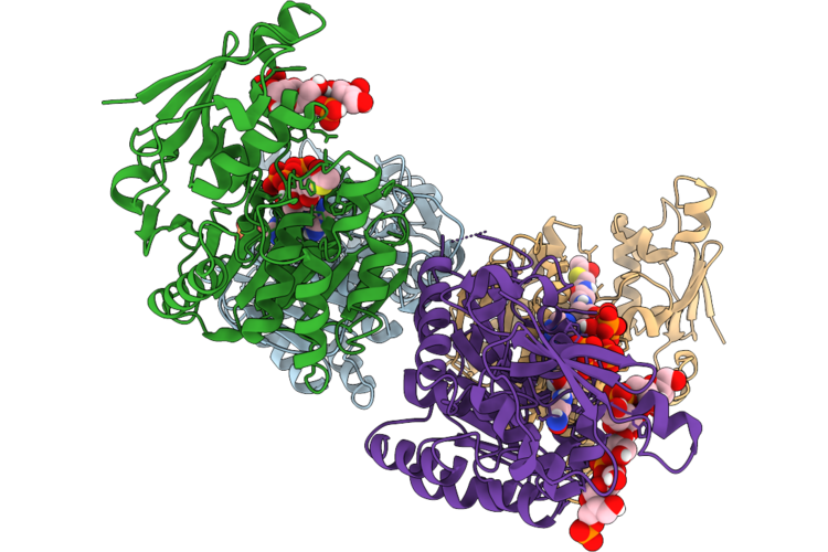
A. Niger Mana In Covalent Complex With Pseudotrisaccharide Inhibitor
Organism: Aspergillus niger
Method: X-RAY DIFFRACTION Resolution:1.42 Å Release Date: 2026-07-22 Classification: HYDROLASE Ligands: SO4, YLL |
Organism: Aspergillus niger
Method: X-RAY DIFFRACTION
Release Date: 2026-07-22
Ligands: SO4, YLL
 |
Bacterial Protein - 4_5
Organism: Actinobacillus pleuropneumoniae serovar 11 str. 56153
Method: X-RAY DIFFRACTION Resolution:2.15 Å Release Date: 2026-07-15 Classification: TRANSFERASE Ligands: ACO, PO4 |
Organism: Actinobacillus pleuropneumoniae serovar 11 str. 56153
Method: X-RAY DIFFRACTION
Release Date: 2026-07-15
Ligands: ACO, PO4
 |
Bacterial Protein - Unliganded
Organism: Actinobacillus pleuropneumoniae serovar 11 str. 56153
Method: X-RAY DIFFRACTION Resolution:1.85 Å Release Date: 2026-07-15 Classification: TRANSFERASE Ligands: AU, PO4 |
Organism: Actinobacillus pleuropneumoniae serovar 11 str. 56153
Method: X-RAY DIFFRACTION
Release Date: 2026-07-15
Ligands: AU, PO4
 |
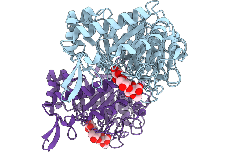
Bacterial Protein - Acoa
Organism: Actinobacillus pleuropneumoniae serovar 11 str. 56153
Method: X-RAY DIFFRACTION Resolution:2.50 Å Release Date: 2026-07-15 Classification: TRANSFERASE Ligands: ACO, COA, PO4 |
Organism: Actinobacillus pleuropneumoniae serovar 11 str. 56153
Method: X-RAY DIFFRACTION
Release Date: 2026-07-15
Ligands: ACO, COA, PO4
 |
Glucuronoxylan-Specific Gh30_8 Family Xylanase Ctxyn30A From Clostridium Thermocellum Complex With Glucuronic Acid Epoxide Inhibitor
Organism: Acetivibrio thermocellus dsm 2360
Method: X-RAY DIFFRACTION Resolution:1.50 Å Release Date: 2026-06-24 Classification: HYDROLASE Ligands: HQ8 |
Organism: Acetivibrio thermocellus dsm 2360
Method: X-RAY DIFFRACTION
Release Date: 2026-06-24
Ligands: HQ8
 |
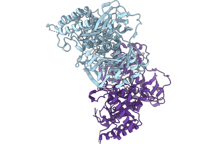
Gh10 Family Xylanase Xyna From Bacillus Sp. Kw1 Complex With Xylobiosyl-Configured Cyclophellitol Probe Bearing An Alpha-1,3 - Araf Decoration
Organism: Bacillus sp. (in: firmicutes)
Method: X-RAY DIFFRACTION Resolution:2.02 Å Release Date: 2026-06-24 Classification: HYDROLASE Ligands: A1J01 |
Organism: Bacillus sp. (in: firmicutes)
Method: X-RAY DIFFRACTION
Release Date: 2026-06-24
Ligands: A1J01
 |
Pel Polysaccharide Deacetylase Pela From Bacillus Cereus Atcc 10987
Organism: Bacillus cereus atcc 10987
Method: X-RAY DIFFRACTION Resolution:2.51 Å Release Date: 2026-05-20 Classification: MEMBRANE PROTEIN Ligands: ZN |
Organism: Bacillus cereus atcc 10987
Method: X-RAY DIFFRACTION
Release Date: 2026-05-20
Ligands: ZN
 |
Influenza Neuraminidase In Complex With N-Acyl Oseltamivir Inhibitor
Organism: Influenza a virus
Method: ELECTRON MICROSCOPY Release Date: 2025-12-17 Classification: VIRAL PROTEIN Ligands: A1IVV, NAG, CA |
Organism: Influenza a virus
Method: ELECTRON MICROSCOPY
Release Date: 2025-12-17
Ligands: A1IVV, NAG, CA
 |
Influenza Neuraminidase In Complex With N-Acyl Oseltamivir Inhibitor
Organism: Influenza a virus
Method: ELECTRON MICROSCOPY Release Date: 2025-12-17 Classification: VIRAL PROTEIN Ligands: A1IVW, NAG, CA |
Organism: Influenza a virus
Method: ELECTRON MICROSCOPY
Release Date: 2025-12-17
Ligands: A1IVW, NAG, CA
 |
Influenza Neuraminidase In Complex With N-Acyl Oseltamivir Inhibitor
Organism: Influenza a virus
Method: ELECTRON MICROSCOPY Release Date: 2025-12-17 Classification: VIRAL PROTEIN Ligands: A1IVX, NAG, CA |
Organism: Influenza a virus
Method: ELECTRON MICROSCOPY
Release Date: 2025-12-17
Ligands: A1IVX, NAG, CA
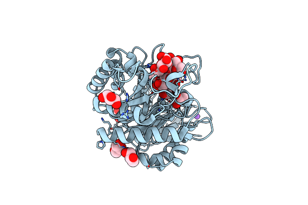 |
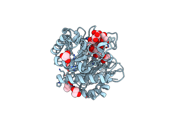
Structure Of Pslg With A Covalently- Bound Pentasaccharide
Organism: Pseudomonas aeruginosa
Method: X-RAY DIFFRACTION Resolution:1.55 Å Release Date: 2025-03-05 Classification: HYDROLASE Ligands: CL, NA, GOL, YLL |
Organism: Pseudomonas aeruginosa
Method: X-RAY DIFFRACTION
Release Date: 2025-03-05
Ligands: CL, NA, GOL, YLL
 |
Structure Of Pslg With An Iminosugar Inhibitor
Organism: Pseudomonas aeruginosa
Method: X-RAY DIFFRACTION Resolution:1.50 Å Release Date: 2025-03-05 Classification: HYDROLASE Ligands: GOL, CL |
Organism: Pseudomonas aeruginosa
Method: X-RAY DIFFRACTION
Release Date: 2025-03-05
Ligands: GOL, CL
 |
Crystal Structure Of The L. Monocytogenes Rmlt In Complex With 3-Rbop-(Ch2)6Nh2
Organism: Listeria monocytogenes
Method: X-RAY DIFFRACTION Resolution:2.34 Å Release Date: 2024-12-18 Classification: TRANSFERASE Ligands: A1IRZ, MG, GOL |
Organism: Listeria monocytogenes
Method: X-RAY DIFFRACTION
Release Date: 2024-12-18
Ligands: A1IRZ, MG, GOL
 |
Crystal Structure Of The L. Monocytogenes Rmlt In Complex With 4-Rbop-(Ch2)6Nh2
Organism: Listeria monocytogenes
Method: X-RAY DIFFRACTION Resolution:2.52 Å Release Date: 2024-12-18 Classification: TRANSFERASE Ligands: A1IR6, MG |
Organism: Listeria monocytogenes
Method: X-RAY DIFFRACTION
Release Date: 2024-12-18
Ligands: A1IR6, MG
 |
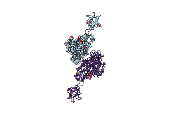
Crystal Structure Of Susa Amylase From Bacteroides Thetaiotaomicron Covalently Bound To Alpha-1,6 Branched Pseudo-Trisaccharide Activity-Based Probe
Organism: Bacteroides thetaiotaomicron
Method: X-RAY DIFFRACTION Resolution:2.43 Å Release Date: 2024-12-11 Classification: HYDROLASE Ligands: PBW, OC9, IMD, CA |
Organism: Bacteroides thetaiotaomicron
Method: X-RAY DIFFRACTION
Release Date: 2024-12-11
Ligands: PBW, OC9, IMD, CA
 |
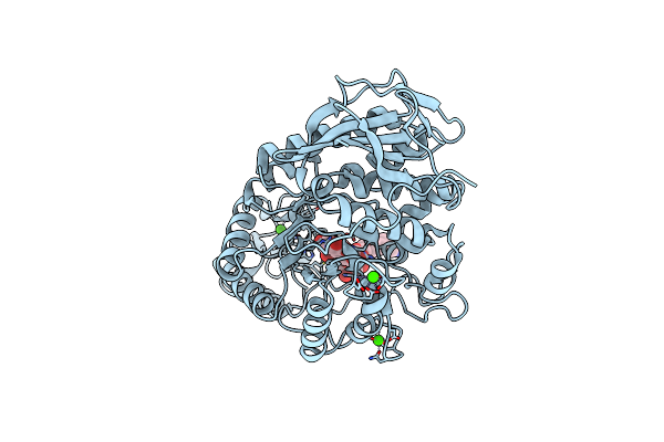
Crystal Structure Of Susg From Bacteroides Thetaiotaomicron Covalently Bound To Alpha-1,6 Branched Pseudo-Trisaccharide Activity-Based Probe
Organism: Bacteroides thetaiotaomicron
Method: X-RAY DIFFRACTION Resolution:2.65 Å Release Date: 2024-12-11 Classification: HYDROLASE Ligands: PBW, OC9, A1ILG, CA, IMD, ACT |
Organism: Bacteroides thetaiotaomicron
Method: X-RAY DIFFRACTION
Release Date: 2024-12-11
Ligands: PBW, OC9, A1ILG, CA, IMD, ACT
 |
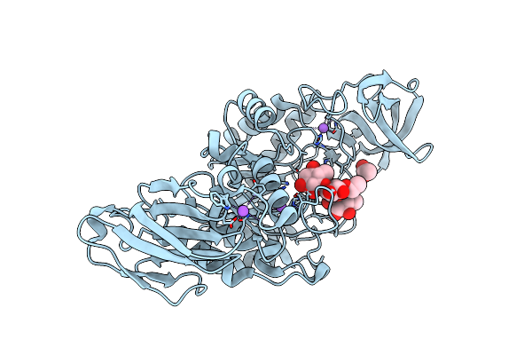
Crystal Structure Of Amylase 5 (Amy5) From Ruminococcus Bromii Covalently Bound To Alpha-1,6 Branched Pseudo-Trisaccharide Activity-Based Probe
Organism: Ruminococcus bromii
Method: X-RAY DIFFRACTION Resolution:1.40 Å Release Date: 2024-12-11 Classification: HYDROLASE Ligands: PBW, A1IHI, CA |
Organism: Ruminococcus bromii
Method: X-RAY DIFFRACTION
Release Date: 2024-12-11
Ligands: PBW, A1IHI, CA
 |
Crystal Structure Of K38 Amylase From Bacillus Sp. Strain Ksm-K38 Covalently Bound To Alpha-1,6 Branched Pseudo-Trisaccharide Activity-Based Probe
Organism: Bacillus sp. ksm-k38
Method: X-RAY DIFFRACTION Resolution:2.02 Å Release Date: 2024-12-11 Classification: HYDROLASE Ligands: PBW, OC9, ACT, NA |
Organism: Bacillus sp. ksm-k38
Method: X-RAY DIFFRACTION
Release Date: 2024-12-11
Ligands: PBW, OC9, ACT, NA