

Search Count: 20,651
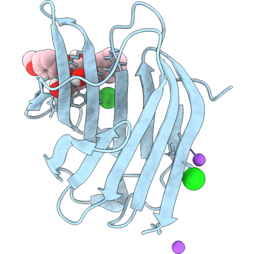 |
Crystal Structure Of The Human Galectin-3 Carbohydrate-Recognition Domain In Complex With P-Methoxyphenyl 3-O-Benzyl-Beta-D-Galactopyranoside
Organism: Homo sapiens
Method: X-RAY DIFFRACTION Resolution:1.10 Å Release Date: 2026-09-16 Classification: SUGAR BINDING PROTEIN Ligands: A1JZ9, CL, NA |
Organism: Homo sapiens
Method: X-RAY DIFFRACTION
Release Date: 2026-09-16
Ligands: A1JZ9, CL, NA
 |
Crystal Structure Of The Human Galectin-3 Carbohydrate-Recognition Domain In Complex With P-Methoxyphenyl 3-O-Benzyl-Beta-D-Galactopyranoside
Organism: Homo sapiens
Method: X-RAY DIFFRACTION Resolution:1.10 Å Release Date: 2026-09-16 Classification: SUGAR BINDING PROTEIN Ligands: A1J0D, CL, NA |
Organism: Homo sapiens
Method: X-RAY DIFFRACTION
Release Date: 2026-09-16
Ligands: A1J0D, CL, NA
 |
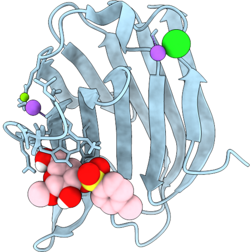
Crystal Structure Of The Human Galectin-3 Carbohydrate-Recognition Domain In Complex With P-Methoxyphenyl 3-O-Benzonyl-Beta-D-Galactopyranoside
Organism: Homo sapiens
Method: X-RAY DIFFRACTION Resolution:1.13 Å Release Date: 2026-09-16 Classification: SUGAR BINDING PROTEIN Ligands: A1J0B, CL, NA, MG |
Organism: Homo sapiens
Method: X-RAY DIFFRACTION
Release Date: 2026-09-16
Ligands: A1J0B, CL, NA, MG
 |
Crystal Structure Of The Human Galectin-3 Carbohydrate-Recognition Domain In Complex With Methyl 3-O-Toluensulfonyl-Beta-D-Galactopyranoside
Organism: Homo sapiens
Method: X-RAY DIFFRACTION Resolution:1.13 Å Release Date: 2026-09-16 Classification: SUGAR BINDING PROTEIN Ligands: A1J0A, CL, MG, NA |
Organism: Homo sapiens
Method: X-RAY DIFFRACTION
Release Date: 2026-09-16
Ligands: A1J0A, CL, MG, NA
 |
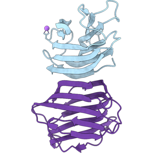
Crystal Structure Of The Human Galectin-1 In Complex With P-Methoxyphenyl 3-O-Benzyl-Beta-D-Galactopyranoside
Organism: Homo sapiens
Method: X-RAY DIFFRACTION Resolution:2.00 Å Release Date: 2026-09-16 Classification: SUGAR BINDING PROTEIN Ligands: CL |
Organism: Homo sapiens
Method: X-RAY DIFFRACTION
Release Date: 2026-09-16
Ligands: CL
 |
Crystal Structure Of The Human Galectin-1 In Complex With P-Methoxyphenyl 3-O-Toluensulfonyl-Beta-D- Galactopyranoside
Organism: Homo sapiens
Method: X-RAY DIFFRACTION Resolution:1.73 Å Release Date: 2026-09-16 Classification: SUGAR BINDING PROTEIN Ligands: CL, NA |
Organism: Homo sapiens
Method: X-RAY DIFFRACTION
Release Date: 2026-09-16
Ligands: CL, NA
 |
Crystal Structure Of The Carbohydrate-Recognition Domain Of R144S Galectin-3 Mutant In Apo Form
Organism: Homo sapiens
Method: X-RAY DIFFRACTION Resolution:1.20 Å Release Date: 2026-09-16 Classification: SUGAR BINDING PROTEIN Ligands: TRS, CL, NA |
Organism: Homo sapiens
Method: X-RAY DIFFRACTION
Release Date: 2026-09-16
Ligands: TRS, CL, NA
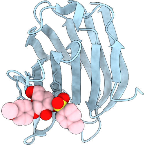 |
Crystal Structure Of The Carbohydrate-Recognition Domain Of R144S Galectin-3 Mutant In Complex With P-Methoxyphenyl 3-O-Toluensulfonyl-Beta-D- Galactopyranoside
Organism: Homo sapiens
Method: X-RAY DIFFRACTION Resolution:1.30 Å Release Date: 2026-09-16 Classification: SUGAR BINDING PROTEIN Ligands: CL, A1J0D |
Organism: Homo sapiens
Method: X-RAY DIFFRACTION
Release Date: 2026-09-16
Ligands: CL, A1J0D
 |
Crystal Structure Of The Carbohydrate-Recognition Domain Of R144S Galectin-3 Mutant In Complex With Methyl 3-O-Toluensulfonyl-Beta-D-Galactopyranoside
Organism: Homo sapiens
Method: X-RAY DIFFRACTION Resolution:1.40 Å Release Date: 2026-09-16 Classification: SUGAR BINDING PROTEIN Ligands: A1J0A, CL |
Organism: Homo sapiens
Method: X-RAY DIFFRACTION
Release Date: 2026-09-16
Ligands: A1J0A, CL
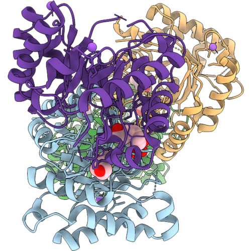 |
Crystal Structure Of L-Erythrulose-1-Phosphate Isomerase From Brucella Melitensis In Complex With Ethyl Dihydrogen Phosphate
Organism: Brucella abortus 2308
Method: X-RAY DIFFRACTION Resolution:1.50 Å Release Date: 2026-09-16 Classification: ISOMERASE Ligands: EFS, HEZ, NA, CL, GOL |
Organism: Brucella abortus 2308
Method: X-RAY DIFFRACTION
Release Date: 2026-09-16
Ligands: EFS, HEZ, NA, CL, GOL
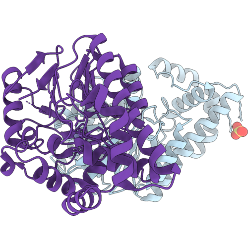 |
Crystal Structure Of A Beta-Lactamase From Burkholderia Ambifaria
Organism: Burkholderia ambifaria mc40-6
Method: X-RAY DIFFRACTION Resolution:3.00 Å Release Date: 2026-09-16 Classification: TRANSFERASE Ligands: CL, SO4, ZN |
Organism: Burkholderia ambifaria mc40-6
Method: X-RAY DIFFRACTION
Release Date: 2026-09-16
Ligands: CL, SO4, ZN
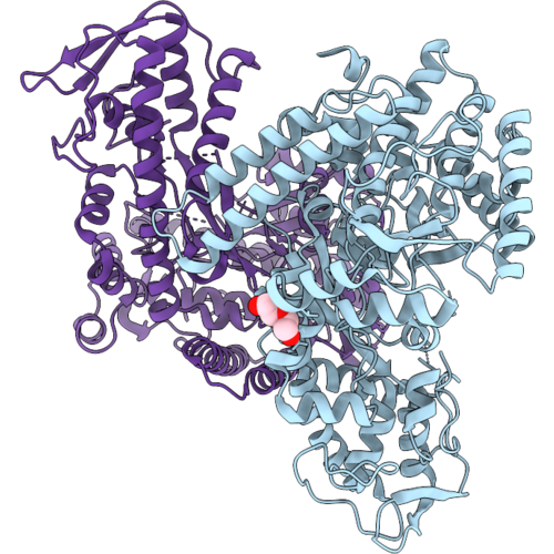 |
Crystal Structure Of A Formate Acetyltransferase (Pfl) From Amygdalobacter Indicium
Organism: Amygdalobacter indicium
Method: X-RAY DIFFRACTION Resolution:2.29 Å Release Date: 2026-09-16 Classification: TRANSFERASE Ligands: CL, PEG |
Organism: Amygdalobacter indicium
Method: X-RAY DIFFRACTION
Release Date: 2026-09-16
Ligands: CL, PEG
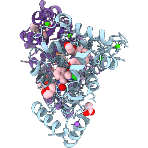 |
Ncs-1 Bound To Xchem Ligand 16
Organism: Homo sapiens
Method: X-RAY DIFFRACTION Resolution:2.26 Å Release Date: 2026-09-16 Classification: METAL BINDING PROTEIN Ligands: CA, P6G, NA, ACT, 1PE, A1I7U, CL |
Organism: Homo sapiens
Method: X-RAY DIFFRACTION
Release Date: 2026-09-16
Ligands: CA, P6G, NA, ACT, 1PE, A1I7U, CL
 |
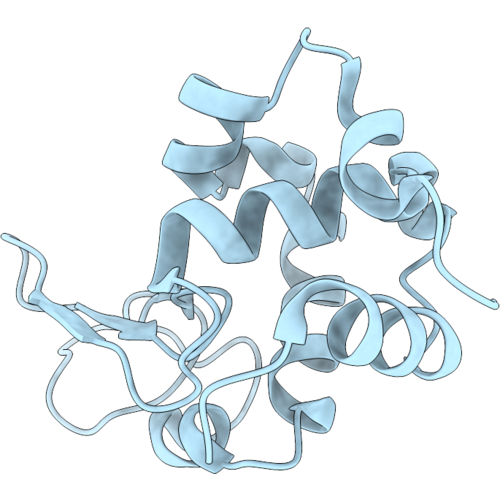
Acoustofluidic Serial Crystallography - On, Same Number Of Indexed Crystals As Off
Organism: Gallus gallus
Method: X-RAY DIFFRACTION Resolution:1.96 Å Release Date: 2026-09-16 Classification: HYDROLASE Ligands: CL |
Organism: Gallus gallus
Method: X-RAY DIFFRACTION
Release Date: 2026-09-16
Ligands: CL
 |
Acoustofluidic Serial Crystallography - Off, No Acoustic Focusing
Organism: Gallus gallus
Method: X-RAY DIFFRACTION Resolution:1.96 Å Release Date: 2026-09-16 Classification: HYDROLASE Ligands: CL |
Organism: Gallus gallus
Method: X-RAY DIFFRACTION
Release Date: 2026-09-16
Ligands: CL
 |
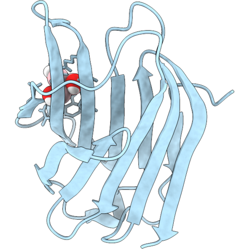
Oxa-48: Q5 Mutant In An Acyl Enzyme Complex With Piperacillin
Organism: Klebsiella pneumoniae
Method: X-RAY DIFFRACTION Resolution:1.50 Å Release Date: 2026-09-16 Classification: ANTIBIOTIC Ligands: EDO, JPP, YPP, CL |
Organism: Klebsiella pneumoniae
Method: X-RAY DIFFRACTION
Release Date: 2026-09-16
Ligands: EDO, JPP, YPP, CL
 |
Crystal Structure Of The Human Galectin-3 Carbohydrate-Recognition Domain In The Apo Form
Organism: Homo sapiens
Method: X-RAY DIFFRACTION Resolution:1.10 Å Release Date: 2026-09-16 Classification: SUGAR BINDING PROTEIN Ligands: TRS, CL |
Organism: Homo sapiens
Method: X-RAY DIFFRACTION
Release Date: 2026-09-16
Ligands: TRS, CL
 |
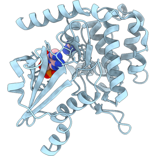
Crystal Structure Of The Human Galectin-3 Carbohydrate-Recognition Domain In Complex With Sedg-Bn
Organism: Homo sapiens
Method: X-RAY DIFFRACTION Resolution:1.20 Å Release Date: 2026-09-16 Classification: SUGAR BINDING PROTEIN Ligands: A1J1N, CL |
Organism: Homo sapiens
Method: X-RAY DIFFRACTION
Release Date: 2026-09-16
Ligands: A1J1N, CL
 |
Crystal Structure Of Nucleotide-Bound Pak2 Kinase Domain Containing K278R Mutant
Organism: Homo sapiens
Method: X-RAY DIFFRACTION Resolution:1.90 Å Release Date: 2026-09-16 Classification: TRANSFERASE Ligands: ANP, GAI, CL |
Organism: Homo sapiens
Method: X-RAY DIFFRACTION
Release Date: 2026-09-16
Ligands: ANP, GAI, CL
 |
Crystal Structure Of Phosphorylated Pak2 Kinase Domain Containing K278R Mutant
Organism: Homo sapiens
Method: X-RAY DIFFRACTION Resolution:1.85 Å Release Date: 2026-09-16 Classification: TRANSFERASE Ligands: GAI, CL |
Organism: Homo sapiens
Method: X-RAY DIFFRACTION
Release Date: 2026-09-16
Ligands: GAI, CL