

Search Count: 884
 |
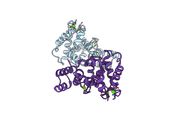
Structure Of A Mitochondrial Calcium Uniporter Holocomplex (Micu1, Micu2, Mcu, Emre) In Low Ca2+
Organism: Tribolium castaneum, Homo sapiens
Method: ELECTRON MICROSCOPY Release Date: 2020-07-29 Classification: TRANSPORT PROTEIN/CALCIUM BINDING PROTEIN Ligands: CA |
Organism: Tribolium castaneum, Homo sapiens
Method: ELECTRON MICROSCOPY
Release Date: 2020-07-29
Ligands: CA
 |
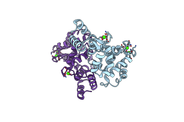
Crystal Structure Of A190T Mutant Of Human Hippocalcin At 2.5 A Resolution
Organism: Homo sapiens
Method: X-RAY DIFFRACTION Resolution:2.54 Å Release Date: 2017-05-03 Classification: CALCIUM BINDING PROTEIN Ligands: CA |
Organism: Homo sapiens
Method: X-RAY DIFFRACTION
Release Date: 2017-05-03
Ligands: CA
 |
Crystal Structure Of Human Hippocalcin At 2.4 A Resolution
Organism: Homo sapiens
Method: X-RAY DIFFRACTION Resolution:2.42 Å Release Date: 2017-05-03 Classification: CALCIUM BINDING PROTEIN Ligands: CA |
Organism: Homo sapiens
Method: X-RAY DIFFRACTION
Release Date: 2017-05-03
Ligands: CA
 |
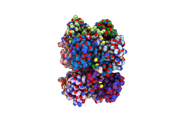
Structure Of The Human Micu1-Micu2 Heterodimer, Calcium Bound, In Association With A Lipid Nanodisc
Organism: Homo sapiens
Method: ELECTRON MICROSCOPY Release Date: 2020-07-29 Classification: CALCIUM BINDING PROTEIN Ligands: CA |
Organism: Homo sapiens
Method: ELECTRON MICROSCOPY
Release Date: 2020-07-29
Ligands: CA
 |
Structure Of S100A4 With Pcp
Organism: Homo sapiens
Method: X-RAY DIFFRACTION Resolution:2.80 Å Release Date: 2010-05-12 Classification: Calcium Binding Protein Ligands: P77, CA, DIO |
Organism: Homo sapiens
Method: X-RAY DIFFRACTION
Release Date: 2010-05-12
Ligands: P77, CA, DIO
 |
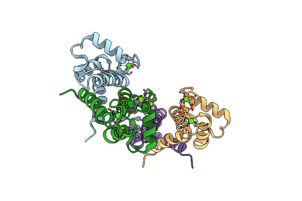
Crystal Structure Of N-Terminal Calmodulin-Like Calcium Sensor Of Human Mitochondrial Atp-Mg/Pi Carrier Scamc1
Organism: Homo sapiens
Method: X-RAY DIFFRACTION Resolution:2.10 Å Release Date: 2013-12-18 Classification: CALCIUM-BINDING PROTEIN Ligands: CA, PEG |
Organism: Homo sapiens
Method: X-RAY DIFFRACTION
Release Date: 2013-12-18
Ligands: CA, PEG
 |
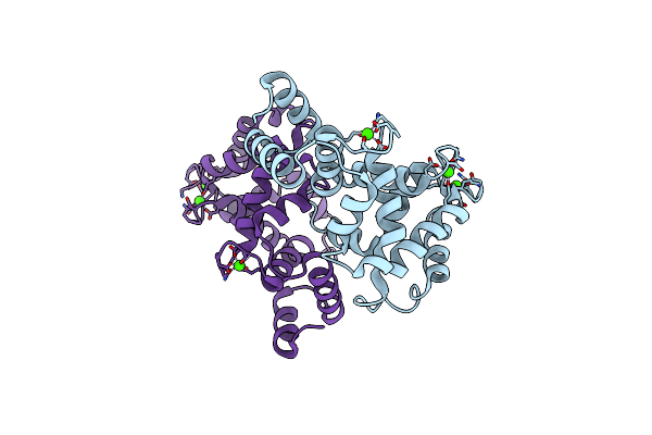
The 1.5 A Crystal Structure Of Ca2+-Bound S100A4
Organism: Homo sapiens
Method: X-RAY DIFFRACTION Resolution:1.50 Å Release Date: 2008-04-01 Classification: CALCIUM BINDING PROTEIN Ligands: CA |
Organism: Homo sapiens
Method: X-RAY DIFFRACTION
Release Date: 2008-04-01
Ligands: CA
 |
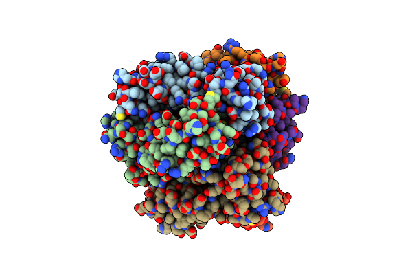
Crystal Structure Of T71N Mutant Of Human Hippocalcin
Organism: Homo sapiens
Method: X-RAY DIFFRACTION Resolution:3.00 Å Release Date: 2017-04-12 Classification: CALCIUM BINDING PROTEIN Ligands: CA |
Organism: Homo sapiens
Method: X-RAY DIFFRACTION
Release Date: 2017-04-12
Ligands: CA
 |
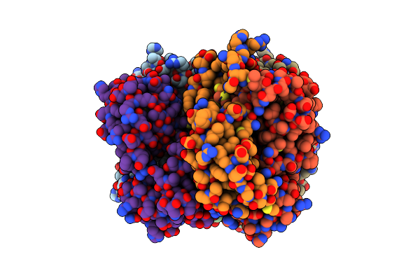
Crystal Structure Of The Calcium Sensor Calcium-Binding Protein 1 (Cabp1)
Organism: Homo sapiens
Method: X-RAY DIFFRACTION Resolution:2.90 Å Release Date: 2010-12-22 Classification: Calcium binding protein Ligands: CA |
Organism: Homo sapiens
Method: X-RAY DIFFRACTION
Release Date: 2010-12-22
Ligands: CA
 |
Crystal Structure Of The Calcium Sensor Calcium-Binding Protein 1 (Cabp1)
Organism: Homo sapiens
Method: X-RAY DIFFRACTION Resolution:2.40 Å Release Date: 2010-12-22 Classification: calcium binding protein Ligands: CA, HEZ |
Organism: Homo sapiens
Method: X-RAY DIFFRACTION
Release Date: 2010-12-22
Ligands: CA, HEZ
 |
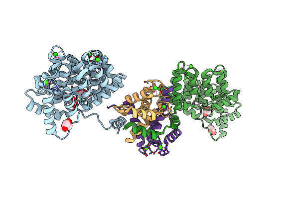
Fasciola Hepatica Calcium Binding Protein Fhcabp2: Structure Of The Dynein Light Chain-Like Domain. P41212 Mercury Derivative.
Organism: Fasciola hepatica
Method: X-RAY DIFFRACTION Resolution:2.30 Å Release Date: 2016-04-27 Classification: CALCIUM-BINDING PROTEIN Ligands: HG, CL |
Organism: Fasciola hepatica
Method: X-RAY DIFFRACTION
Release Date: 2016-04-27
Ligands: HG, CL
 |
Crystal Structure Of Annexin A2 Complexed With S100A4
Organism: Homo sapiens
Method: X-RAY DIFFRACTION Resolution:2.10 Å Release Date: 2017-07-05 Classification: calcium-binding protein Ligands: CA, GOL |
Organism: Homo sapiens
Method: X-RAY DIFFRACTION
Release Date: 2017-07-05
Ligands: CA, GOL
 |
Crystal Structure Of Cbara1 In The Ca2+ Binding Form
Organism: Homo sapiens
Method: X-RAY DIFFRACTION Resolution:2.70 Å Release Date: 2014-02-26 Classification: Calcium Binding protein Ligands: CA, CL, MPD |
Organism: Homo sapiens
Method: X-RAY DIFFRACTION
Release Date: 2014-02-26
Ligands: CA, CL, MPD
 |
The Refined Structure Of Vitamin D-Dependent Calcium-Binding Protein From Bovine Intestine. Molecular Details, Ion Binding, And Implications For The Structure Of Other Calcium-Binding Proteins
Organism: Bos taurus
Method: X-RAY DIFFRACTION Resolution:2.30 Å Release Date: 1986-10-24 Classification: CALCIUM-BINDING PROTEIN Ligands: CA, SO4 |
Organism: Bos taurus
Method: X-RAY DIFFRACTION
Release Date: 1986-10-24
Ligands: CA, SO4
 |
Crystal Structure Of Mn(Ii) Ca(Ii) Na(I) Bound Calprotectin
Organism: Homo sapiens
Method: X-RAY DIFFRACTION Resolution:1.76 Å Release Date: 2015-04-15 Classification: METAL BINDING PROTEIN Ligands: CA, NA, MN |
Organism: Homo sapiens
Method: X-RAY DIFFRACTION
Release Date: 2015-04-15
Ligands: CA, NA, MN
 |
Crystal Structure Of Metastasis-Associated Protein S100A4 In The Active, Calcium-Bound Form
Organism: Homo sapiens
Method: X-RAY DIFFRACTION Resolution:2.03 Å Release Date: 2008-09-30 Classification: METAL BINDING PROTEIN Ligands: CA |
Organism: Homo sapiens
Method: X-RAY DIFFRACTION
Release Date: 2008-09-30
Ligands: CA
 |
Murine S100A7/S100A15 In Presence Of Calcium
Organism: Mus musculus
Method: X-RAY DIFFRACTION Resolution:1.69 Å Release Date: 2024-02-07 Classification: METAL BINDING PROTEIN Ligands: GOL, CA, SCN, NA, ACT |
Organism: Mus musculus
Method: X-RAY DIFFRACTION
Release Date: 2024-02-07
Ligands: GOL, CA, SCN, NA, ACT
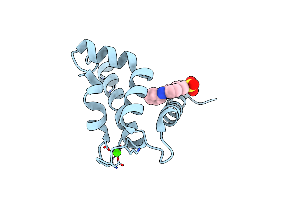 |
Structure Of Human S100A7 In Complex With 2,6 Ans
Organism: Homo sapiens
Method: X-RAY DIFFRACTION Resolution:1.70 Å Release Date: 2009-10-20 Classification: CALCIUM-BINDING PROTEIN Ligands: CA, 6AN, ZN |
Organism: Homo sapiens
Method: X-RAY DIFFRACTION
Release Date: 2009-10-20
Ligands: CA, 6AN, ZN
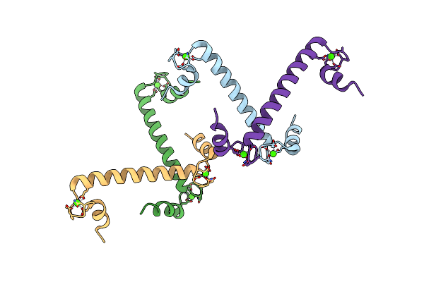 |
Crystal Structure And Trimer-Monomer Transition Of N-Terminal Domain Of Ehcabp1 From Entamoeba Histolytica
Organism: Entamoeba histolytica
Method: X-RAY DIFFRACTION Resolution:2.50 Å Release Date: 2010-02-02 Classification: METAL BINDING PROTEIN Ligands: CA |
Organism: Entamoeba histolytica
Method: X-RAY DIFFRACTION
Release Date: 2010-02-02
Ligands: CA
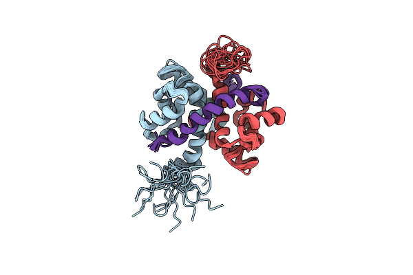 |
Solution Structure Of Ca-Bound S100A4 In Complex With Non-Muscle Myosin Iia
Organism: Homo sapiens
Method: SOLUTION NMR Release Date: 2012-04-25 Classification: CALCIUM BINDING PROTEIN |
Organism: Homo sapiens
Method: SOLUTION NMR
Release Date: 2012-04-25