Deposition Date
2008-03-05
Release Date
2008-09-30
Last Version Date
2024-02-21
Entry Detail
PDB ID:
3CGA
Keywords:
Title:
Crystal structure of metastasis-associated protein S100A4 in the active, calcium-bound form
Biological Source:
Source Organism(s):
Homo sapiens (Taxon ID: )
Expression System(s):
Method Details:
Experimental Method:
Resolution:
2.03 Å
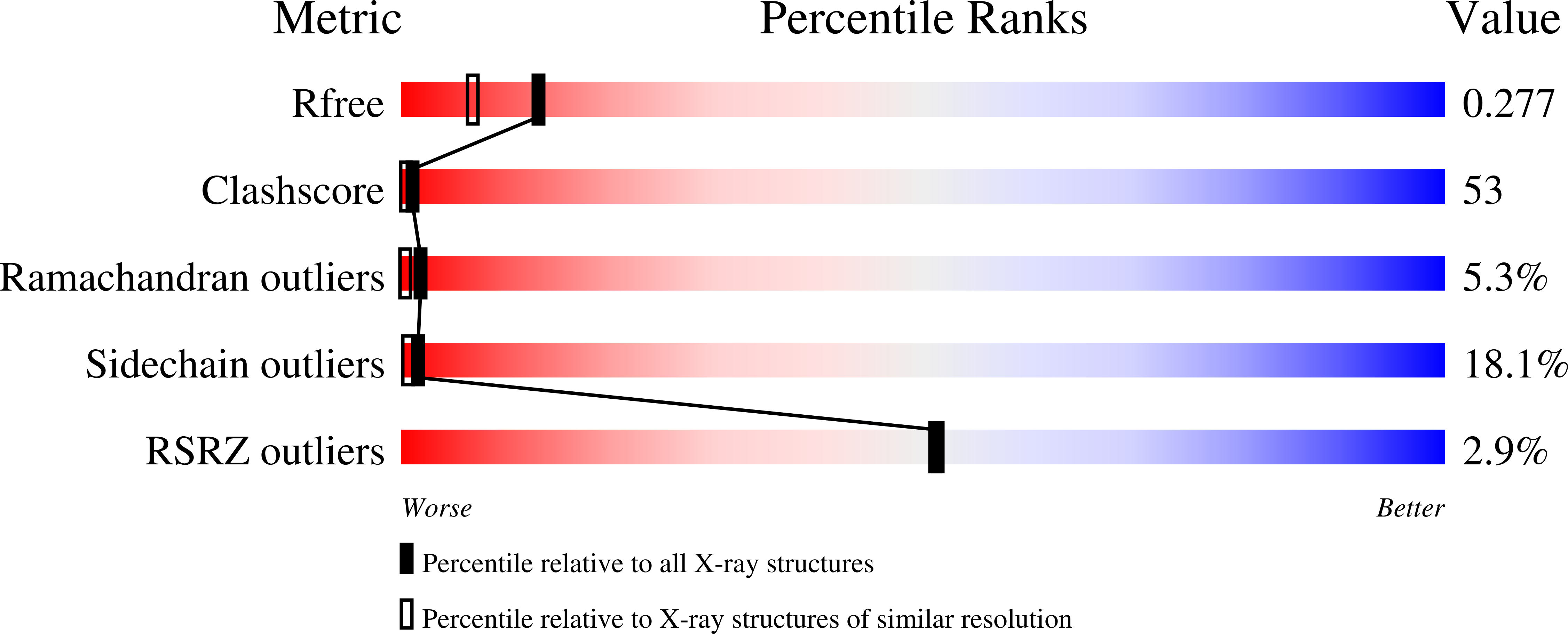
R-Value Free:
0.33
R-Value Observed:
0.25
Space Group:
P 65