3C1V image
Deposition Date 2008-01-24
Release Date 2008-04-01
Last Version Date 2023-11-01
Entry Detail
PDB ID:
3C1V
Title:
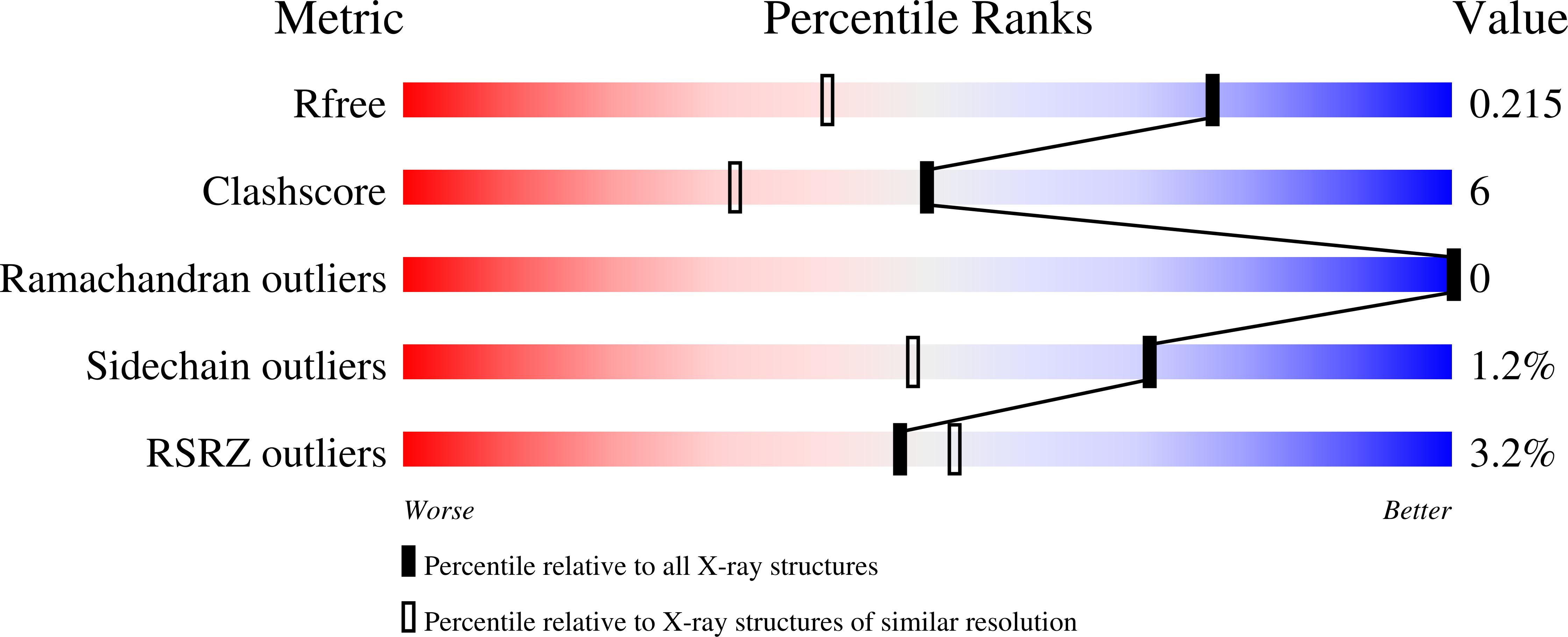
The 1.5 A Crystal structure of Ca2+-bound S100A4
Biological Source:
Source Organism(s):
Homo sapiens (Taxon ID: )
Expression System(s):
Method Details:
Experimental Method:
Resolution:
1.50 Å
R-Value Free:
0.21
R-Value Work:
0.19
R-Value Observed:
0.19
Space Group:
P 32
Macromolecular Entities
Structures with similar UniProt ID
Protein Blast
Polymer Type:polypeptide(L)
Molecule:Protein S100-A4
Gene (Uniprot):S100A4
Chain IDs:A, B, C, D
Chain Length:113
Number of Molecules:4
Biological Source:Homo sapiens
Ligand Molecules
Primary Citation
Crystal structure of the Ca(2+)-form and Ca(2+)-binding kinetics of metastasis-associated protein, S100A4
FEBS Lett. 582 1651 1656 (2008)
PMID: 18435928 DOI: 10.1016/j.febslet.2008.04.017

Abstact

S100A4 takes part in control of tumour cell migration and contributes to metastatic spread in in vivo models. In the active dimeric Ca(2+)-bound state it interacts with multiple intracellular targets. Conversely, oligomeric forms of S100A4 are linked with the extracellular function of this protein. We report the 1.5A X-ray crystal structure of Ca(2+)-bound S100A4 and use it to identify the residues involved in target recognition and to derive a model of the oligomeric state. We applied stopped-flow analysis of tyrosine fluorescence to derive kinetics of S100A4 activation by Ca(2+) (k(on)=3.5 microM(-1)s(-1), k(off)=20s(-1)).

Legend

Protein

Chemical

Disease

Primary Citation of related structures
Feedback Form
Name
Email
Institute
Feedback