

Search Count: 168
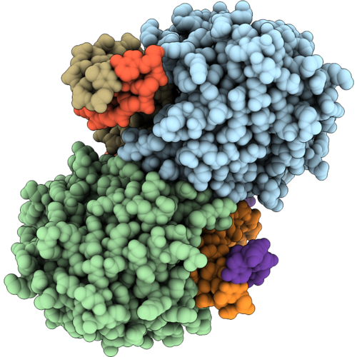 |
Mouse Cgas In Complex With Dna And Atp, Crystallized In Presence Of Ap4A.
Organism: Mus musculus, Synthetic construct
Method: X-RAY DIFFRACTION Resolution:2.70 Å Release Date: 2026-09-02 Classification: DNA BINDING PROTEIN Ligands: ZN, MG, ATP |
Organism: Mus musculus, Synthetic construct
Method: X-RAY DIFFRACTION
Release Date: 2026-09-02
Ligands: ZN, MG, ATP
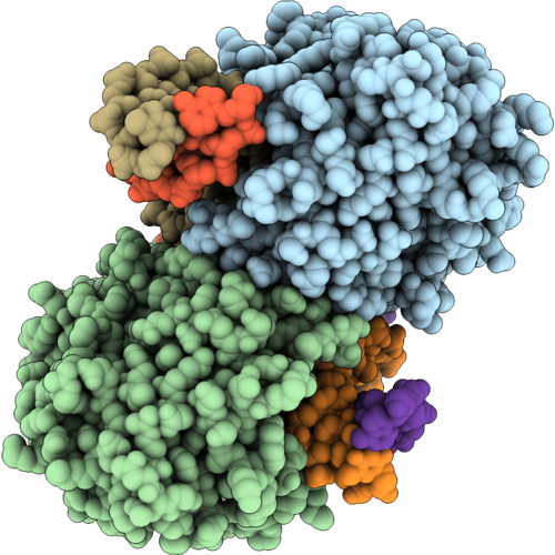 |
Mouse Cgas In Complex With Dna And Pppgpa, Crystallized In Presence Of Ap4G.
Organism: Mus musculus, Synthetic construct
Method: X-RAY DIFFRACTION Resolution:2.56 Å Release Date: 2026-09-02 Classification: DNA BINDING PROTEIN Ligands: ZN, MG, A1J67 |
Organism: Mus musculus, Synthetic construct
Method: X-RAY DIFFRACTION
Release Date: 2026-09-02
Ligands: ZN, MG, A1J67
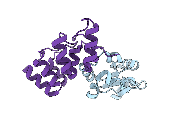 |
Crystal Structure Of The Acl1 Ankyrin Repeat Domain In Complex With The Second Rpl1 Domain
Organism: Saccharomyces cerevisiae
Method: X-RAY DIFFRACTION Resolution:2.55 Å Release Date: 2026-04-08 Classification: RIBOSOMAL PROTEIN |
Organism: Saccharomyces cerevisiae
Method: X-RAY DIFFRACTION
Release Date: 2026-04-08
 |
Crystal Structure Of An Atypical Fungal Aspartate/Glutamate Racemase Family Member Opae
Organism: Aspergillus ustus
Method: X-RAY DIFFRACTION Resolution:2.65 Å Release Date: 2026-03-18 Classification: ISOMERASE Ligands: SO4 |
Organism: Aspergillus ustus
Method: X-RAY DIFFRACTION
Release Date: 2026-03-18
Ligands: SO4
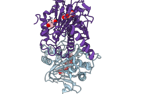 |
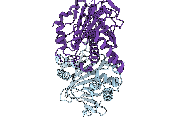
Fe/Alpha-Kg-Dependent Decarboxylase Trah In Complexes With Mn2+, Alpha-Kg, And Substrate
Organism: Penicillium crustosum
Method: X-RAY DIFFRACTION Resolution:2.89 Å Release Date: 2026-03-04 Classification: OXIDOREDUCTASE Ligands: AKG, MN, A1I30, PEG |
Organism: Penicillium crustosum
Method: X-RAY DIFFRACTION
Release Date: 2026-03-04
Ligands: AKG, MN, A1I30, PEG
 |
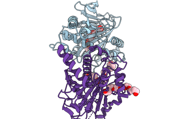
Crystal Structure Of Fe/Alphakg-Dependent Decarboxylase Trah In Complexes With Mn2+
Organism: Penicillium crustosum
Method: X-RAY DIFFRACTION Resolution:2.10 Å Release Date: 2026-03-04 Classification: OXIDOREDUCTASE Ligands: MN |
Organism: Penicillium crustosum
Method: X-RAY DIFFRACTION
Release Date: 2026-03-04
Ligands: MN
 |
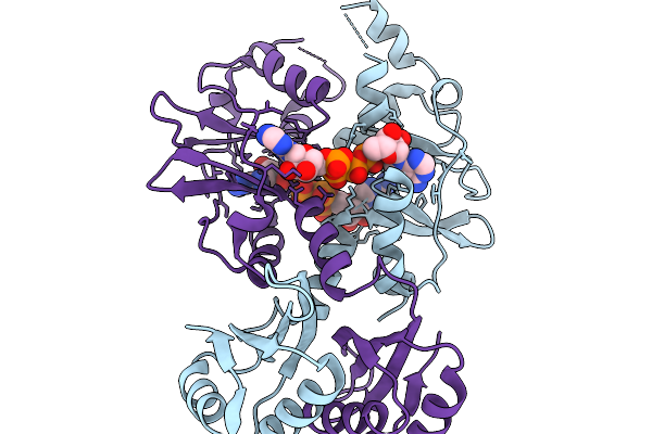
Fe/Alpha-Kg-Dependent Decarboxylase Trah In Complexes With Mn2+, Alpha-Kg, And Substrate
Organism: Penicillium crustosum
Method: X-RAY DIFFRACTION Resolution:2.40 Å Release Date: 2026-03-04 Classification: OXIDOREDUCTASE Ligands: MN, AKG, A1I31, 1PG, PGE |
Organism: Penicillium crustosum
Method: X-RAY DIFFRACTION
Release Date: 2026-03-04
Ligands: MN, AKG, A1I31, 1PG, PGE
 |
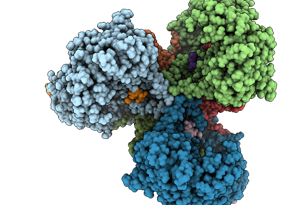
Acub,Acetoin Utilization Protein
Organism: Bacillus subtilis subsp. subtilis str. 168
Method: X-RAY DIFFRACTION Resolution:2.41 Å Release Date: 2026-02-18 Classification: BIOSYNTHETIC PROTEIN Ligands: B4P |
Organism: Bacillus subtilis subsp. subtilis str. 168
Method: X-RAY DIFFRACTION
Release Date: 2026-02-18
Ligands: B4P
 |
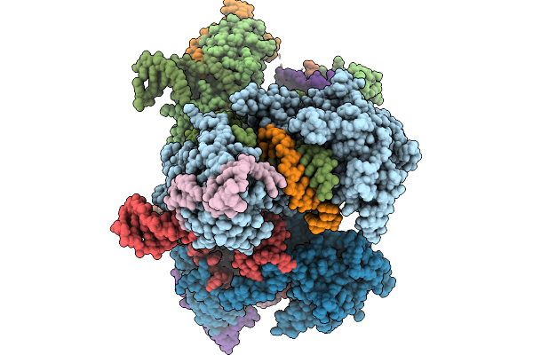
Cryo-Em Structure Of Activated Retron Eco2 (Ec67)
Organism: Escherichia coli nctc 86
Method: ELECTRON MICROSCOPY Resolution:2.90 Å Release Date: 2026-02-18 Classification: ANTIVIRAL PROTEIN Ligands: MG |
Organism: Escherichia coli nctc 86
Method: ELECTRON MICROSCOPY
Release Date: 2026-02-18
Ligands: MG
 |
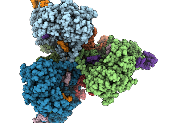
Cryo-Em Structure Of Retron Eco2 (Ec67)
Organism: Escherichia coli nctc 86
Method: ELECTRON MICROSCOPY Resolution:2.80 Å Release Date: 2026-02-04 Classification: ANTIVIRAL PROTEIN |
Organism: Escherichia coli nctc 86
Method: ELECTRON MICROSCOPY
Release Date: 2026-02-04
 |
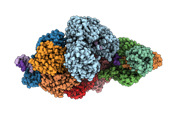
Cryo-Em Structure Of Retron Eco2 (Ec67) In Presence Of Mg Ions
Organism: Escherichia coli nctc 86
Method: ELECTRON MICROSCOPY Resolution:3.00 Å Release Date: 2026-02-04 Classification: ANTIVIRAL PROTEIN Ligands: MG |
Organism: Escherichia coli nctc 86
Method: ELECTRON MICROSCOPY
Release Date: 2026-02-04
Ligands: MG
 |
Structure Of The Minimal Type I-F2 Crispr-Cas Dna-Interference Complex.
Organism: Shewanella putrefaciens cn-32
Method: ELECTRON MICROSCOPY Release Date: 2026-01-21 Classification: ANTIVIRAL PROTEIN Ligands: MG |
Organism: Shewanella putrefaciens cn-32
Method: ELECTRON MICROSCOPY
Release Date: 2026-01-21
Ligands: MG
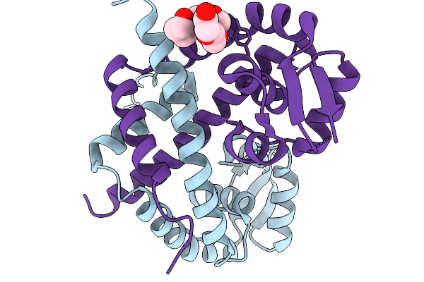 |
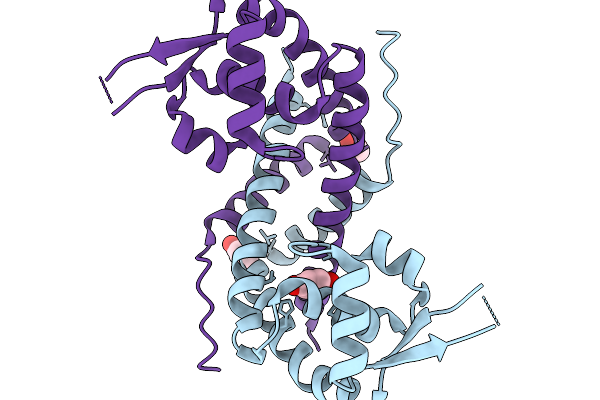
Structure Of Staphylococcus Aureus Marr-Type Repressor Mhqr
Organism: Staphylococcus aureus
Method: X-RAY DIFFRACTION Resolution:1.55 Å Release Date: 2026-01-14 Classification: DNA BINDING PROTEIN Ligands: PGE |
Organism: Staphylococcus aureus
Method: X-RAY DIFFRACTION
Release Date: 2026-01-14
Ligands: PGE
 |
Structure Of Staphylococcus Aureus Marr-Type Repressor Mhqr Bound To Mbq
Organism: Staphylococcus aureus
Method: X-RAY DIFFRACTION Resolution:2.28 Å Release Date: 2026-01-14 Classification: DNA BINDING PROTEIN Ligands: YMR |
Organism: Staphylococcus aureus
Method: X-RAY DIFFRACTION
Release Date: 2026-01-14
Ligands: YMR
 |
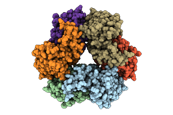
Crystal Structure Of A Lectin-Like Protein From Ustilago Maydis
Organism: Mycosarcoma maydis
Method: X-RAY DIFFRACTION Resolution:2.01 Å Release Date: 2025-08-20 Classification: UNKNOWN FUNCTION Ligands: SO4 |
Organism: Mycosarcoma maydis
Method: X-RAY DIFFRACTION
Release Date: 2025-08-20
Ligands: SO4
 |
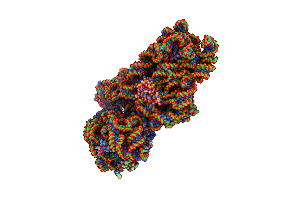
Cryo-Em Structure Of Yham
Organism: Bacillus subtilis
Method: ELECTRON MICROSCOPY Release Date: 2025-08-06 Classification: DNA BINDING PROTEIN Ligands: MG |
Organism: Bacillus subtilis
Method: ELECTRON MICROSCOPY
Release Date: 2025-08-06
Ligands: MG
 |
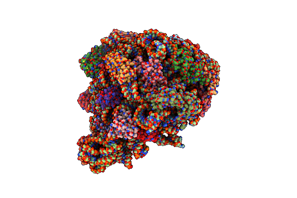
Cryo-Em Structure Of The Vibrio Natrigens 30S Ribosomal Subunit In Complex With Spectinomycin.
Organism: Vibrio natriegens
Method: ELECTRON MICROSCOPY Resolution:2.80 Å Release Date: 2025-05-21 Classification: RIBOSOME Ligands: SCM |
Organism: Vibrio natriegens
Method: ELECTRON MICROSCOPY
Release Date: 2025-05-21
Ligands: SCM
 |
Cryo-Em Structure Of The Vibrio Natrigens 50S Ribosomal Subunit In Complex With The Proline-Rich Antimicrobial Peptide Bac5(1-17).
Organism: Bos taurus, Vibrio natriegens
Method: ELECTRON MICROSCOPY Resolution:2.70 Å Release Date: 2025-05-21 Classification: RIBOSOME Ligands: ZN |
Organism: Bos taurus, Vibrio natriegens
Method: ELECTRON MICROSCOPY
Release Date: 2025-05-21
Ligands: ZN
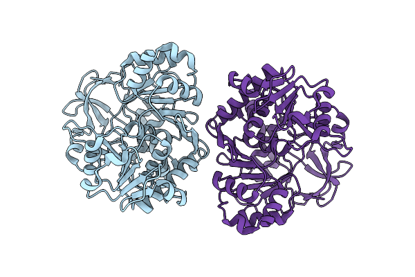 |
Cryo-Em Structure Of Acetyl-Coenzyme A Synthetase (Acsa) Dimer
Organism: Bacillus subtilis
Method: ELECTRON MICROSCOPY Release Date: 2025-04-16 Classification: BIOSYNTHETIC PROTEIN |
Organism: Bacillus subtilis
Method: ELECTRON MICROSCOPY
Release Date: 2025-04-16
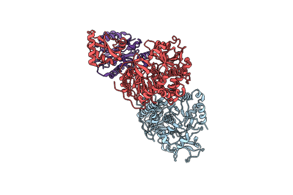 |
Cryo-Em Structure Of Acetyl-Coenzyme A Synthetase (Acsa) Dimer
Organism: Bacillus subtilis
Method: ELECTRON MICROSCOPY Release Date: 2025-04-16 Classification: BIOSYNTHETIC PROTEIN |
Organism: Bacillus subtilis
Method: ELECTRON MICROSCOPY
Release Date: 2025-04-16