

Search Count: 220
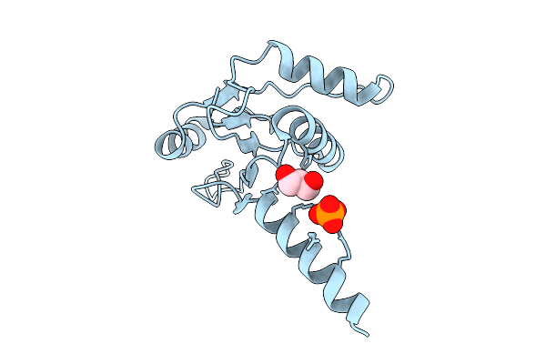 |
Oligomerisation Of Type Iii Crispr-Associated Csx15 To Regulate Antiviral Signalling
Organism: Pseudomonas fluorescens
Method: X-RAY DIFFRACTION Resolution:1.88 Å Release Date: 2026-04-15 Classification: VIRAL PROTEIN Ligands: GOL, PO4 |
Organism: Pseudomonas fluorescens
Method: X-RAY DIFFRACTION
Release Date: 2026-04-15
Ligands: GOL, PO4
 |
Crystal Structure Of Pseudomonas Fluorescens Peroxidase Efeb
Organism: Pseudomonas fluorescens
Method: X-RAY DIFFRACTION Resolution:2.11 Å Release Date: 2026-04-01 Classification: OXYGEN BINDING Ligands: HEM, PG4, EDO, SO4, PEG, TRS, OXY, PGE |
Organism: Pseudomonas fluorescens
Method: X-RAY DIFFRACTION
Release Date: 2026-04-01
Ligands: HEM, PG4, EDO, SO4, PEG, TRS, OXY, PGE
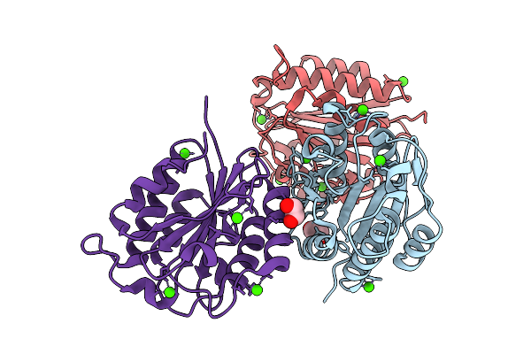 |
Crystal Structure Of Vwfa Domain From Lapa Adhesin Of Pseudomonas Fluorescens
Organism: Pseudomonas fluorescens
Method: X-RAY DIFFRACTION Resolution:1.45 Å Release Date: 2026-04-01 Classification: CELL ADHESION Ligands: CA, EDO |
Organism: Pseudomonas fluorescens
Method: X-RAY DIFFRACTION
Release Date: 2026-04-01
Ligands: CA, EDO
 |
Crystal Structure Of Antitoxin Hipb From Pseudomonas Fluorescens 2P24
Organism: Pseudomonas fluorescens
Method: X-RAY DIFFRACTION Resolution:2.15 Å Release Date: 2026-04-01 Classification: ANTITOXIN |
Organism: Pseudomonas fluorescens
Method: X-RAY DIFFRACTION
Release Date: 2026-04-01
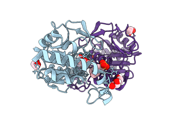 |
Pseudomonas Fluorescens Isocyanide Hydratase, Ph=6.5
Organism: Pseudomonas fluorescens
Method: X-RAY DIFFRACTION Resolution:0.89 Å Release Date: 2025-10-29 Classification: LYASE Ligands: EDO, MES |
Organism: Pseudomonas fluorescens
Method: X-RAY DIFFRACTION
Release Date: 2025-10-29
Ligands: EDO, MES
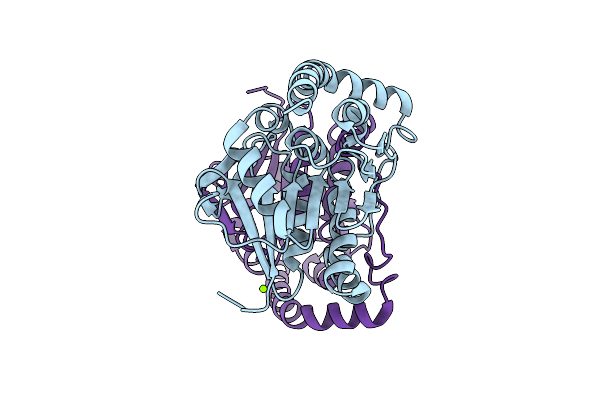 |
Pseudomonas Fluorescens Isocyanide Hydratase D17N Mutant, Ph=8.8
Organism: Pseudomonas fluorescens
Method: X-RAY DIFFRACTION Resolution:1.20 Å Release Date: 2025-10-29 Classification: LYASE Ligands: MG, CL |
Organism: Pseudomonas fluorescens
Method: X-RAY DIFFRACTION
Release Date: 2025-10-29
Ligands: MG, CL
 |
Pseudomonas Fluorescens Isocyanide Hydratase D17N Mutant, Ph=6.5
Organism: Pseudomonas fluorescens
Method: X-RAY DIFFRACTION Resolution:1.15 Å Release Date: 2025-10-29 Classification: LYASE Ligands: CL |
Organism: Pseudomonas fluorescens
Method: X-RAY DIFFRACTION
Release Date: 2025-10-29
Ligands: CL
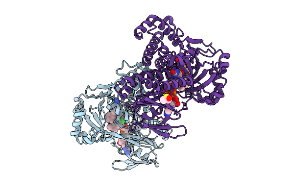 |
Kynurenine Monooxygenase From Pseudomonas Fluorescens Complexed With 4-Cyclohexylphenylacetyltetrazole
Organism: Pseudomonas fluorescens
Method: X-RAY DIFFRACTION Resolution:1.95 Å Release Date: 2025-08-27 Classification: FLAVOPROTEIN/INHIBITOR Ligands: A1AIN, FAD, CL, DMS |
Organism: Pseudomonas fluorescens
Method: X-RAY DIFFRACTION
Release Date: 2025-08-27
Ligands: A1AIN, FAD, CL, DMS
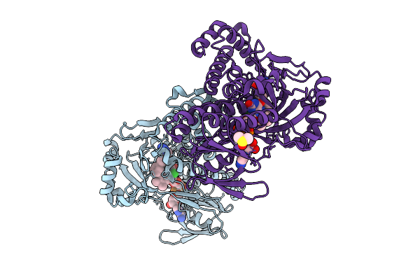 |
Kynurenine Monooxygenase From Pseudomonas Fluorescens Complexed With Biphenylacetyltetrazole
Organism: Pseudomonas fluorescens
Method: X-RAY DIFFRACTION Resolution:1.88 Å Release Date: 2025-08-27 Classification: FLAVOPROTEIN/INHIBITOR Ligands: FAD, CL, A1AIU, DMS |
Organism: Pseudomonas fluorescens
Method: X-RAY DIFFRACTION
Release Date: 2025-08-27
Ligands: FAD, CL, A1AIU, DMS
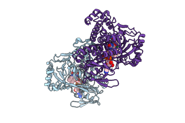 |
Kynurenine Monooxygenase From Pseudomonas Fluorescens Complexed With 4-Chlorophenylacetyltetrazole
Organism: Pseudomonas fluorescens
Method: X-RAY DIFFRACTION Resolution:2.05 Å Release Date: 2025-08-27 Classification: FLAVOPROTEIN/INHIBITOR Ligands: FAD, CL, A1AI5 |
Organism: Pseudomonas fluorescens
Method: X-RAY DIFFRACTION
Release Date: 2025-08-27
Ligands: FAD, CL, A1AI5
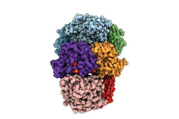 |
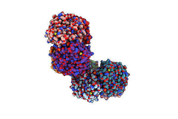
Crystal Structure Of Trans-O-Hydroxybenzylidenepyruvate Hydratase-Aldolase From Pseudomonas Fluorescens N3 Bound To Substrate Intermediate
Organism: Pseudomonas fluorescens
Method: X-RAY DIFFRACTION Resolution:1.76 Å Release Date: 2025-07-23 Classification: LYASE Ligands: KRN, PO4, GOL, NK |
Organism: Pseudomonas fluorescens
Method: X-RAY DIFFRACTION
Release Date: 2025-07-23
Ligands: KRN, PO4, GOL, NK
 |
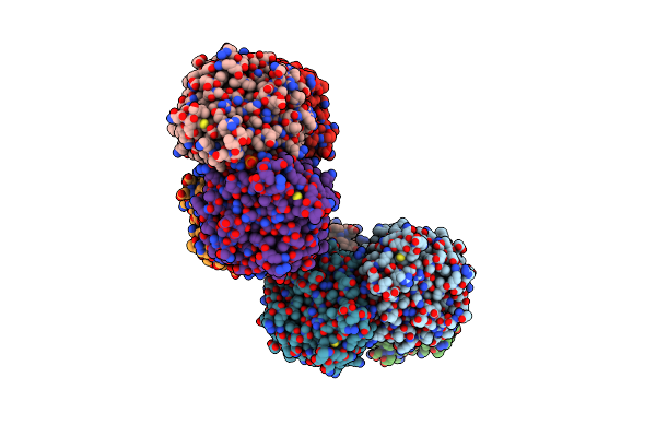
Crystal Structure Of Trans-O-Hydroxybenzylidenepyruvate Hydratase-Aldolase From Pseudomonas Fluorescens N3 Bound To Pyruvate
Organism: Pseudomonas fluorescens
Method: X-RAY DIFFRACTION Resolution:2.30 Å Release Date: 2025-07-02 Classification: LYASE Ligands: PYR, PO4, GOL |
Organism: Pseudomonas fluorescens
Method: X-RAY DIFFRACTION
Release Date: 2025-07-02
Ligands: PYR, PO4, GOL
 |
Crystal Structure Of Trans-O-Hydroxybenzylidenepyruvate Hydratase-Aldolase From Pseudomonas Fluorescens N3
Organism: Pseudomonas fluorescens
Method: X-RAY DIFFRACTION Resolution:1.96 Å Release Date: 2025-06-25 Classification: LYASE Ligands: GOL, PO4 |
Organism: Pseudomonas fluorescens
Method: X-RAY DIFFRACTION
Release Date: 2025-06-25
Ligands: GOL, PO4
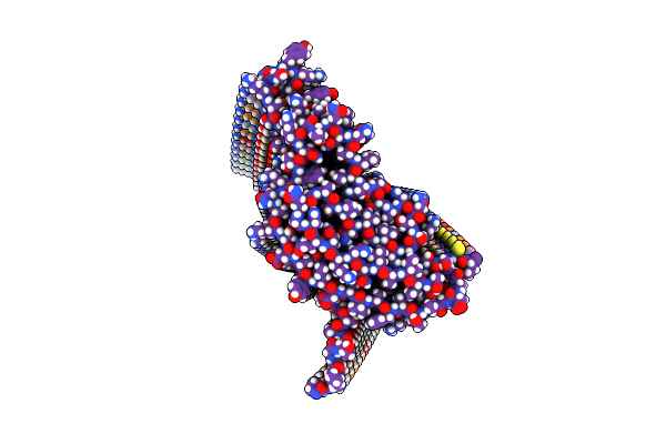 |
Structure Of Pseudomonas Fapc Biofilm-Forming Functional Amyloid
Organism: Pseudomonas fluorescens
Method: ELECTRON MICROSCOPY Release Date: 2025-06-25 Classification: STRUCTURAL PROTEIN |
Organism: Pseudomonas fluorescens
Method: ELECTRON MICROSCOPY
Release Date: 2025-06-25
 |
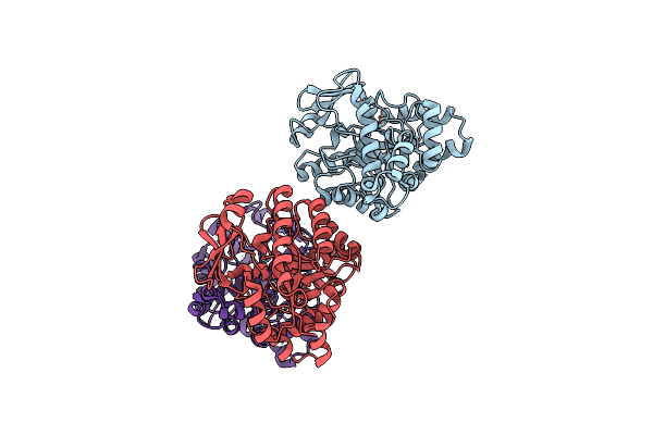
Crystal Structure Of Acmsd In Complex With Malonate
Organism: Pseudomonas fluorescens
Method: X-RAY DIFFRACTION Resolution:2.22 Å Release Date: 2024-10-16 Classification: LYASE Ligands: ZN, MLA |
Organism: Pseudomonas fluorescens
Method: X-RAY DIFFRACTION
Release Date: 2024-10-16
Ligands: ZN, MLA
 |
Crystal Structure Of Acmsd Mutant W194A
Organism: Pseudomonas fluorescens
Method: X-RAY DIFFRACTION Resolution:2.02 Å Release Date: 2024-10-16 Classification: LYASE Ligands: ZN |
Organism: Pseudomonas fluorescens
Method: X-RAY DIFFRACTION
Release Date: 2024-10-16
Ligands: ZN
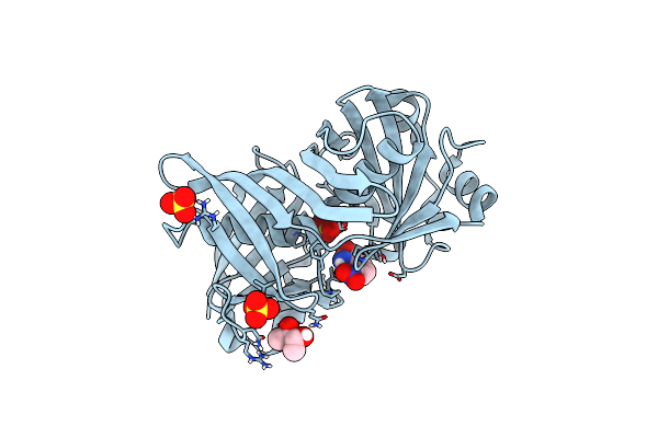 |
Complex Of Phenazine Biosynthesis Enzyme Phzf With 2-Amino-3-Nitrobenzoic Acid
Organism: Pseudomonas fluorescens
Method: X-RAY DIFFRACTION Resolution:1.32 Å Release Date: 2024-10-09 Classification: ISOMERASE Ligands: SO4, A1IAX, BU3 |
Organism: Pseudomonas fluorescens
Method: X-RAY DIFFRACTION
Release Date: 2024-10-09
Ligands: SO4, A1IAX, BU3
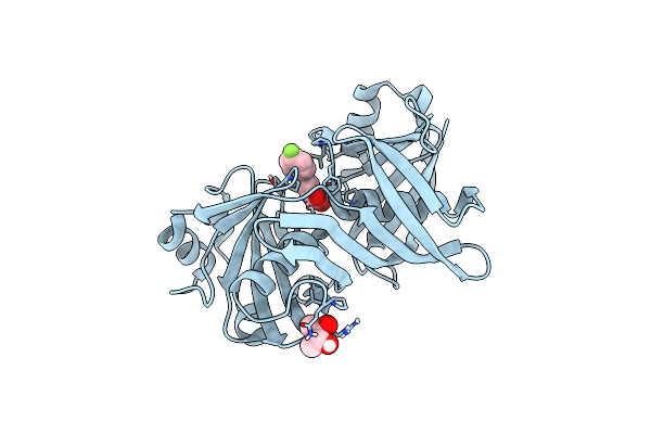 |
Complex Of Phenazine Biosynthesis Enzyme Phzf With 2-Amino-5-(4-Fluorophenyl)Benzoic Acid
Organism: Pseudomonas fluorescens
Method: X-RAY DIFFRACTION Resolution:1.54 Å Release Date: 2024-10-09 Classification: ISOMERASE Ligands: A1IAV, BU3 |
Organism: Pseudomonas fluorescens
Method: X-RAY DIFFRACTION
Release Date: 2024-10-09
Ligands: A1IAV, BU3
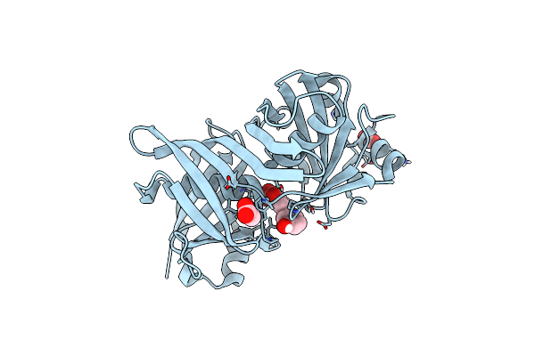 |
Complex Of Phenazine Biosynthesis Enzyme Phzf With 2-Amino-5-(3-Hydroxyphenyl)Benzoic Acid
Organism: Pseudomonas fluorescens
Method: X-RAY DIFFRACTION Resolution:1.32 Å Release Date: 2024-10-09 Classification: ISOMERASE Ligands: FMT, ACT, A1IAY |
Organism: Pseudomonas fluorescens
Method: X-RAY DIFFRACTION
Release Date: 2024-10-09
Ligands: FMT, ACT, A1IAY
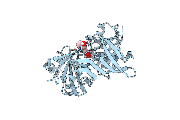 |
Complex Of Phenazine Biosynthesis Enzyme Phzf With 2-Amino-3-Hydroxy-5-(3-Hydroxyphenyl)Benzoic Acid
Organism: Pseudomonas fluorescens
Method: X-RAY DIFFRACTION Resolution:1.46 Å Release Date: 2024-10-09 Classification: ISOMERASE Ligands: A1IAU |
Organism: Pseudomonas fluorescens
Method: X-RAY DIFFRACTION
Release Date: 2024-10-09
Ligands: A1IAU