

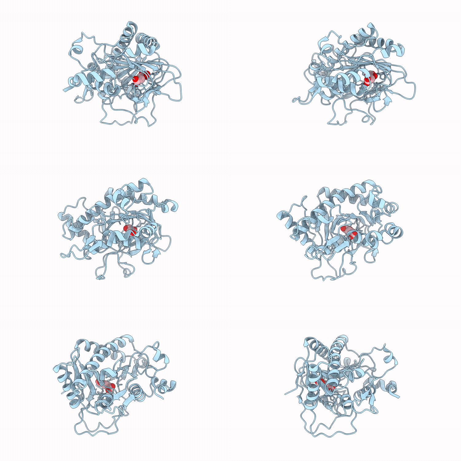
9SCF image
Deposition Date 2025-08-10
Release Date 2026-03-18
Last Version Date 2026-03-18
Entry Detail
PDB ID:
9SCF
Keywords:
Title:
Fe(II)-2-oxoglutarate-dependent Pseudomonas savastanoi pv phaseolicola 1449B in complex with 2-oxoglutarate
Biological Source:
Source Organism(s):
Expression System(s):
Method Details:
Experimental Method:
Resolution:
1.65 Å
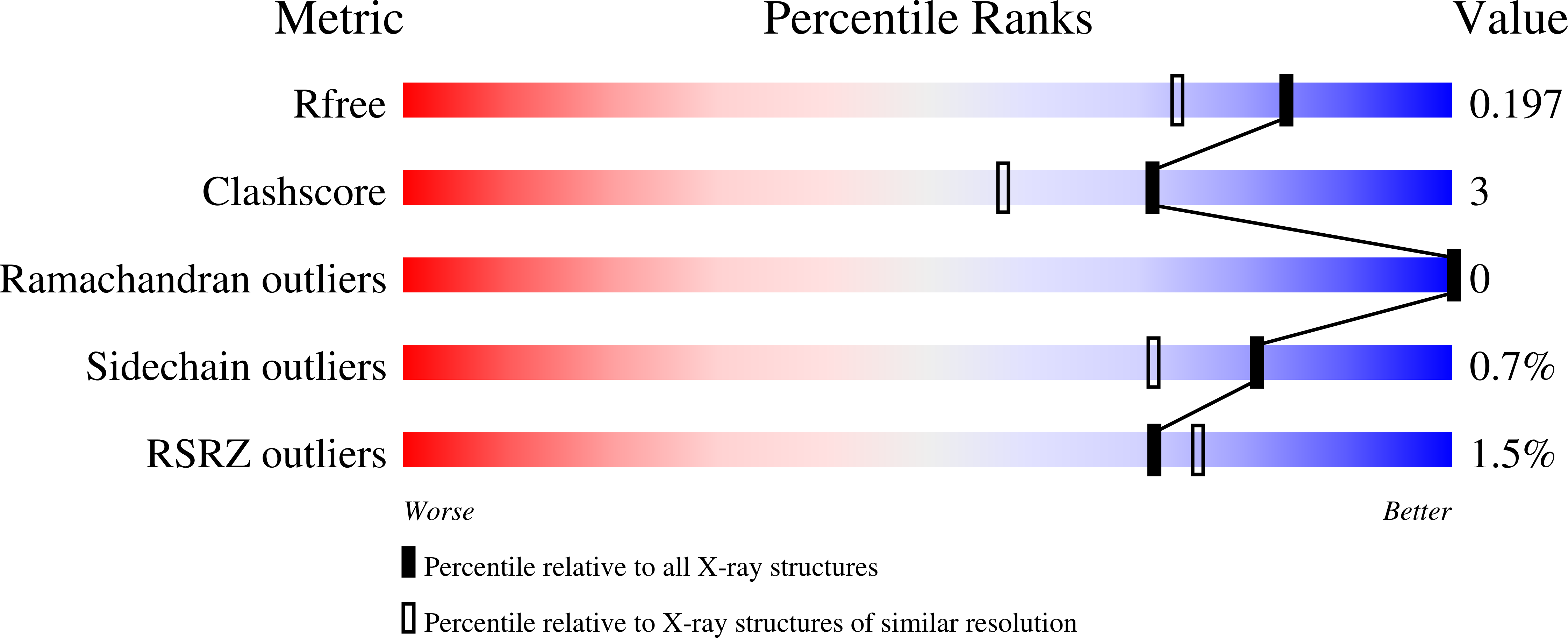
R-Value Free:
0.19
R-Value Work:
0.17
R-Value Observed:
0.17
Space Group:
P 43
Macromolecular Entities
Structures with similar UniProt ID
Protein Blast
Polymer Type:polypeptide(L)
Molecule:2-oxoglutarate-dependent ethy
Chain IDs:A
Chain Length:337
Number of Molecules:1
Biological Source:Pseudomonas savastanoi
Primary Citation
Structural and Functional Validation of Pseudomonas Savastanoi Ethylene Forming Enzymes Reveals Flexibility in 2-Oxoglutarate Binding Mode and Conformation.
Chembiochem 27 e202500702 e202500702 (2026)
PMID: 41797450 DOI: 10.1002/cbic.202500702

Abstact

Pseudomonas savastanoi pv. phaseolicola PK2 employs an Fe(II)-dependent ethylene/succinate-forming enzyme (PK2 PsEFE) to produce ethylene from 2-oxoglutarate (2OG). Here we report NMR-based assays showing that the putative P. savastanoi pv. glycinea PsEFE, which differs from PK2 PsEFE by a single residue, and the P. savastanoi pv.1449B PsEFE, which differs from PK2 PsEFE by 28 residues and a C-terminal 13-residue truncation, catalyze ethylene production from 2OG. Like the PK2 PsEFE, they catalyze oxidation of naturally occurring 2OG derivatives to give alcohol and diacid products. Crystallographic analysis demonstrates that the overall fold and active site of 1449B PsEFE is similar to that of PK2 PsEFE. Interestingly, 2OG was observed to adopt an atypical inverse metal ion binding mode in complex with 1449B PsEFE:Mn in which its 2-oxoacid group is positioned to interact with the guanidinium group of R277, but not the Mn ion, which substitutes for catalytically active Fe(II). Together with reported crystallographic results, this observation indicates that 2OG metal ion binding modes and conformations at the active sites of 2OG oxygenases can vary, possibly in a functionally or disease relevant manner.

Legend

Protein

Chemical

Disease

Primary Citation of related structures
Feedback Form
Name
Email
Institute
Feedback