
Search Count: 15,373
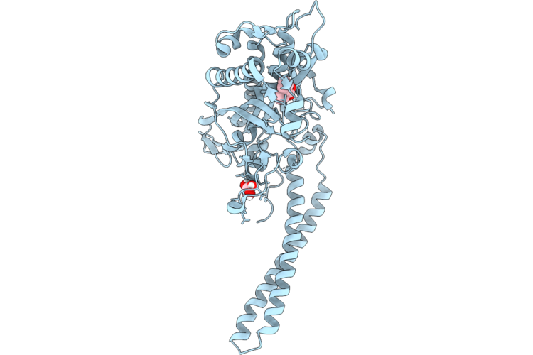 |
Seryl-Trna Synthetase In Complex With A Fragment-Sized Inhibitor (3-Cyclopropyl Benzoic Acid)
Organism: Escherichia coli
Method: X-RAY DIFFRACTION Resolution:1.47 Å Release Date: 2026-06-24 Classification: LIGASE Ligands: A1J8R, GOL |
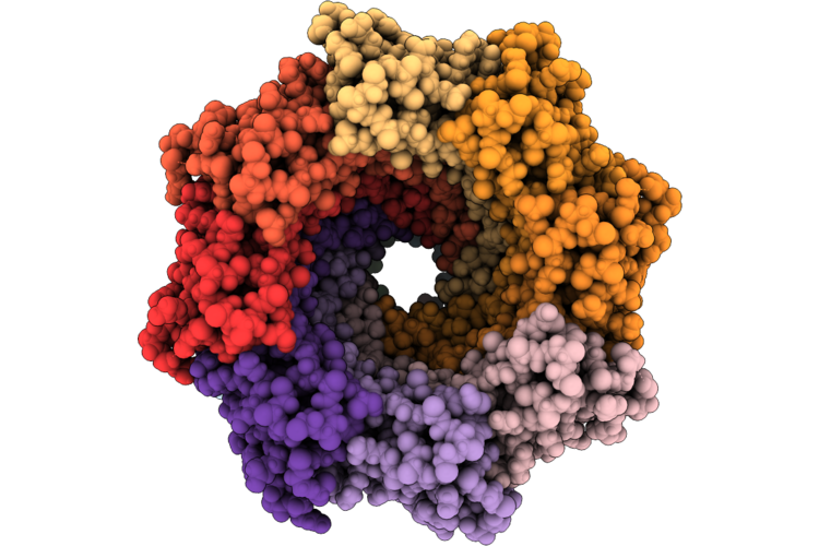 |
C1 Symmetry Cryoem Structure Of The Soluble-Wraped Membranous Portion Of Mspa (Mycobacterium Smegmatis Porin), Dimerized Along The Native Interface.
Organism: Mycolicibacterium smegmatis mc2 155
Method: ELECTRON MICROSCOPY Resolution:3.34 Å Release Date: 2026-06-24 Classification: DE NOVO PROTEIN |
 |
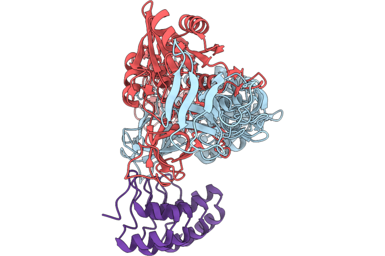
Cryo-Em Structure Of Gephyrin In Complex With Darpin 27F3, Revealing Linker-E Domain Interactions
Organism: Rattus norvegicus, Synthetic construct
Method: ELECTRON MICROSCOPY Release Date: 2026-06-24 Classification: STRUCTURAL PROTEIN |
 |
Organism: Rattus norvegicus, Synthetic construct
Method: ELECTRON MICROSCOPY Release Date: 2026-06-24 Classification: STRUCTURAL PROTEIN |
 |
Organism: Rattus norvegicus
Method: ELECTRON MICROSCOPY Release Date: 2026-06-24 Classification: STRUCTURAL PROTEIN |
 |
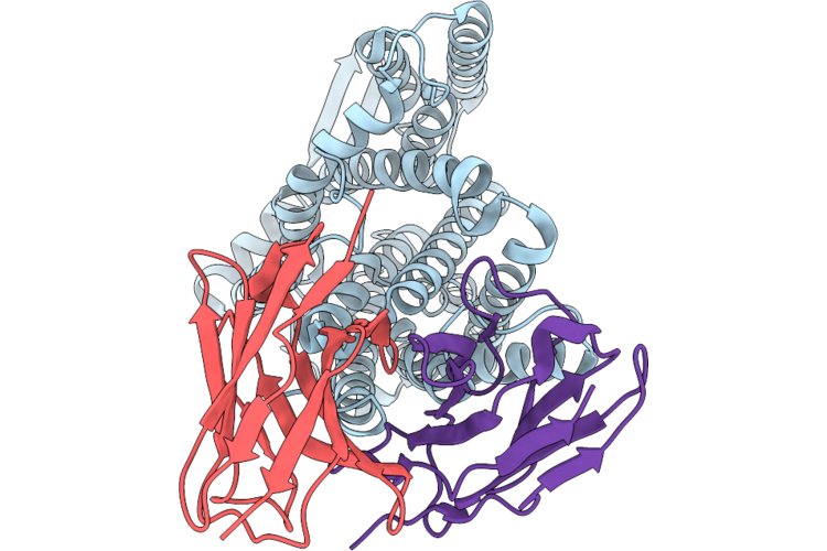
Organism: Homo sapiens
Method: ELECTRON MICROSCOPY Release Date: 2026-06-24 Classification: BLOOD CLOTTING Ligands: NAG |
 |
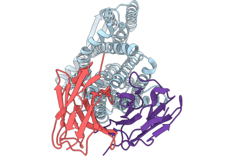
Organism: Homo sapiens
Method: ELECTRON MICROSCOPY Release Date: 2026-06-24 Classification: BLOOD CLOTTING Ligands: NAG |
 |
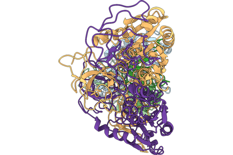
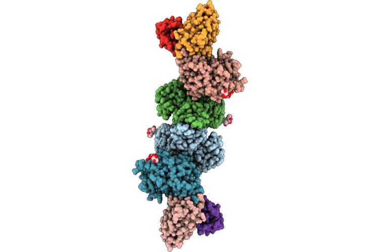
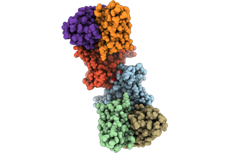
Cryo-Em Structure Of Human C3 Pro-Convertase Bound To The Compstatin Analog Cp60, Ted Conformation 1
Organism: Synthetic construct, Homo sapiens
Method: ELECTRON MICROSCOPY Resolution:2.88 Å Release Date: 2026-06-17 Classification: IMMUNE SYSTEM Ligands: NI, NAG |
 |
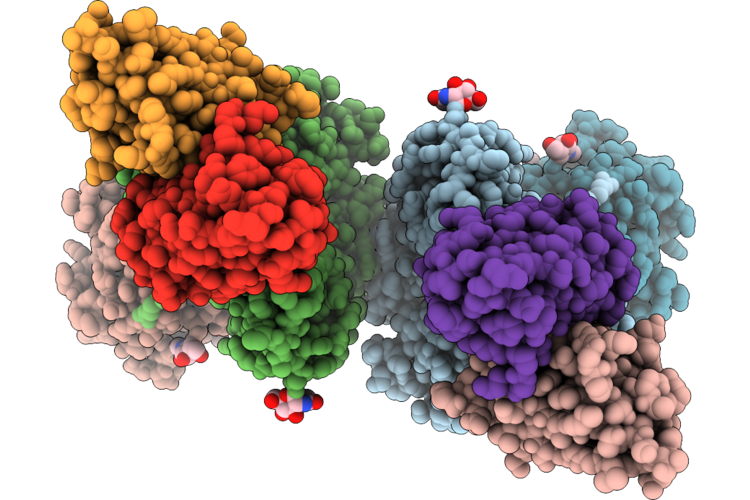
Cryo-Em Structure Of Human C3 Pro-Convertase Bound To The Compstatin Analog Cp60, Ted Conformation 2
Organism: Synthetic construct, Homo sapiens
Method: ELECTRON MICROSCOPY Resolution:3.00 Å Release Date: 2026-06-17 Classification: IMMUNE SYSTEM Ligands: NI, NAG |
 |
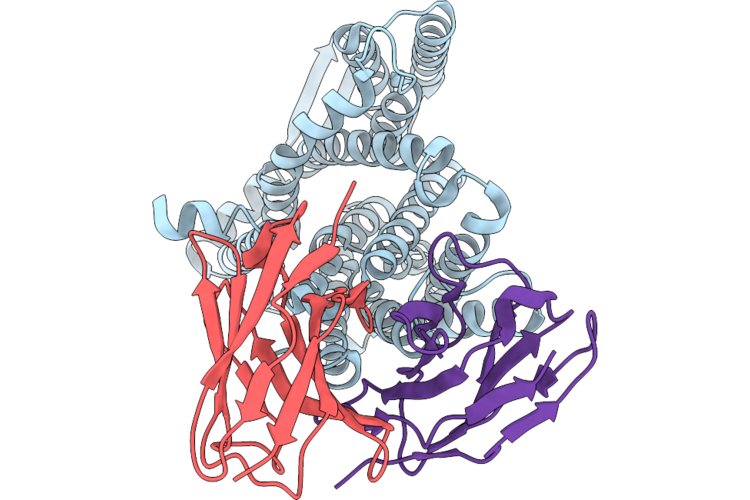
Response Regulator Domains Of Bt4124 Hybrid Two-Component System From Bacteroides Thetaiotaomicron
Organism: Bacteroides thetaiotaomicron
Method: X-RAY DIFFRACTION Resolution:2.90 Å Release Date: 2026-06-17 Classification: DNA BINDING PROTEIN |
 |
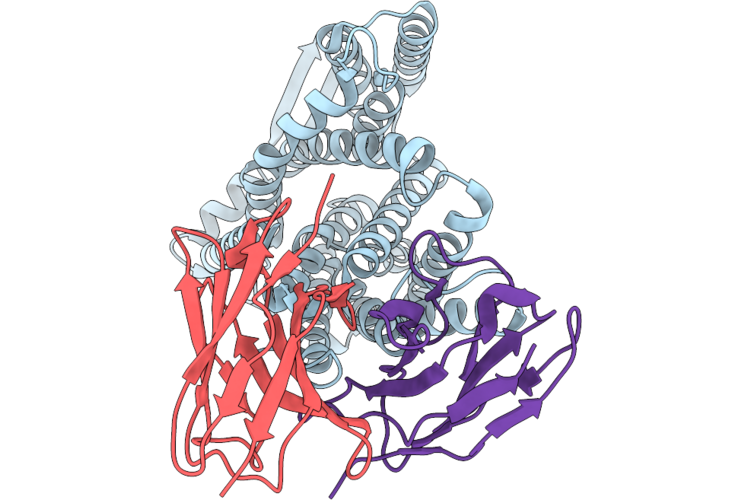
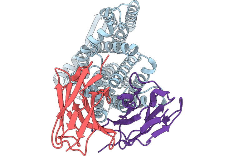
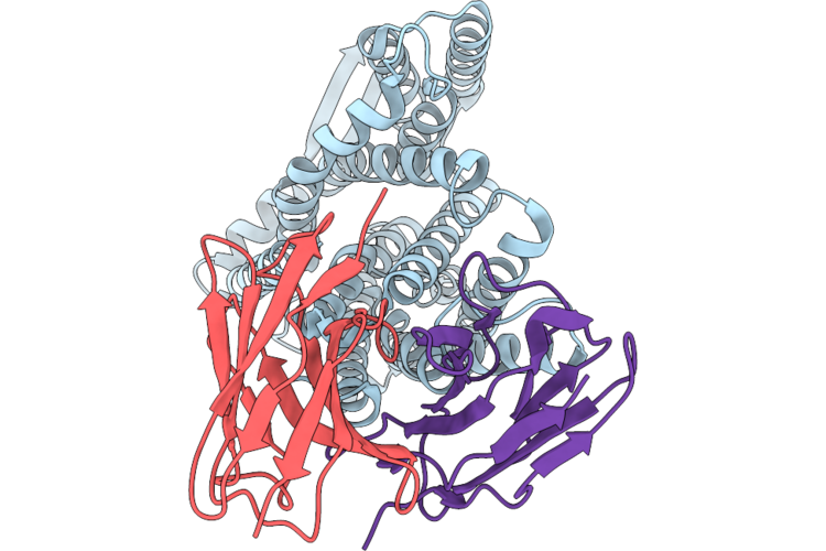
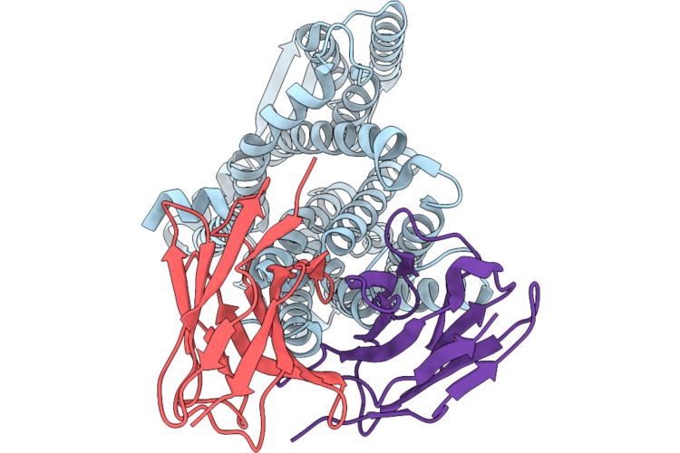
Organism: Escherichia coli, Mus musculus
Method: ELECTRON MICROSCOPY Resolution:2.70 Å Release Date: 2026-06-17 Classification: TRANSPORT PROTEIN Ligands: CDL, PGT |
 |
Cryo-Em Structure Of The Inward-Facing Apo Nhaa In The Unplugged State At Ph 7.5
Organism: Escherichia coli, Mus musculus
Method: ELECTRON MICROSCOPY Release Date: 2026-06-17 Classification: TRANSPORT PROTEIN |
 |
Cryo-Em Structure Of The Inward-Facing Apo Nhaa In The Plugged State At Ph 7.5
Organism: Escherichia coli, Mus musculus
Method: ELECTRON MICROSCOPY Release Date: 2026-06-17 Classification: TRANSPORT PROTEIN |
 |
Cryo-Em Structure Of The Inward-Facing Apo Nhaa With Flexible N-Terminus At Ph 7.5
Organism: Escherichia coli, Mus musculus
Method: ELECTRON MICROSCOPY Release Date: 2026-06-17 Classification: TRANSPORT PROTEIN |
 |
Cryo-Em Structure Of The Inward-Facing Apo Nhaa In The Unplugged State At Ph 6.3
Organism: Escherichia coli, Mus musculus
Method: ELECTRON MICROSCOPY Release Date: 2026-06-17 Classification: TRANSPORT PROTEIN |
 |
Cryo-Em Structure Of The Inward-Facing Apo Nhaa In The Plugged State At Ph 6.3
Organism: Escherichia coli, Mus musculus
Method: ELECTRON MICROSCOPY Release Date: 2026-06-17 Classification: TRANSPORT PROTEIN |
 |
Cryo-Em Structure Of The Inward-Facing Apo Nhaa With Flexible N-Terminus At Ph 6.3
Organism: Escherichia coli, Mus musculus
Method: ELECTRON MICROSCOPY Release Date: 2026-06-17 Classification: TRANSPORT PROTEIN |
 |
Cryo-Em Structure Of The Inward-Facing Apo Nhaa In The Unplugged State At Ph 5.5
Organism: Escherichia coli, Mus musculus
Method: ELECTRON MICROSCOPY Release Date: 2026-06-17 Classification: TRANSPORT PROTEIN |
 |
Cryo-Em Structure Of The Inward-Facing Apo Nhaa In The Plugged State At Ph 5.5
Organism: Escherichia coli, Mus musculus
Method: ELECTRON MICROSCOPY Release Date: 2026-06-17 Classification: TRANSPORT PROTEIN |
 |
Cryo-Em Structure Of The Inward-Facing Apo Nhaa With Flexible N-Terminus At Ph 5.5
Organism: Escherichia coli, Mus musculus
Method: ELECTRON MICROSCOPY Release Date: 2026-06-17 Classification: TRANSPORT PROTEIN |