

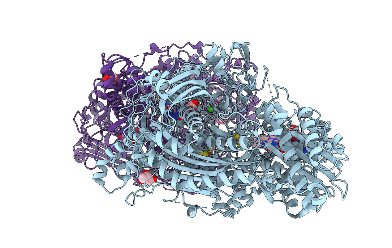
3AN1 image
Deposition Date 2010-08-27
Release Date 2010-12-01
Last Version Date 2026-08-12
Entry Detail
PDB ID:
3AN1
Keywords:
Title:
Crystal structure of rat D428A mutant, urate bound form
Biological Source:
Source Organism(s):
Expression System(s):
Method Details:
Experimental Method:
Resolution:
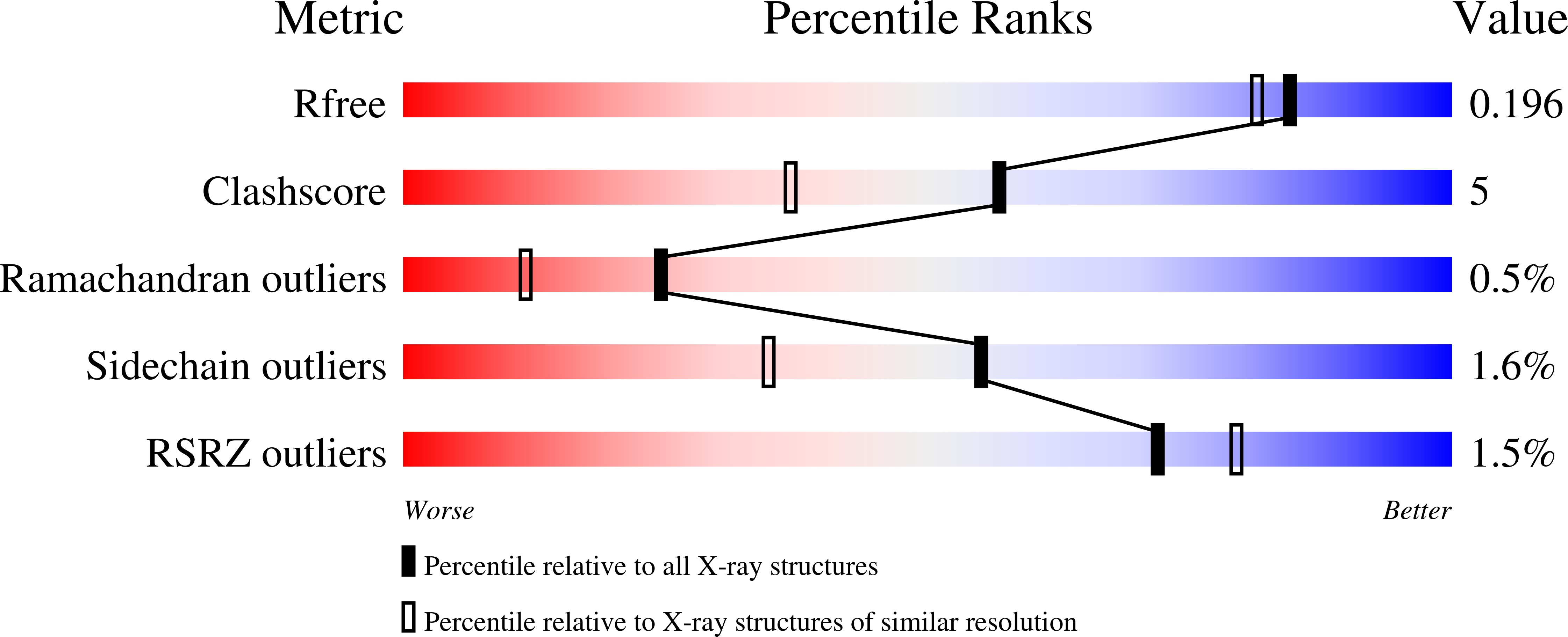
1.73 Å
R-Value Free:
0.19
R-Value Work:
0.16
R-Value Observed:
0.16
Space Group:
P 21 21 21
Macromolecular Entities
Polymer Type:polypeptide(L)
Molecule:Xanthine dehydrogenase/oxidas
Gene (Uniprot):Xdh
Mutagens:D428A
Chain IDs:A, B
Chain Length:1331
Number of Molecules:2
Biological Source:Rattus norvegicus
Primary Citation
Crystal Structures of Urate Bound Form of Xanthine Oxidoreductase: Substrate Orientation and Structure of the Key Reaction Intermediate
J.Am.Chem.Soc. 132 17080 17083 (2010)
PMID: 21077683 DOI: 10.1021/ja1077574

Abstact

Two contradictory models have been proposed for the binding mode of the substrate xanthine to and its activation mechanism by xanthine oxidoreductase. In an effort to distinguish between the two models, we determined the crystal structures of the urate complexes of the demolybdo-form of the D428A mutant of rat xanthine oxidoreductase at 1.7 Å and of the reduced bovine milk enzyme at 2.1 Å, the latter representing a reaction intermediate. The results clearly indicate the catalytically relevant binding mode of the substrate xanthine.

Legend

Protein

Chemical

Disease

Primary Citation of related structures
Feedback Form
Name
Email
Institute
Feedback