
Search Count: 120
All
Selected
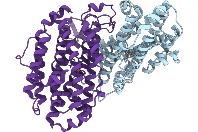 |
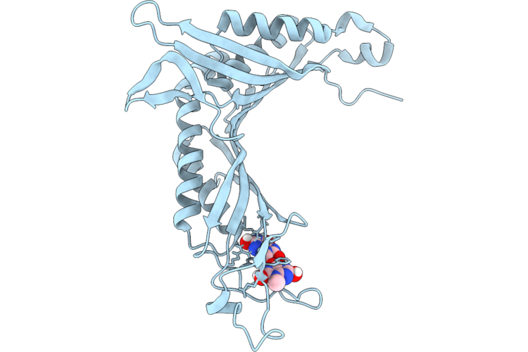
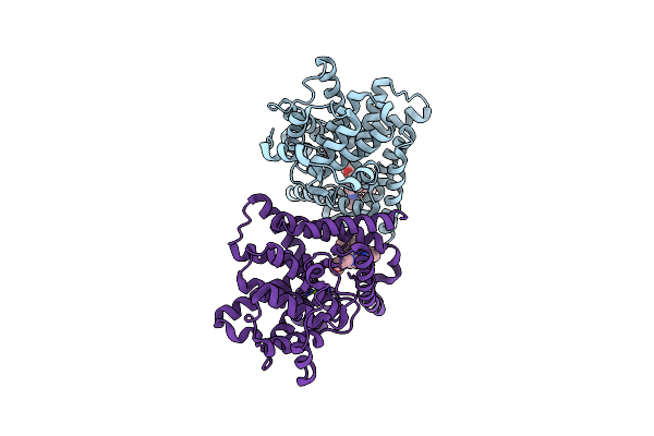
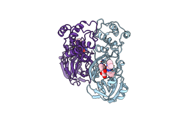
Xfel Structure Of Oxidised Ribonucleotide Reductase R2A Y122F Mutant From E. Coli
Organism: Escherichia coli
Method: X-RAY DIFFRACTION Resolution:1.90 Å Release Date: 2026-05-13 Classification: OXIDOREDUCTASE Ligands: FE |
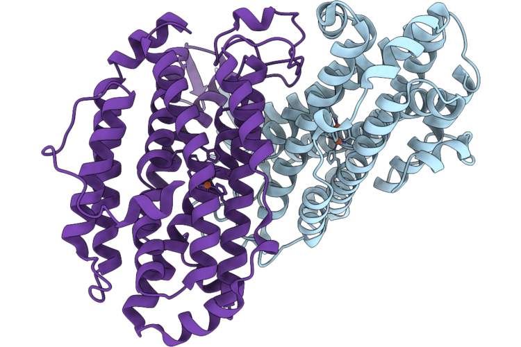 |
Xfel Structure Of Ribonucleotide Reductase R2A Y122F Mutant From E. Coli,Reduced Form
Organism: Escherichia coli
Method: X-RAY DIFFRACTION Resolution:1.70 Å Release Date: 2026-05-13 Classification: OXIDOREDUCTASE Ligands: FE2 |
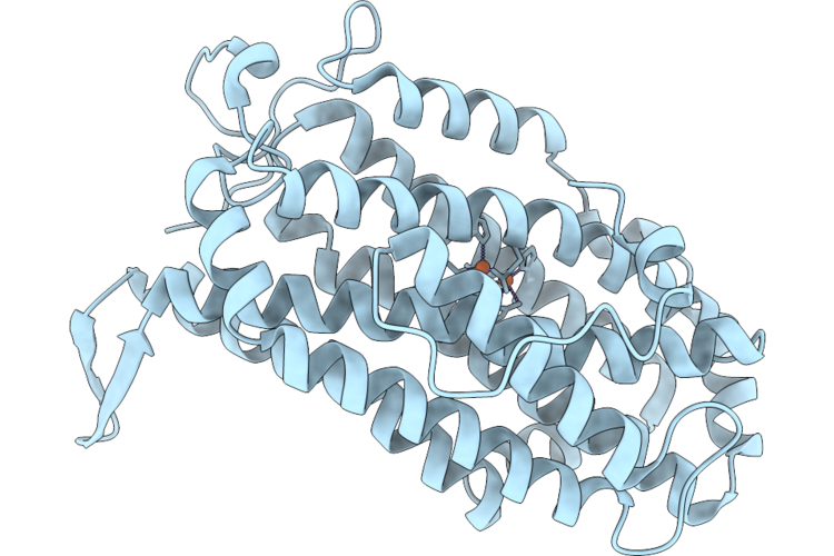 |
Xfel Structure Of Oxidised Ribonucleotide Reductase R2A Y122F Mutant From E. Coli, Hexagonal P6122 Form
Organism: Escherichia coli
Method: X-RAY DIFFRACTION Resolution:2.70 Å Release Date: 2026-05-13 Classification: OXIDOREDUCTASE Ligands: FE |
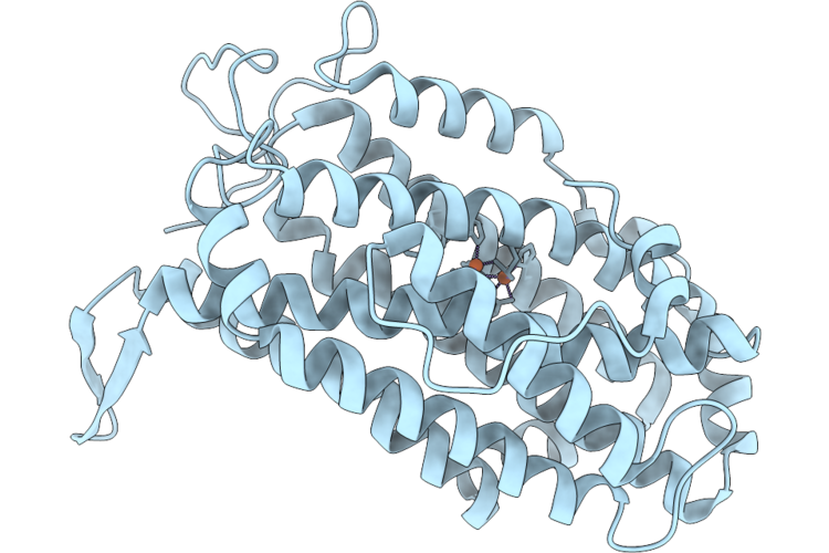 |
Xfel Structure Of Ribonucleotide Reductase R2A Y122F Mutant From E. Coli,Reduced Form, Hexagonal P6122
Organism: Escherichia coli
Method: X-RAY DIFFRACTION Resolution:2.70 Å Release Date: 2026-05-13 Classification: OXIDOREDUCTASE Ligands: FE2 |
 |
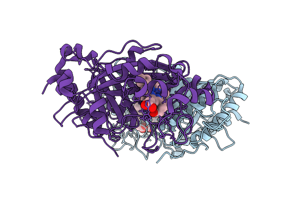
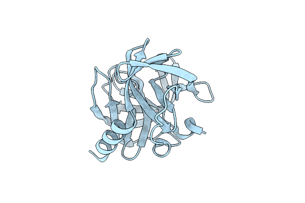
Urate Oxidase From Aspergillus Flavus With Its Inhibitor 9-Methyl Uric Acid By Continuous Serial Electron Diffraction (Serialed)
Organism: Aspergillus flavus
Method: ELECTRON CRYSTALLOGRAPHY Resolution:1.25 Å Release Date: 2026-04-29 Classification: OXIDOREDUCTASE Ligands: MUA |
 |
Urate Oxidase From Aspergillus Flavus With Its Substrate Uric Acid By Continuous Serial Electron Diffraction (Serialed)
Organism: Aspergillus flavus
Method: ELECTRON CRYSTALLOGRAPHY Resolution:1.75 Å Release Date: 2026-04-29 Classification: OXIDOREDUCTASE Ligands: OXY, URC |
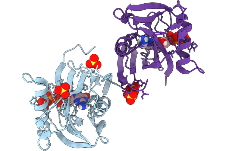 |
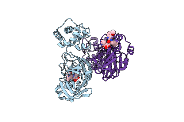
Structure Of Human Mth1 In Complex With 8Dg By Continuous Serial Electron Diffraction (Serialed)
Organism: Homo sapiens
Method: ELECTRON CRYSTALLOGRAPHY Resolution:1.66 Å Release Date: 2026-04-22 Classification: HYDROLASE Ligands: SO4, 8DG |
 |
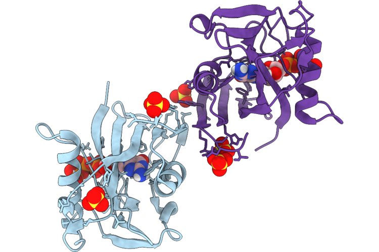
Structure Of Human Mth1 In Complex With 8Dg By Microed Using High Electron Fluence
Organism: Homo sapiens
Method: ELECTRON CRYSTALLOGRAPHY Resolution:2.32 Å Release Date: 2026-04-22 Classification: HYDROLASE Ligands: 8DG, SO4 |
 |
Structure Of Human Mth1 In Complex With 8Dg By Microed Using Low Electron Fluence
Organism: Homo sapiens
Method: ELECTRON CRYSTALLOGRAPHY Resolution:2.86 Å Release Date: 2026-04-22 Classification: HYDROLASE Ligands: 8DG, SO4 |
 |
Organism: Gallus gallus
Method: ELECTRON CRYSTALLOGRAPHY Resolution:0.83 Å Release Date: 2026-04-22 Classification: HYDROLASE Ligands: ACT, CL |
 |
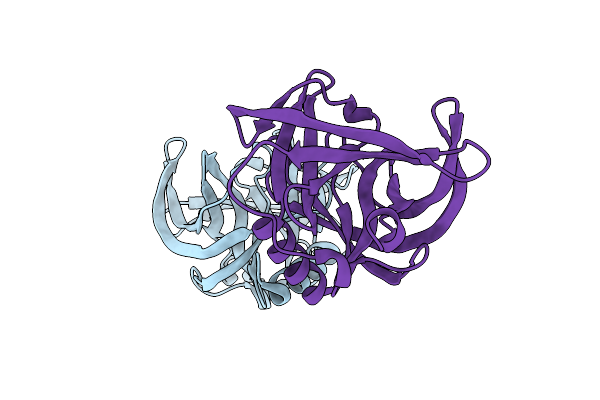
Organism: Lama glama, Homo sapiens
Method: ELECTRON MICROSCOPY Resolution:3.06 Å Release Date: 2025-12-17 Classification: LIGASE |
 |
Dye Type Peroxidase Aa From Streptomyces Lividans With N3 Ligand By Serial Electron Diffraction (Serialed)
Organism: Streptomyces lividans
Method: ELECTRON CRYSTALLOGRAPHY Resolution:1.10 Å Release Date: 2025-07-16 Classification: OXIDOREDUCTASE Ligands: AZI, HEM |
 |
Dye Type Peroxidase Aa From Streptomyces Lividans By Microcrystal Electron Diffraction (Microed/3D Ed)
Organism: Streptomyces lividans
Method: ELECTRON CRYSTALLOGRAPHY Resolution:2.40 Å Release Date: 2025-07-16 Classification: OXIDOREDUCTASE Ligands: HEM |
 |
Dye Type Peroxidase Aa From Streptomyces Lividans By Serial Electron Diffraction (Serialed)
Organism: Streptomyces lividans
Method: ELECTRON CRYSTALLOGRAPHY Resolution:1.30 Å Release Date: 2025-07-16 Classification: OXIDOREDUCTASE Ligands: HEM |
 |
Organism: Homo sapiens
Method: X-RAY DIFFRACTION Resolution:2.30 Å Release Date: 2025-06-25 Classification: HYDROLASE Ligands: ZN, MG, A1EBE |
 |
Organism: Coxsackievirus b3
Method: X-RAY DIFFRACTION Resolution:2.10 Å Release Date: 2024-08-14 Classification: VIRAL PROTEIN |
 |
Organism: Coxsackievirus b4
Method: X-RAY DIFFRACTION Resolution:2.01 Å Release Date: 2024-08-14 Classification: VIRAL PROTEIN |
 |
Crystal Structure Of Sars-Cov-2 Main Protease M49I Mutant In Complex With Pf00835231
Organism: Severe acute respiratory syndrome coronavirus 2
Method: X-RAY DIFFRACTION Resolution:2.21 Å Release Date: 2024-05-01 Classification: VIRAL PROTEIN/INHIBITOR Ligands: V2M |
 |
Organism: Severe acute respiratory syndrome coronavirus 2
Method: X-RAY DIFFRACTION Resolution:2.21 Å Release Date: 2024-04-17 Classification: VIRAL PROTEIN/INHIBITOR Ligands: V2M |
 |
Organism: Middle east respiratory syndrome-related coronavirus
Method: X-RAY DIFFRACTION Resolution:2.30 Å Release Date: 2024-04-17 Classification: VIRAL PROTEIN/INHIBITOR Ligands: V2M |