
Search Count: 46
 |
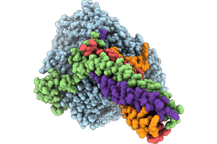
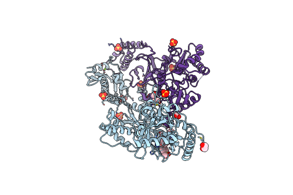
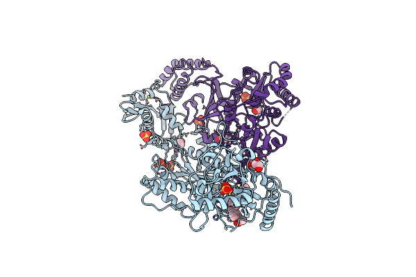
Cryo-Em Structure Of Nipah Virus Polymerase In Complex With G671
Organism: Henipavirus nipahense
Method: ELECTRON MICROSCOPY Resolution:2.92 Å Release Date: 2026-04-29 Classification: VIRAL PROTEIN Ligands: A1E3H, ZN |
 |
Cryo-Em Structure Of Nipah Virus Polymerase In Complex With Erdrp-0519
Organism: Henipavirus nipahense
Method: ELECTRON MICROSCOPY Resolution:2.66 Å Release Date: 2026-04-29 Classification: REPLICATION Ligands: ZN, A1EF9 |
 |
Cryo-Em Structure Of Measles Virus Polymerase In Complex With Erdrp-0519
Organism: Measles morbillivirus, Measles virus genotype b3
Method: ELECTRON MICROSCOPY Resolution:2.73 Å Release Date: 2026-04-29 Classification: REPLICATION Ligands: ZN, A1EF9 |
 |
Cryo-Em Structure Of Nipah Virus Polymerase In Complex With Gl22
Organism: Henipavirus nipahense
Method: ELECTRON MICROSCOPY Resolution:2.87 Å Release Date: 2026-04-29 Classification: REPLICATION Ligands: ZN, A1ET4 |
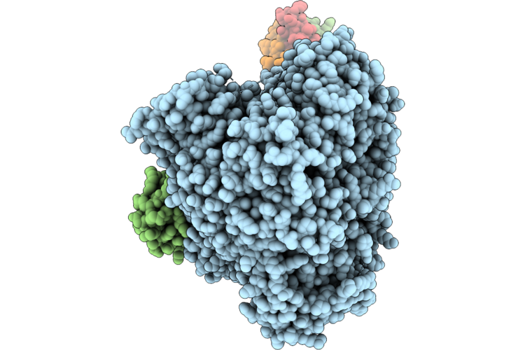 |
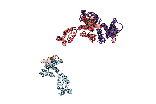
Cryo-Em Structure Of Measles Virus L Protein Bound By Phosphoprotein Tetramer
Organism: Measles morbillivirus
Method: ELECTRON MICROSCOPY Resolution:2.68 Å Release Date: 2026-04-29 Classification: VIRAL PROTEIN Ligands: ZN |
 |
Cryo-Em Structure Of Peste Des Petits Ruminants Virus Polymerase In Complex With Erdrp-0519
Organism: Peste-des-petits-ruminants virus
Method: ELECTRON MICROSCOPY Resolution:2.71 Å Release Date: 2026-04-29 Classification: VIRAL PROTEIN Ligands: ZN, A1EF9 |
 |
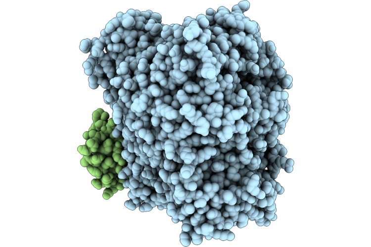
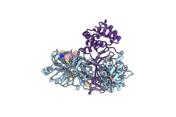
Cryo-Em Structure Of Peste Des Petits Ruminants Virus L Protein Bound By Phosphoprotein Tetramer
Organism: Peste-des-petits-ruminants virus
Method: ELECTRON MICROSCOPY Resolution:2.40 Å Release Date: 2026-04-29 Classification: VIRAL PROTEIN |
 |
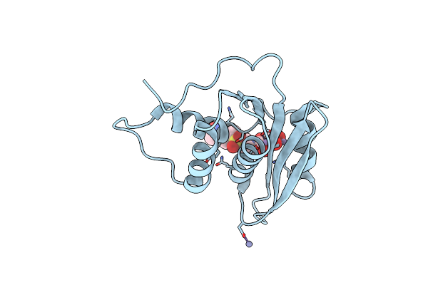
Hiv-Ca Disulfide Linked Hexamer Bound To 4-Quinazolinone Scaffold Inhibitor
Organism: Human immunodeficiency virus 1
Method: X-RAY DIFFRACTION Resolution:1.80 Å Release Date: 2025-07-23 Classification: VIRAL PROTEIN Ligands: A1ADQ, IOD |
 |
Crystal Structure Of Sars-Cov-2 Main Protease (Mpro) In Complex With The Noncovalently Bound Inhibitor C5N17A
Organism: Severe acute respiratory syndrome coronavirus 2
Method: X-RAY DIFFRACTION Resolution:1.65 Å Release Date: 2025-07-02 Classification: VIRAL PROTEIN Ligands: DMS, A1IHT, CL, MG |
 |
Crystal Structure Of Sars-Cov-2 Main Protease (Mpro) In Complex With The Noncovalently Bound Inhibitor C5N17B
Organism: Severe acute respiratory syndrome coronavirus 2
Method: X-RAY DIFFRACTION Resolution:1.67 Å Release Date: 2025-07-02 Classification: VIRAL PROTEIN Ligands: DMS, A1IHV, IMD |
 |
Structure Of Urat1 With No External Ligand Added
Organism: Homo sapiens
Method: ELECTRON MICROSCOPY Release Date: 2025-06-18 Classification: TRANSPORT PROTEIN |
 |
Structure Of Urat1 In Complex With Benzbromarone
Organism: Homo sapiens
Method: ELECTRON MICROSCOPY Release Date: 2025-06-18 Classification: TRANSPORT PROTEIN Ligands: R75 |
 |
Structure Of Urat1 In Complex With Lesinurad
Organism: Homo sapiens
Method: ELECTRON MICROSCOPY Release Date: 2025-06-18 Classification: TRANSPORT PROTEIN Ligands: A1AIL |
 |
Structure Of Urat1 In Complex With Td-3
Organism: Homo sapiens
Method: ELECTRON MICROSCOPY Release Date: 2025-06-18 Classification: TRANSPORT PROTEIN Ligands: A1A45 |
 |
Crystal Structure Of Gh9 (K101P, K103N, V108I) Hiv-1 Reverse Transcriptase In Complex With Non-Nucleoside Inhibitor 5I3
Organism: Human immunodeficiency virus 1
Method: X-RAY DIFFRACTION Resolution:2.36 Å Release Date: 2025-05-28 Classification: VIRAL PROTEIN,TRANSFERASE/INHIBITOR Ligands: SO4, WB3, EDO, DMS, MG |
 |
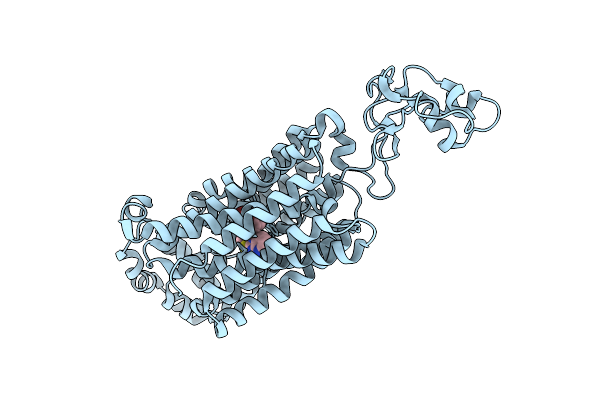
Cryo-Em Structure Of A Truncated Nipah Virus L Protein Bound By Phosphoprotein Tetramer
Organism: Henipavirus nipahense
Method: ELECTRON MICROSCOPY Resolution:2.31 Å Release Date: 2025-05-21 Classification: VIRAL PROTEIN Ligands: ZN |
 |
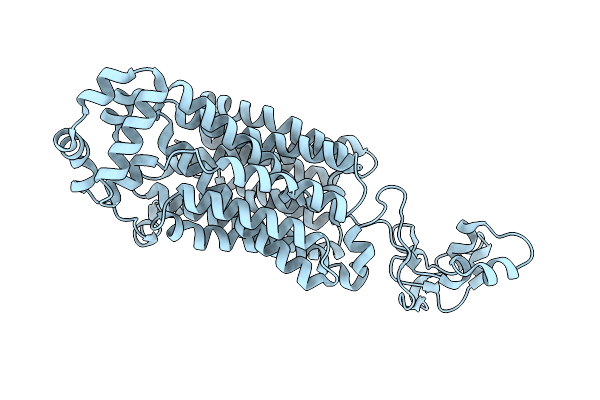
Cryo-Em Structure Of The Full-Length Nipah Virus L Protein Bound By Phosphoprotein Tetramer
Organism: Henipavirus nipahense
Method: ELECTRON MICROSCOPY Resolution:2.52 Å Release Date: 2025-05-21 Classification: VIRAL PROTEIN Ligands: ZN |
 |
Crystal Structure Of Hiv-1 Reverse Transcriptase Rnase H Domain Complexed With A Galloyl Inhibitor
Organism: Human immunodeficiency virus 1
Method: X-RAY DIFFRACTION Resolution:1.96 Å Release Date: 2025-05-14 Classification: VIRAL PROTEIN Ligands: MN, ZN, A1L9C |
 |
Crystal Structure Of Wild-Type Hiv-1 Reverse Transcriptase In Complex With Non-Nucleoside Inhibitor 5I3
Organism: Human immunodeficiency virus type 1 bh10
Method: X-RAY DIFFRACTION Resolution:1.99 Å Release Date: 2025-05-07 Classification: VIRAL PROTEIN,TRANSFERASE/INHIBITOR Ligands: WB3, SO4, EDO, MG |
 |
Crystal Structure Of Wild-Type Hiv-1 Reverse Transcriptase In Complex With Non-Nucleoside Inhibitor 5E2
Organism: Human immunodeficiency virus 1
Method: X-RAY DIFFRACTION Resolution:2.18 Å Release Date: 2025-04-30 Classification: VIRAL PROTEIN,TRANSFERASE/INHIBITOR Ligands: PKX, SO4, EDO, MG |