
Search Count: 115
All
Selected
 |
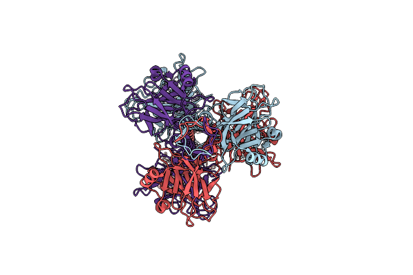
Organism: Mus musculus
Method: ELECTRON MICROSCOPY Release Date: 2025-09-17 Classification: DNA BINDING PROTEIN/DNA Ligands: ANP, MG |
 |
Organism: Mus musculus
Method: ELECTRON MICROSCOPY Release Date: 2025-09-10 Classification: DNA BINDING PROTEIN/DNA Ligands: ANP, MG |
 |
Organism: Mus musculus
Method: ELECTRON MICROSCOPY Release Date: 2025-09-10 Classification: DNA BINDING PROTEIN/DNA Ligands: MG, ANP |
 |
Organism: Mus musculus
Method: ELECTRON MICROSCOPY Release Date: 2025-09-10 Classification: DNA BINDING PROTEIN/DNA Ligands: ANP, MG |
 |
Organism: Mus musculus
Method: ELECTRON MICROSCOPY Release Date: 2025-09-10 Classification: DNA BINDING PROTEIN/DNA Ligands: ANP, MG |
 |
Cryo-Em Structure Of Mycobacteriophage Douge Genome-Packed Vertex (Gp8 And Gp113)
Organism: Mycolicibacterium smegmatis mc2 155
Method: ELECTRON MICROSCOPY Release Date: 2025-07-16 Classification: VIRAL PROTEIN |
 |
Organism: Mycolicibacterium smegmatis mc2 155
Method: ELECTRON MICROSCOPY Release Date: 2025-07-16 Classification: VIRAL PROTEIN |
 |
Cryo-Em Structure Of Mycobacteriophage Douge Genome-Packed Capsid (Gp8 And Gp113)
Organism: Mycolicibacterium smegmatis mc2 155
Method: ELECTRON MICROSCOPY Release Date: 2025-06-25 Classification: VIRUS |
 |
Organism: Mycolicibacterium smegmatis mc2 155
Method: ELECTRON MICROSCOPY Release Date: 2025-06-25 Classification: VIRUS |
 |
Cryo-Em Structure Of Mycobacteriophage Douge Genome-Packed Connector (Gp5, Gp9, Gp10, Gp12 And Gp13)
Organism: Mycolicibacterium smegmatis mc2 155
Method: ELECTRON MICROSCOPY Release Date: 2025-06-25 Classification: VIRAL PROTEIN |
 |
Cryo-Em Structure Of Mycobacteriophage Douge Genome-Free Connector (Gp5, Gp9, Gp10, Gp12 And Gp13)
Organism: Mycolicibacterium smegmatis mc2 155
Method: ELECTRON MICROSCOPY Release Date: 2025-06-25 Classification: VIRAL PROTEIN |
 |
Organism: Mycolicibacterium smegmatis mc2 155
Method: ELECTRON MICROSCOPY Release Date: 2025-06-25 Classification: VIRAL PROTEIN |
 |
Cryo-Em Structure Of Mycobacteriophage Douge Baseplate (Gp13, Gp17, Gp23, Gp16, Gp18 And Gp20)
Organism: Mycolicibacterium smegmatis mc2 155
Method: ELECTRON MICROSCOPY Release Date: 2025-06-25 Classification: VIRAL PROTEIN |
 |
Organism: Mycolicibacterium smegmatis mc2 155
Method: ELECTRON MICROSCOPY Release Date: 2025-06-25 Classification: VIRAL PROTEIN |
 |
Cryo-Em Structure Of Mycobacteriophage Douge Complete Baseplate (Gp13, Gp17, Gp23, Gp16, Gp18 And Gp20)
Organism: Mycolicibacterium smegmatis mc2 155
Method: ELECTRON MICROSCOPY Release Date: 2025-06-25 Classification: VIRAL PROTEIN |
 |
Cryo-Em Structure Of Mycobacteriophage Douge Genome-Packed Tail Tube (Gp13)
Organism: Mycolicibacterium smegmatis mc2 155
Method: ELECTRON MICROSCOPY Release Date: 2025-06-25 Classification: VIRAL PROTEIN |
 |
Organism: Chlamydomonas reinhardtii cc3269
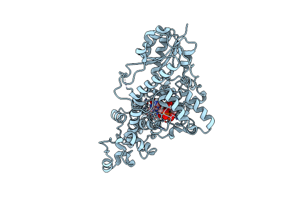
Method: X-RAY DIFFRACTION Resolution:1.64 Å Release Date: 2025-05-14 Classification: FLAVOPROTEIN Ligands: FAD, CL |
 |
Sfx Structure Of Cracry 10 Ns After Photoexcitation Of The Oxidized Protein
Organism: Chlamydomonas reinhardtii
Method: X-RAY DIFFRACTION Resolution:1.95 Å Release Date: 2025-05-14 Classification: FLAVOPROTEIN Ligands: FAD, CL |
 |
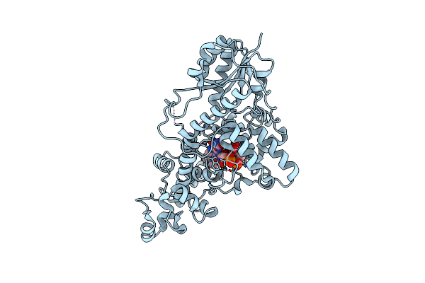
Sfx Structure Of Cracry 30 Ns After Photoexcitation Of The Oxidized Protein
Organism: Chlamydomonas reinhardtii
Method: X-RAY DIFFRACTION Resolution:2.00 Å Release Date: 2025-05-14 Classification: FLAVOPROTEIN Ligands: FAD, CL |
 |
Sfx Structure Of Cracry 100 Ns After Photoexcitation Of The Oxidized Protein
Organism: Chlamydomonas reinhardtii
Method: X-RAY DIFFRACTION Resolution:2.00 Å Release Date: 2025-05-14 Classification: FLAVOPROTEIN Ligands: FAD, CL |