
Search Count: 396
All
Selected
 |
Organism: Pseudomonas phage phiyy
Method: ELECTRON MICROSCOPY Release Date: 2026-04-01 Classification: VIRUS |
 |
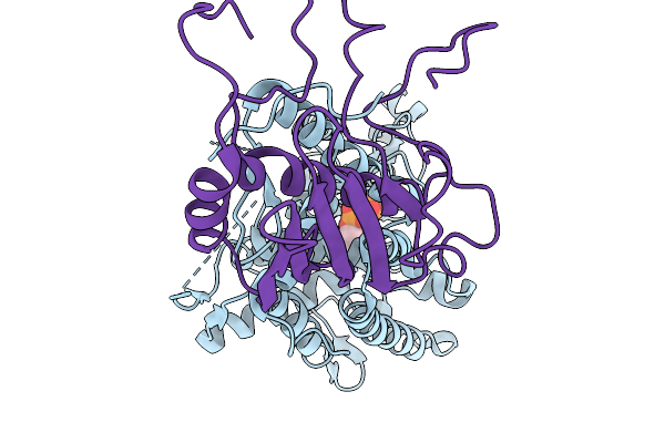
Functional Characterization Of Type Iii Polyketide Synthases Involved In Tropane Alkaloid Biosynthesis In Brassicaceae
Organism: Cochlearia officinalis
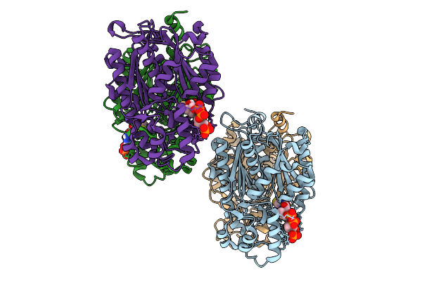
Method: X-RAY DIFFRACTION Resolution:2.15 Å Release Date: 2026-02-25 Classification: BIOSYNTHETIC PROTEIN Ligands: COA |
 |
Organism: Roseiflexus castenholzii
Method: ELECTRON MICROSCOPY Resolution:2.28 Å Release Date: 2026-02-18 Classification: STRUCTURAL PROTEIN Ligands: 07D |
 |
Organism: Rhodospirillum rubrum
Method: ELECTRON MICROSCOPY Resolution:2.74 Å Release Date: 2026-02-18 Classification: STRUCTURAL PROTEIN Ligands: 07D, 08I, U10, PGV, LMT, 8K6, PEF, FE, CRT, RQ0, CDL |
 |
The Local Refined Map Of Sars-Cov-2 Eg.5.1 Variant Spike Protein Complexed With Antibody Xgi-171
Organism: Homo sapiens, Severe acute respiratory syndrome coronavirus 2
Method: ELECTRON MICROSCOPY Release Date: 2025-12-10 Classification: VIRAL PROTEIN/IMMUNE SYSTEM |
 |
The Local Refined Map Of Sars-Cov-2 Eg.5.1 Variant Spike Protein Complexed With Antibody Xgi-183
Organism: Homo sapiens, Severe acute respiratory syndrome coronavirus 2
Method: ELECTRON MICROSCOPY Release Date: 2025-12-10 Classification: VIRAL PROTEIN/IMMUNE SYSTEM |
 |
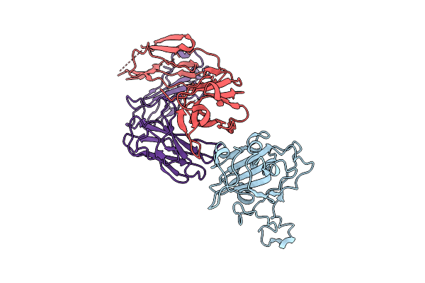
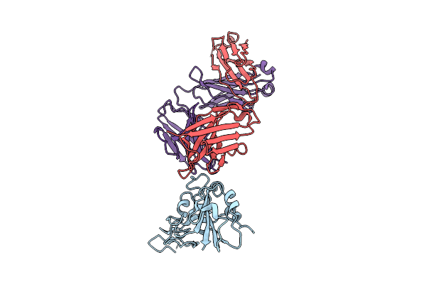
Structure Of Sars-Cov-2 Eg.5.1 Variant Spike Protein Complexed With Antibody Xgi-171
Organism: Homo sapiens, Severe acute respiratory syndrome coronavirus 2
Method: ELECTRON MICROSCOPY Release Date: 2025-12-10 Classification: VIRAL PROTEIN/IMMUNE SYSTEM Ligands: NAG |
 |
The Local Refined Map Of Sars-Cov-2 Eg.5.1 Variant Spike Protein Complexed With Antibody Xgi-198
Organism: Homo sapiens, Severe acute respiratory syndrome coronavirus 2
Method: ELECTRON MICROSCOPY Release Date: 2025-12-10 Classification: VIRAL PROTEIN/IMMUNE SYSTEM |
 |
The Local Refined Map Of Sars-Cov-2 Eg.5.1 Variant Spike Protein Complexed With Antibody Xgi-203
Organism: Severe acute respiratory syndrome coronavirus 2, Homo sapiens
Method: ELECTRON MICROSCOPY Release Date: 2025-12-10 Classification: VIRAL PROTEIN/IMMUNE SYSTEM |
 |
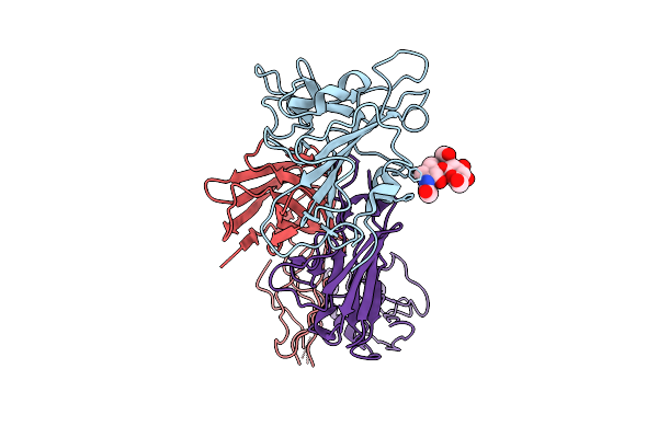
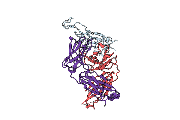
Structure Of Sars-Cov-2 Eg.5.1 Variant Spike Protein Complexed With Antibody Xgi-203
Organism: Homo sapiens, Severe acute respiratory syndrome coronavirus 2
Method: ELECTRON MICROSCOPY Release Date: 2025-12-10 Classification: VIRAL PROTEIN/IMMUNE SYSTEM Ligands: NAG |
 |
Structure Of Sars-Cov-2 Eg.5.1 Variant Spike Protein Complexed With Antibody Xgi-198
Organism: Severe acute respiratory syndrome coronavirus 2, Homo sapiens
Method: ELECTRON MICROSCOPY Release Date: 2025-12-10 Classification: VIRAL PROTEIN/IMMUNE SYSTEM Ligands: NAG |
 |
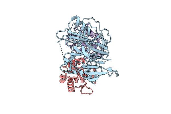
Organism: Serratia marcescens
Method: ELECTRON MICROSCOPY Release Date: 2025-11-19 Classification: ANTIVIRAL PROTEIN Ligands: GTP |
 |
Organism: Homo sapiens
Method: X-RAY DIFFRACTION Resolution:1.90 Å Release Date: 2025-10-22 Classification: CHAPERONE Ligands: MG |
 |
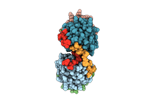
Cryo-Em Structure Of The Agr2 Dimer In Complex With The Monomeric Ire1Beta Luminal Domain
Organism: Homo sapiens
Method: ELECTRON MICROSCOPY Resolution:2.90 Å Release Date: 2025-10-22 Classification: CHAPERONE |
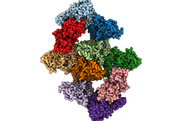 |
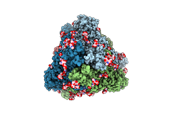
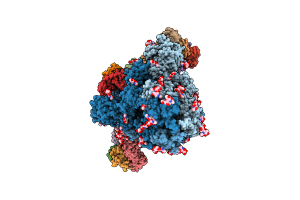
The Cryo-Em Structure Of The Human Dna Methyltransferases Dnmt3A2 And Dnmt3L Dodecamer
Organism: Homo sapiens
Method: ELECTRON MICROSCOPY Release Date: 2025-10-08 Classification: DNA BINDING PROTEIN Ligands: ZN, SAH |
 |
The Cryo-Em Structure Of The Human Dna Methyltransferase Dnmt3A2 And Dnmt3L Octamer
Organism: Homo sapiens
Method: ELECTRON MICROSCOPY Release Date: 2025-10-08 Classification: DNA BINDING PROTEIN Ligands: ZN, SAH |
 |
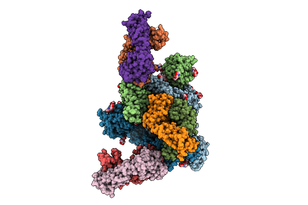
The Cryo-Em Structure Of Nucleosome-Bound Dna Methyltransferases Dnmt3A2 And Dnmt3L
Organism: Xenopus laevis, Synthetic construct, Homo sapiens
Method: ELECTRON MICROSCOPY Release Date: 2025-10-08 Classification: DNA BINDING PROTEIN Ligands: ZN, SAH |
 |
Organism: Homo sapiens, Synthetic construct
Method: ELECTRON MICROSCOPY Release Date: 2025-09-03 Classification: DNA BINDING PROTEIN/DNA |
 |
Organism: Homo sapiens, Synthetic construct, Sus scrofa
Method: ELECTRON MICROSCOPY Release Date: 2025-09-03 Classification: TRANSCRIPTION Ligands: ZN, MG |
 |
Organism: Streptomyces albogriseolus 1-36
Method: X-RAY DIFFRACTION Resolution:2.19 Å Release Date: 2025-02-19 Classification: LIGASE |