

Search Count: 69
 |
Cryo-Em Structure Of The Lh1 Complex From Roseiflexus Castenholzii
Organism: Roseiflexus castenholzii
Method: ELECTRON MICROSCOPY Resolution:2.28 Å Release Date: 2026-02-18 Classification: STRUCTURAL PROTEIN Ligands: 07D |
Organism: Roseiflexus castenholzii
Method: ELECTRON MICROSCOPY
Release Date: 2026-02-18
Ligands: 07D
 |
Cryo-Em Structure Of The Rc Complex From Rhodospirillum Rubrum
Organism: Rhodospirillum rubrum
Method: ELECTRON MICROSCOPY Resolution:2.74 Å Release Date: 2026-02-18 Classification: STRUCTURAL PROTEIN Ligands: 07D, 08I, U10, PGV, LMT, 8K6, PEF, FE, CRT, RQ0, CDL |
Organism: Rhodospirillum rubrum
Method: ELECTRON MICROSCOPY
Release Date: 2026-02-18
Ligands: 07D, 08I, U10, PGV, LMT, 8K6, PEF, FE, CRT, RQ0, CDL
 |
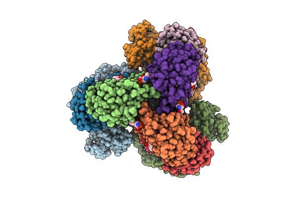
Structure Of Phage T4 Topoisomerase Ii Central Domain
Organism: Enterobacteria phage t4
Method: ELECTRON MICROSCOPY Release Date: 2025-11-26 Classification: ISOMERASE |
Organism: Enterobacteria phage t4
Method: ELECTRON MICROSCOPY
Release Date: 2025-11-26
 |
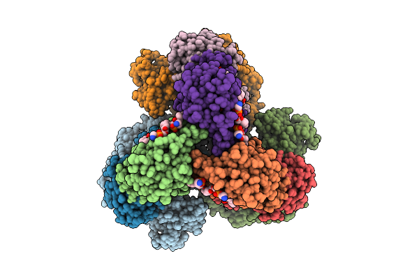
Cryo-Em Structure Of The Apo-Form Succinate Dehydrogenase From Chloroflexus Aurantiacus
Organism: Chloroflexus aurantiacus j-10-fl
Method: ELECTRON MICROSCOPY Release Date: 2025-11-12 Classification: MEMBRANE PROTEIN Ligands: FAD, SIN, SF4, FES, F3S, HEM, LMT, PGV, PEV |
Organism: Chloroflexus aurantiacus j-10-fl
Method: ELECTRON MICROSCOPY
Release Date: 2025-11-12
Ligands: FAD, SIN, SF4, FES, F3S, HEM, LMT, PGV, PEV
 |
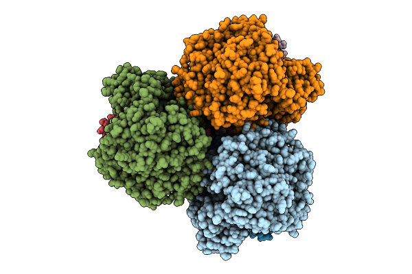
Cryo-Em Structure Of The Lipid-Bound Succiante Dehydrogenase From Chloroflexus Aurantiacus
Organism: Chloroflexus aurantiacus j-10-fl
Method: ELECTRON MICROSCOPY Release Date: 2025-11-12 Classification: MEMBRANE PROTEIN Ligands: FAD, SIN, SF4, FES, F3S, HEM, LMT, PGV, PEV |
Organism: Chloroflexus aurantiacus j-10-fl
Method: ELECTRON MICROSCOPY
Release Date: 2025-11-12
Ligands: FAD, SIN, SF4, FES, F3S, HEM, LMT, PGV, PEV
 |
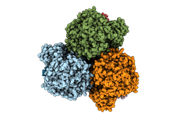
Cryo-Em Structure Of The Mk7-Bound Succinate Dehydrogenase From Chloroflexus Aurantiacus
Organism: Chloroflexus aurantiacus j-10-fl
Method: ELECTRON MICROSCOPY Release Date: 2025-11-12 Classification: MEMBRANE PROTEIN Ligands: FAD, SF4, FES, F3S, HEM, LMT, MQ7 |
Organism: Chloroflexus aurantiacus j-10-fl
Method: ELECTRON MICROSCOPY
Release Date: 2025-11-12
Ligands: FAD, SF4, FES, F3S, HEM, LMT, MQ7
 |
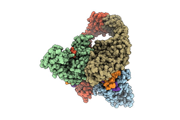
Cryo-Em Structure Of The Mk4-Bound Succinate Dehydrogenase From Chloroflexus Aurantiacus
Organism: Chloroflexus aurantiacus j-10-fl
Method: ELECTRON MICROSCOPY Release Date: 2025-11-12 Classification: MEMBRANE PROTEIN Ligands: FAD, SF4, FES, F3S, HEM, LMT, 1L3 |
Organism: Chloroflexus aurantiacus j-10-fl
Method: ELECTRON MICROSCOPY
Release Date: 2025-11-12
Ligands: FAD, SF4, FES, F3S, HEM, LMT, 1L3
 |
Structure Of Phage T4 Topoisomerase Ii Central Domain Bound With Dna
Organism: Escherichia phage t4, Homo sapiens
Method: ELECTRON MICROSCOPY Release Date: 2025-10-22 Classification: ISOMERASE Ligands: MG |
Organism: Escherichia phage t4, Homo sapiens
Method: ELECTRON MICROSCOPY
Release Date: 2025-10-22
Ligands: MG
 |
Bacteriophage T4 Topoisomerse Ii Bound With A T-Segment Dna
Organism: Escherichia phage t4, Dna molecule
Method: ELECTRON MICROSCOPY Release Date: 2025-10-01 Classification: ISOMERASE/DNA |
Organism: Escherichia phage t4, Dna molecule
Method: ELECTRON MICROSCOPY
Release Date: 2025-10-01
 |
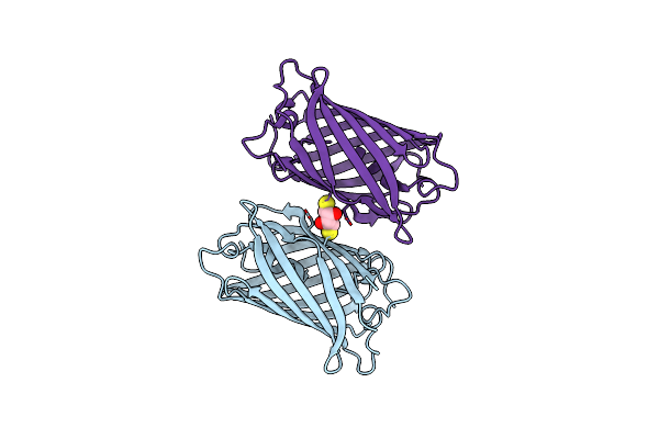
Structure Of Meos3.2 In The Green Fluorescent State
Organism: Lobophyllia hemprichii
Method: X-RAY DIFFRACTION Resolution:1.34 Å Release Date: 2025-04-16 Classification: FLUORESCENT PROTEIN Ligands: SO4 |
Organism: Lobophyllia hemprichii
Method: X-RAY DIFFRACTION
Release Date: 2025-04-16
Ligands: SO4
 |
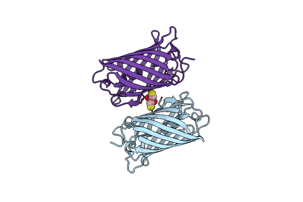
Structure Of Pcstar In The Green Fluorescent State
Organism: Lobophyllia hemprichii
Method: X-RAY DIFFRACTION Resolution:1.66 Å Release Date: 2025-04-16 Classification: FLUORESCENT PROTEIN Ligands: DTT |
Organism: Lobophyllia hemprichii
Method: X-RAY DIFFRACTION
Release Date: 2025-04-16
Ligands: DTT
 |
Structure Of Meos3.2 In The Green Fluorescent State
Organism: Lobophyllia hemprichii
Method: X-RAY DIFFRACTION Resolution:1.85 Å Release Date: 2025-04-16 Classification: FLUORESCENT PROTEIN Ligands: DTT |
Organism: Lobophyllia hemprichii
Method: X-RAY DIFFRACTION
Release Date: 2025-04-16
Ligands: DTT
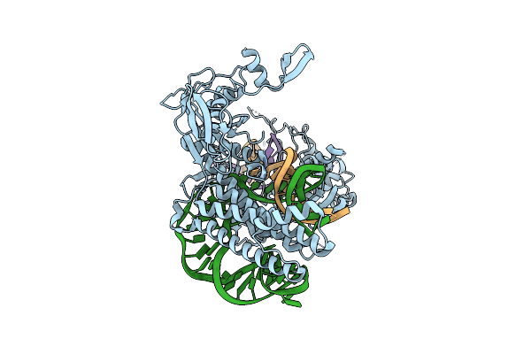 |
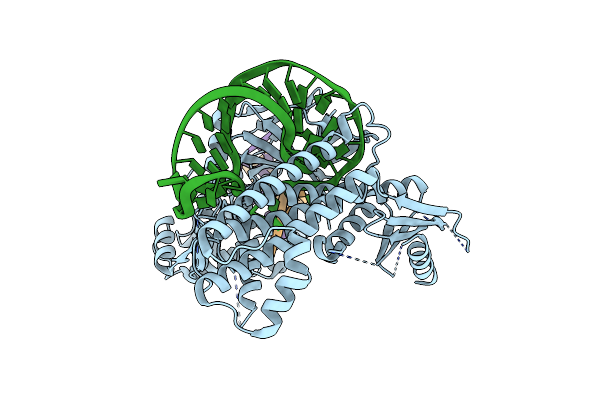
Cryo-Em Structure Of Cas12O1, Crrna And Target Dna Complex
Organism: Unidentified, Unclassified sequences
Method: ELECTRON MICROSCOPY Release Date: 2024-12-11 Classification: RNA BINDING PROTEIN/RNA/DNA |
Organism: Unidentified, Unclassified sequences
Method: ELECTRON MICROSCOPY
Release Date: 2024-12-11
 |
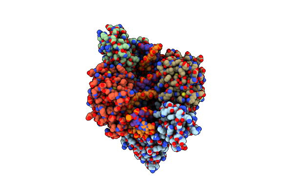
Cryo-Em Structure Of Cas12O1 (E100K), Crrna And Target Dna Complex
Organism: Unclassified sequences
Method: ELECTRON MICROSCOPY Release Date: 2024-12-11 Classification: RNA BINDING PROTEIN/RNA/DNA |
Organism: Unclassified sequences
Method: ELECTRON MICROSCOPY
Release Date: 2024-12-11
 |
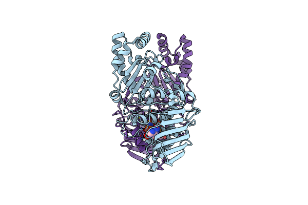
Structure Of Phage T6 Topoisomerase Ii Central Domain Bound With Dna
Organism: Escherichia phage t4, Salmonella phage chi, Dna molecule
Method: ELECTRON MICROSCOPY Release Date: 2024-09-25 Classification: ISOMERASE Ligands: MG |
Organism: Escherichia phage t4, Salmonella phage chi, Dna molecule
Method: ELECTRON MICROSCOPY
Release Date: 2024-09-25
Ligands: MG
 |
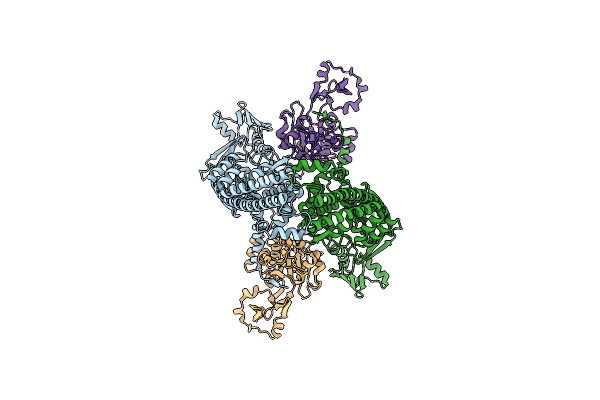
Structure Of Phage T6 Topoisomerase Ii Atpase Domain Bound With Amppnp
Organism: Enterobacteria phage t6
Method: ELECTRON MICROSCOPY Release Date: 2024-09-25 Classification: ISOMERASE Ligands: ANP, MG |
Organism: Enterobacteria phage t6
Method: ELECTRON MICROSCOPY
Release Date: 2024-09-25
Ligands: ANP, MG
 |
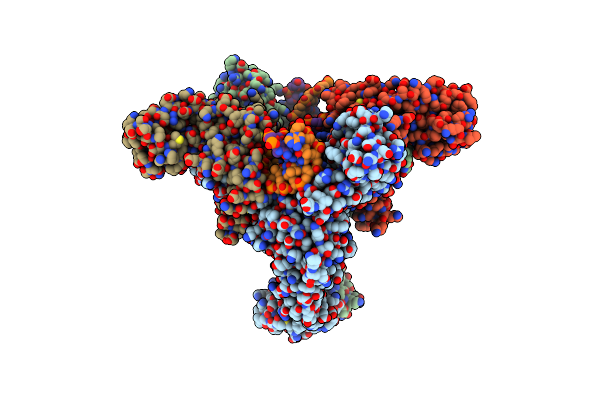
Structure Of Phage T4 Topoisomerase Ii Central Domain
Organism: Escherichia phage t4
Method: ELECTRON MICROSCOPY Release Date: 2024-09-25 Classification: ISOMERASE |
Organism: Escherichia phage t4
Method: ELECTRON MICROSCOPY
Release Date: 2024-09-25
 |
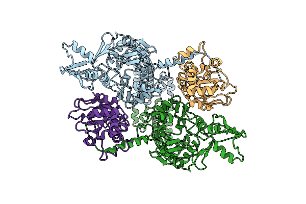
Structure Of Phage T4 Topoisomerase Ii Central Domain Bound With Dna
Organism: Escherichia phage t4, Dna molecule
Method: ELECTRON MICROSCOPY Release Date: 2024-09-25 Classification: ISOMERASE Ligands: MG |
Organism: Escherichia phage t4, Dna molecule
Method: ELECTRON MICROSCOPY
Release Date: 2024-09-25
Ligands: MG
 |
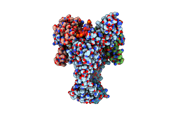
Structure Of Phage T6 Topoisomerase Ii Central Domain
Organism: Escherichia phage t4, Enterobacteria phage t6
Method: ELECTRON MICROSCOPY Release Date: 2024-09-25 Classification: ISOMERASE |
Organism: Escherichia phage t4, Enterobacteria phage t6
Method: ELECTRON MICROSCOPY
Release Date: 2024-09-25
 |
Structure Of Phage T6 Topoisomerase Ii Central Domain Bound With Dna And M-Amsa
Organism: Escherichia phage t4, Enterobacteria phage t6, Dna molecule
Method: ELECTRON MICROSCOPY Release Date: 2024-09-25 Classification: ISOMERASE Ligands: MG, ASW |
Organism: Escherichia phage t4, Enterobacteria phage t6, Dna molecule
Method: ELECTRON MICROSCOPY
Release Date: 2024-09-25
Ligands: MG, ASW