

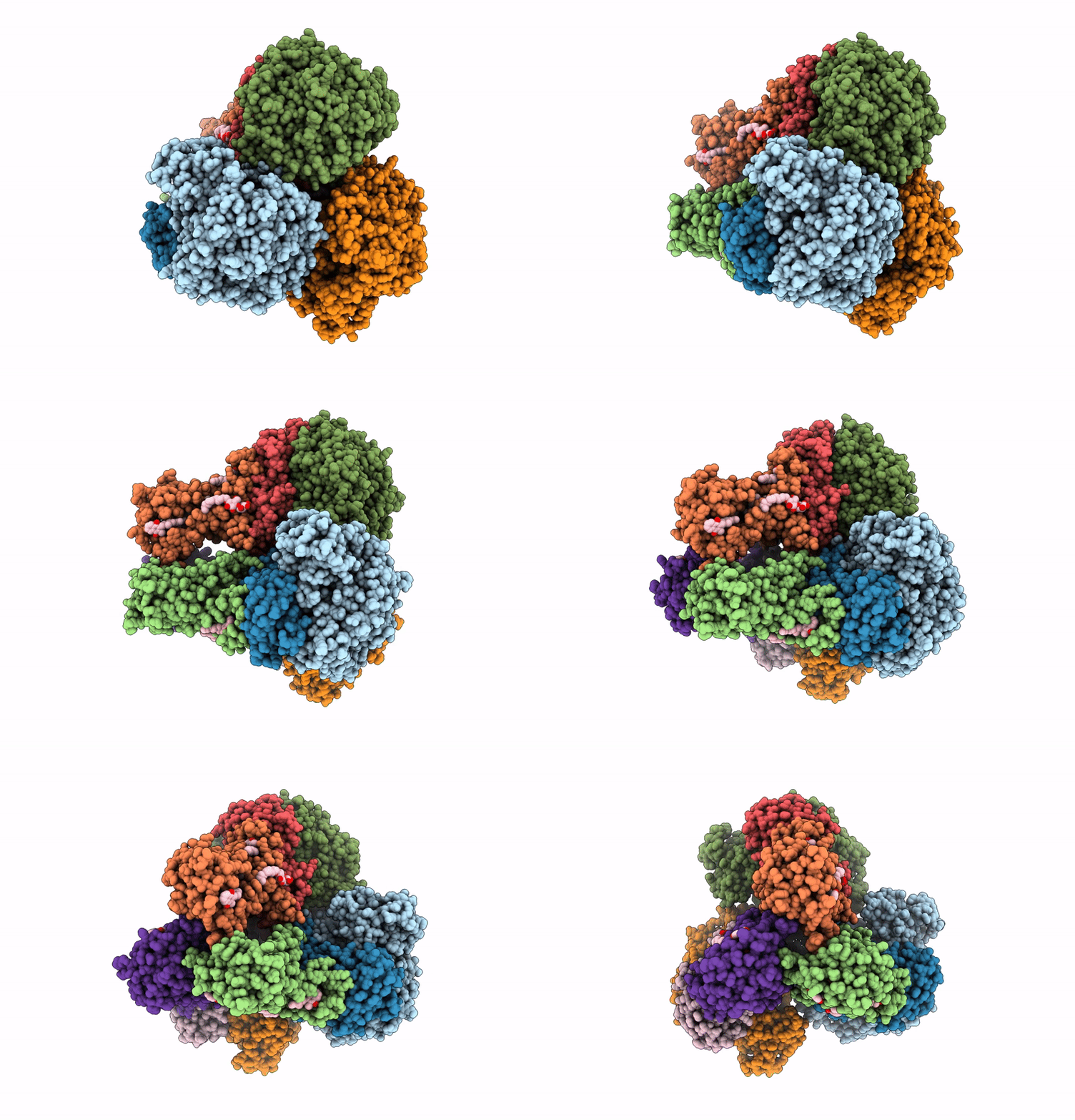
9LB1 image
Deposition Date 2025-01-02
Release Date 2025-11-12
Last Version Date 2026-03-25
Entry Detail
PDB ID:
9LB1
Title:
Cryo-EM structure of the MK4-bound succinate dehydrogenase from Chloroflexus aurantiacus
Biological Source:
Source Organism(s):
Method Details:
Experimental Method:
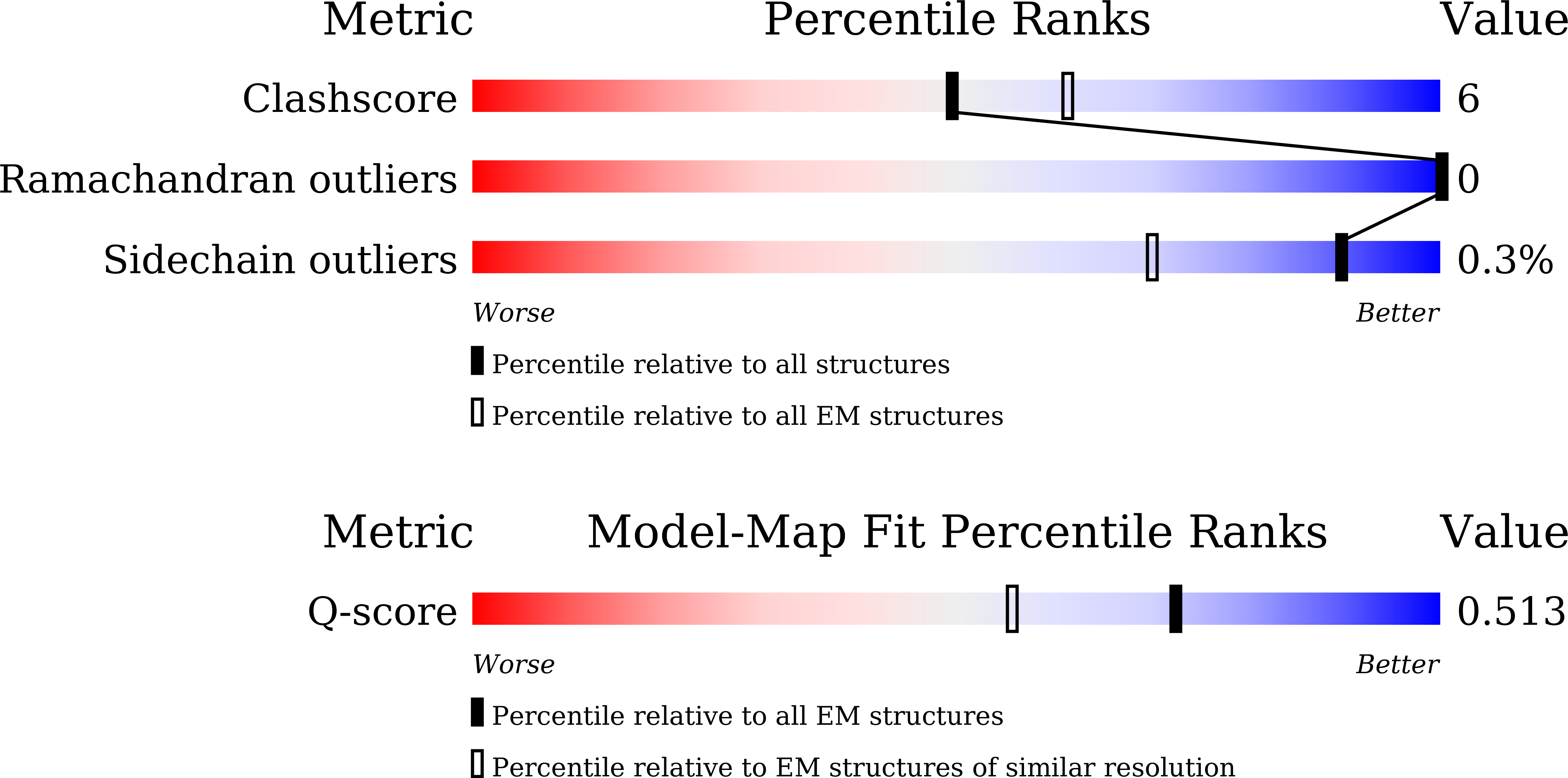
Resolution:
2.90 Å
Aggregation State:
PARTICLE
Reconstruction Method:
SINGLE PARTICLE
Macromolecular Entities
Structures with similar UniProt ID
Protein Blast
Polymer Type:polypeptide(L)
Molecule:Succinate dehydrogenase or fu
Gene (Uniprot):Caur_1881
Chain IDs:A, D, G
Chain Length:657
Number of Molecules:3
Biological Source:Chloroflexus aurantiacus J-10-fl
Structures with similar UniProt ID
Protein Blast
Polymer Type:polypeptide(L)
Molecule:4Fe-4S ferredoxin iron-sulfur
Gene (Uniprot):Caur_1880
Chain IDs:B, E, H
Chain Length:260
Number of Molecules:3
Biological Source:Chloroflexus aurantiacus J-10-fl
Structures with similar UniProt ID
Protein Blast
Polymer Type:polypeptide(L)
Molecule:Succinate dehydrogenase (Or f
Gene (Uniprot):Caur_1882
Chain IDs:C, F, I
Chain Length:239
Number of Molecules:3
Biological Source:Chloroflexus aurantiacus J-10-fl
Primary Citation
Structural basis of menaquinone reduction by succinate dehydrogenase from Chloroflexus aurantiacus.
Nat Commun 16 10782 10782 (2025)
PMID: 41315263 DOI: 10.1038/s41467-025-65828-3

Abstact

Succinate: menaquinone oxidoreductase (SQR) couples the oxidation of succinate with the reduction of menaquinone (MK) as part of the TCA cycle and the aerobic respiratory chain in MK-containing bacteria and archaea. Despite its significance, questions persist regarding the electron and proton transfer mechanisms that drive the endergonic MK reduction by succinate. In this study, we determine cryo-EM structures of succinate dehydrogenase (SDH) from Chloroflexus aurantiacus (CaSDH), a facultative filamentous anoxygenic phototroph (FAP) that forms one of the earliest branches of photosynthetic bacteria. The structures of trimeric CaSDH, resolved in both apo- and MK-bound forms, reveal a single membrane-anchoring subunit containing two b-type hemes, a canonical Q(P) site, and a Q(D) site with atypical location, configuration and specificity, each bound to MK molecules. Using structural analysis, EPR, and enzymatic assays, we uncover electron transfer pathways connecting succinate oxidation to MK reduction at the Q(P) and Q(D) sites. These findings provide structural insights into the electron and proton transfer mechanisms of MK-dependent diheme SQRs and establish a foundation for structure-based inhibitor design and antibacterial drug development targeting these enzymes.

Legend

Protein

Chemical

Disease

Primary Citation of related structures
Feedback Form
Name
Email
Institute
Feedback