
Search Count: 53
All
Selected
 |
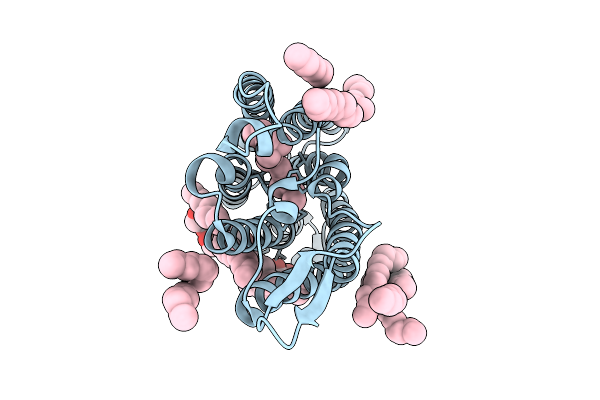
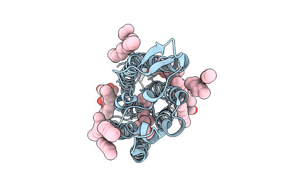
Crystal Structure Of Human Lung Surfactant Protein D Trimeric Fragment With Bound Synthetic Hepi-(1,5)-Kdoi Ligand
Organism: Homo sapiens
Method: X-RAY DIFFRACTION Resolution:1.75 Å Release Date: 2026-03-04 Classification: SURFACTANT PROTEIN Ligands: CA, GMH |
 |
Crystal Structure Of Human Lung Surfactant Protein D Trimeric Fragment With Bound Synthetic Hepii-Hepi-Phosi Ligand
Organism: Homo sapiens
Method: X-RAY DIFFRACTION Resolution:1.92 Å Release Date: 2026-03-04 Classification: SURFACTANT PROTEIN Ligands: CA |
 |
Crystal Structure Of Human Lung Surfactant Protein D Trimeric Fragment With Bound Synthetic Hepiii-Hepii-Hepi Ligand
Organism: Homo sapiens
Method: X-RAY DIFFRACTION Resolution:1.63 Å Release Date: 2026-03-04 Classification: SURFACTANT PROTEIN Ligands: CA, A1JBO |
 |
Crystal Structure Of Human Lung Surfactant Protein D Trimeric Fragment With Bound Synthetic Phosii-Hepii-Hepi Ligand
Organism: Homo sapiens
Method: X-RAY DIFFRACTION Resolution:1.85 Å Release Date: 2026-03-04 Classification: SURFACTANT PROTEIN Ligands: CA, A1JBS, GMH |
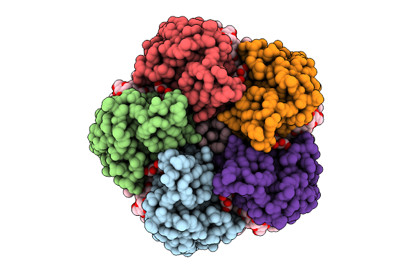 |
Cryo-Em Structure Of The Light-Driven Sodium Pump Ernar In The Pentameric Form
Organism: Erythrobacter
Method: ELECTRON MICROSCOPY Resolution:2.10 Å Release Date: 2025-12-17 Classification: MEMBRANE PROTEIN Ligands: LFA, LMT, RET |
 |
Cryo-Em Structure Of The Light-Driven Sodium Pump Ernar In The Monomeric Form In The K2 State
Organism: Erythrobacter
Method: ELECTRON MICROSCOPY Resolution:2.80 Å Release Date: 2025-12-17 Classification: MEMBRANE PROTEIN Ligands: LFA, LMT, RET |
 |
Cryo-Em Structure Of The Light-Driven Sodium Pump Ernar In The Monomeric Form In The O2 State
Organism: Erythrobacter
Method: ELECTRON MICROSCOPY Resolution:3.30 Å Release Date: 2025-12-17 Classification: MEMBRANE PROTEIN Ligands: LFA, LMT, RET |
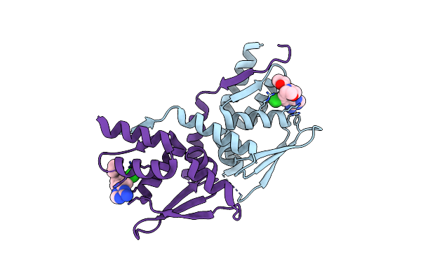 |
Organism: Homo sapiens
Method: X-RAY DIFFRACTION Resolution:1.87 Å Release Date: 2024-10-23 Classification: PROTEIN BINDING Ligands: A1BCT |
 |
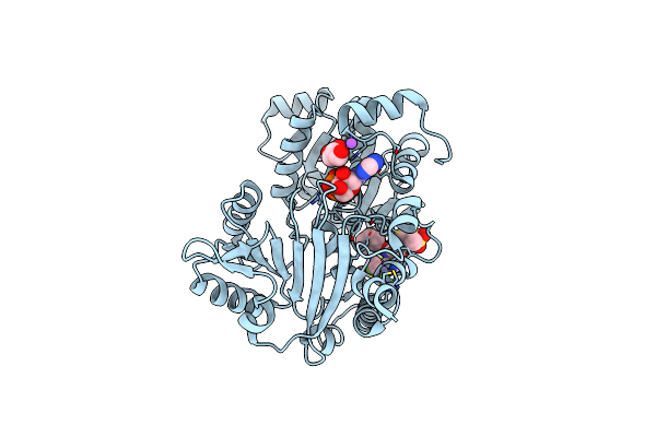
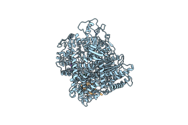
Organism: Homo sapiens
Method: X-RAY DIFFRACTION Resolution:1.57 Å Release Date: 2024-05-01 Classification: HYDROLASE Ligands: ADP, CD, EDO, CL |
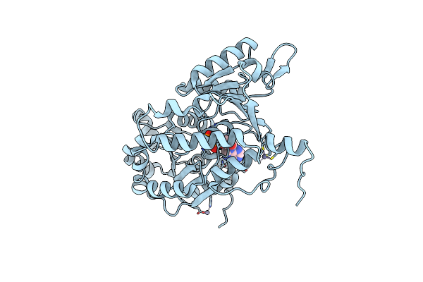 |
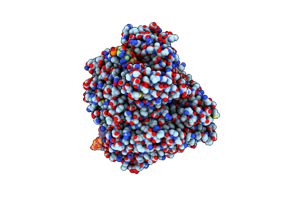
Organism: Homo sapiens
Method: X-RAY DIFFRACTION Resolution:2.21 Å Release Date: 2024-05-01 Classification: HYDROLASE Ligands: ZN, ATP, MG |
 |
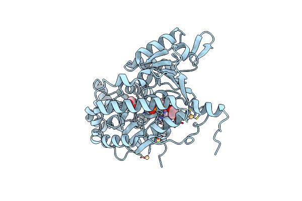
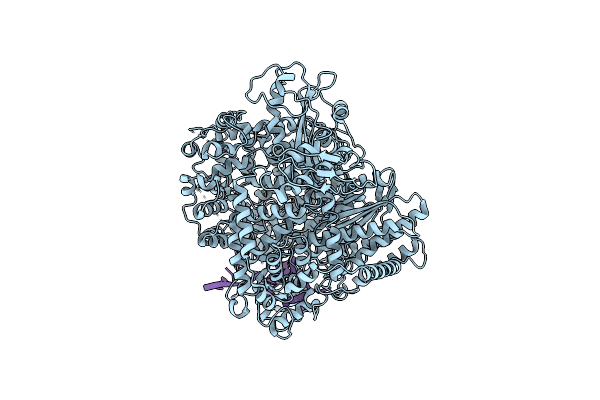
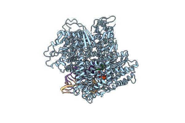
Crystal Structure Of Werner Helicase Fragment 517-945 In Covalent Complex With N-[(E,1S)-1-Cyclopropyl-3-Methylsulfonylprop-2-Enyl]-2-(1,1-Difluoroethyl)-4-Phenoxypyrimidine-5-Carboxamide
Organism: Homo sapiens
Method: X-RAY DIFFRACTION Resolution:1.54 Å Release Date: 2024-05-01 Classification: HYDROLASE Ligands: ZN, ADP, X1L, GOL, NA |
 |
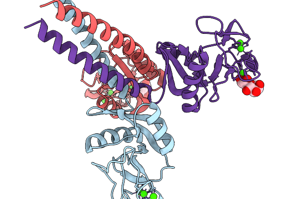
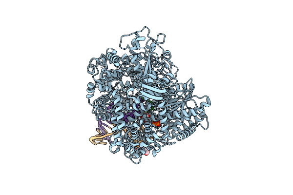
Structure Of The Sftsv L Protein In A Transcription-Priming State Without Capped Rna [Transcription-Priming (In Vitro)]
Organism: Sfts virus ah12
Method: ELECTRON MICROSCOPY Release Date: 2024-04-24 Classification: VIRAL PROTEIN Ligands: MG |
 |
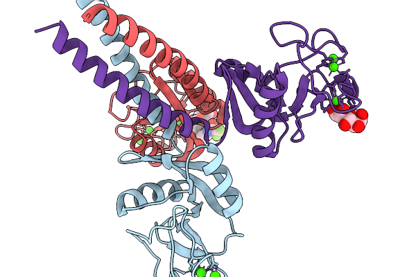
Structure Of The Sftsv L Protein In A Transcription-Priming State With Bound Capped Rna [Transcription-Priming]
Organism: Sfts virus ah12
Method: ELECTRON MICROSCOPY Release Date: 2024-04-24 Classification: VIRAL PROTEIN |
 |
Structure Of The Sftsv L Protein Stalled In A Transcription-Specific Early Elongation State With Bound Capped Rna [Transcription-Early-Elongation]
Organism: Sfts virus ah12
Method: ELECTRON MICROSCOPY Release Date: 2024-04-24 Classification: VIRAL PROTEIN Ligands: 2KH, MG |
 |
Organism: Sfts virus ah12
Method: ELECTRON MICROSCOPY Release Date: 2023-01-18 Classification: VIRAL PROTEIN |
 |
Structure Of The Sftsv L Protein Stalled At Early Elongation [Early-Elongation]
Organism: Sfts virus ah12
Method: ELECTRON MICROSCOPY Release Date: 2023-01-18 Classification: VIRAL PROTEIN Ligands: MG, EPE, 2KH |
 |
Structure Of The Sftsv L Protein Stalled At Early Elongation With The Endonuclease Domain In A Raised Conformation [Early-Elongation-Endo]
Organism: Sfts virus ah12
Method: ELECTRON MICROSCOPY Release Date: 2023-01-18 Classification: VIRAL PROTEIN Ligands: MG, 2KH |
 |
Structure Of The Sftsv L Protein Stalled At Late Elongation [Late-Elongation]
Organism: Sfts virus ah12
Method: ELECTRON MICROSCOPY Release Date: 2023-01-18 Classification: VIRAL PROTEIN Ligands: MG, 2KH |
 |
Organism: Sfts virus ah12
Method: ELECTRON MICROSCOPY Release Date: 2023-01-18 Classification: VIRAL PROTEIN Ligands: MG |
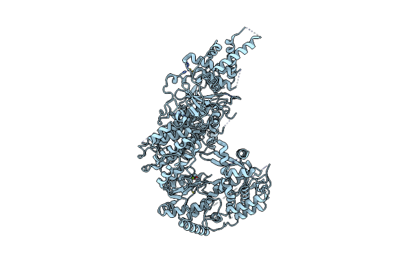 |
Apo-Structure Of Lassa Virus L Protein (Well-Resolved Polymerase Core) [Apo-Core]
Organism: Lassa mammarenavirus
Method: ELECTRON MICROSCOPY Release Date: 2021-12-01 Classification: VIRAL PROTEIN Ligands: ZN, MG |