

Search Count: 115
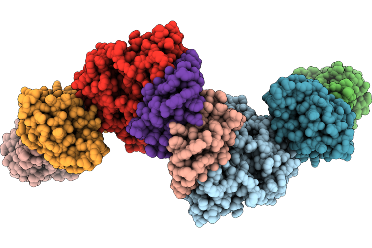 |
Structure Of The Keops Dimer
Organism: Caenorhabditis elegans
Method: ELECTRON MICROSCOPY Resolution:3.10 Å Release Date: 2026-05-27 Classification: TRANSFERASE Ligands: K |
Organism: Caenorhabditis elegans
Method: ELECTRON MICROSCOPY
Release Date: 2026-05-27
Ligands: K
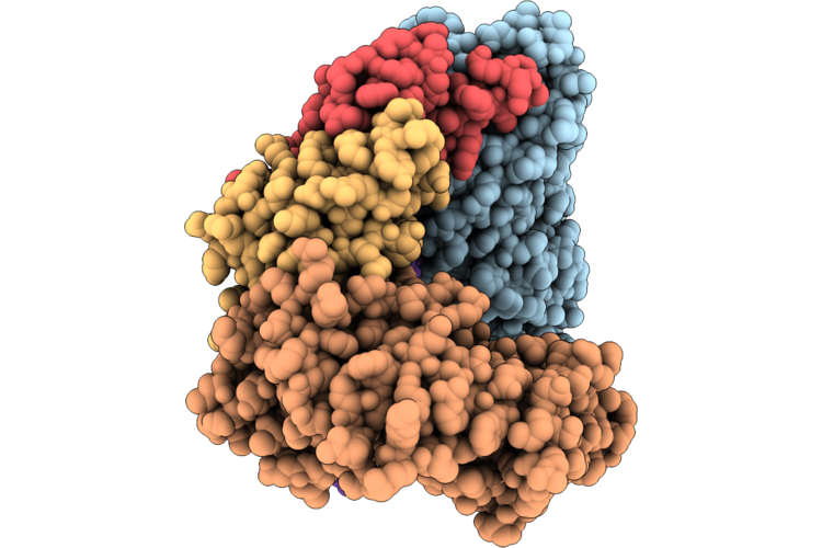 |
Structure Of The Keops-Trna
Organism: Caenorhabditis elegans
Method: ELECTRON MICROSCOPY Resolution:3.36 Å Release Date: 2026-05-27 Classification: TRANSFERASE/RNA Ligands: K |
Organism: Caenorhabditis elegans
Method: ELECTRON MICROSCOPY
Release Date: 2026-05-27
Ligands: K
 |
C. Elegans Pezo-1 Isoform G
Organism: Caenorhabditis elegans
Method: ELECTRON MICROSCOPY Release Date: 2025-12-24 Classification: MEMBRANE PROTEIN |
Organism: Caenorhabditis elegans
Method: ELECTRON MICROSCOPY
Release Date: 2025-12-24
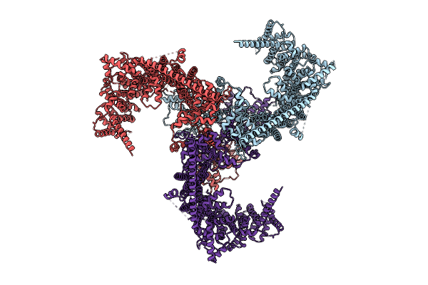 |
C. Elegans Pezo-1 Isoform K
Organism: Caenorhabditis elegans
Method: ELECTRON MICROSCOPY Release Date: 2025-12-24 Classification: MEMBRANE PROTEIN |
Organism: Caenorhabditis elegans
Method: ELECTRON MICROSCOPY
Release Date: 2025-12-24
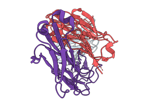 |
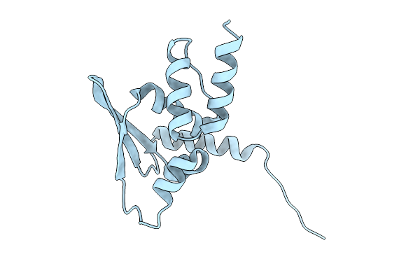
Structure Of Nectin-4 D1 Domain In Complex With The Fab Fragment Of 9Mw2821 Mab
Organism: Mus musculus, Homo sapiens
Method: ELECTRON MICROSCOPY Release Date: 2025-11-19 Classification: IMMUNE SYSTEM |
Organism: Mus musculus, Homo sapiens
Method: ELECTRON MICROSCOPY
Release Date: 2025-11-19
 |
Crystal Structure Of The Btb Domain Mouse Keap1
Organism: Mus musculus
Method: X-RAY DIFFRACTION Resolution:2.80 Å Release Date: 2025-10-22 Classification: TRANSCRIPTION |
Organism: Mus musculus
Method: X-RAY DIFFRACTION
Release Date: 2025-10-22
 |
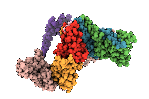
Crystal Structure Of The Btb Domain Mouse Keap1 In Complex With Cddo-Im
Organism: Mus musculus
Method: X-RAY DIFFRACTION Resolution:2.44 Å Release Date: 2025-10-22 Classification: TRANSCRIPTION Ligands: SXJ, CA |
Organism: Mus musculus
Method: X-RAY DIFFRACTION
Release Date: 2025-10-22
Ligands: SXJ, CA
 |
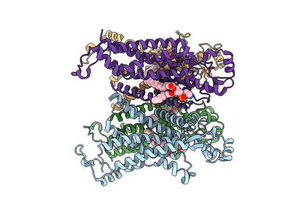
Crystal Structure Of The Btb Domain Mouse Keap1 In Complex With Cddo-Me
Organism: Mus musculus
Method: X-RAY DIFFRACTION Resolution:3.43 Å Release Date: 2025-10-22 Classification: TRANSCRIPTION Ligands: A1L9B |
Organism: Mus musculus
Method: X-RAY DIFFRACTION
Release Date: 2025-10-22
Ligands: A1L9B
 |
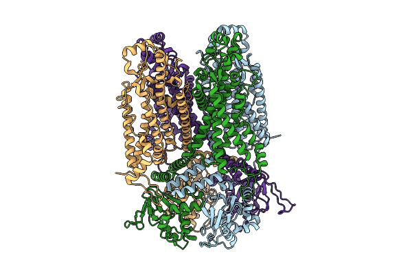
Structure Of The Medicago Truncatula Cngc15B
Organism: Medicago truncatula
Method: ELECTRON MICROSCOPY Release Date: 2025-08-13 Classification: MEMBRANE PROTEIN Ligands: PTY |
Organism: Medicago truncatula
Method: ELECTRON MICROSCOPY
Release Date: 2025-08-13
Ligands: PTY
 |
Structure Of The Medicago Truncatula Cngc15B With Cam
Organism: Medicago truncatula
Method: ELECTRON MICROSCOPY Release Date: 2025-08-13 Classification: MEMBRANE PROTEIN |
Organism: Medicago truncatula
Method: ELECTRON MICROSCOPY
Release Date: 2025-08-13
 |
Structure Of The Pro-Pro Endopeptidase (Ppep-3) E153A Y189F In Complex With Substrate Peptide Ac-Eplpppp-Nh2 From Geobacillus Thermodenitrificans
Organism: Geobacillus thermodenitrificans ng80-2, Synthetic construct
Method: X-RAY DIFFRACTION Resolution:2.00 Å Release Date: 2025-07-30 Classification: HYDROLASE Ligands: ZN, PG4, PGE |
Organism: Geobacillus thermodenitrificans ng80-2, Synthetic construct
Method: X-RAY DIFFRACTION
Release Date: 2025-07-30
Ligands: ZN, PG4, PGE
 |
Structure Of The Pro-Pro Endopeptidase (Ppep-3) E153A Y189F From Geobacillus Thermodenitrificans
Organism: Geobacillus thermodenitrificans ng80-2
Method: X-RAY DIFFRACTION Resolution:1.60 Å Release Date: 2025-07-23 Classification: HYDROLASE Ligands: ZN, BCN, PG4, PEG |
Organism: Geobacillus thermodenitrificans ng80-2
Method: X-RAY DIFFRACTION
Release Date: 2025-07-23
Ligands: ZN, BCN, PG4, PEG
 |
Structure Of The Pro-Pro Endopeptidase (Ppep-3) From Geobacillus Thermodenitrificans
Organism: Geobacillus thermodenitrificans ng80-2
Method: X-RAY DIFFRACTION Resolution:1.55 Å Release Date: 2025-07-16 Classification: HYDROLASE Ligands: ZN, BCN, PEG, EDO |
Organism: Geobacillus thermodenitrificans ng80-2
Method: X-RAY DIFFRACTION
Release Date: 2025-07-16
Ligands: ZN, BCN, PEG, EDO
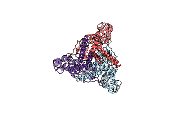 |
The Structure Of Brtnac1 At High Ph
Organism: Scolopendra mutilans
Method: ELECTRON MICROSCOPY Release Date: 2025-03-05 Classification: MEMBRANE PROTEIN |
Organism: Scolopendra mutilans
Method: ELECTRON MICROSCOPY
Release Date: 2025-03-05
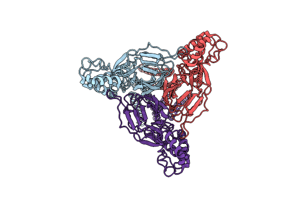 |
The Structure Of Brtnac1 At Low Ph
Organism: Scolopendra mutilans
Method: ELECTRON MICROSCOPY Release Date: 2025-03-05 Classification: MEMBRANE PROTEIN |
Organism: Scolopendra mutilans
Method: ELECTRON MICROSCOPY
Release Date: 2025-03-05
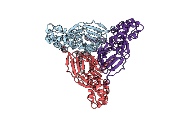 |
Structure Of Brtnac1 At Low Ph With Testosterone
Organism: Scolopendra mutilans
Method: ELECTRON MICROSCOPY Release Date: 2025-03-05 Classification: MEMBRANE PROTEIN |
Organism: Scolopendra mutilans
Method: ELECTRON MICROSCOPY
Release Date: 2025-03-05
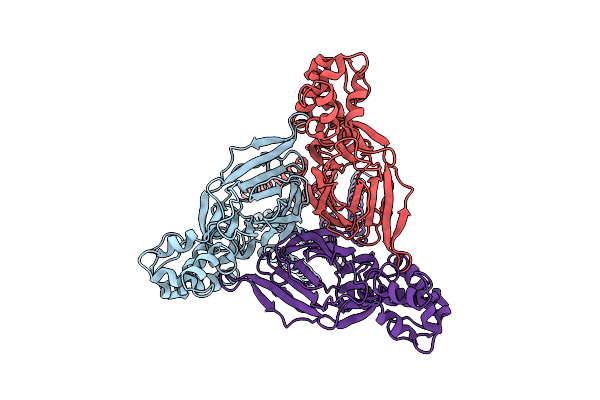 |
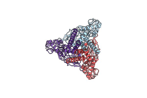
Acid Activated State Of Brtnac1
Organism: Scolopendra mutilans
Method: ELECTRON MICROSCOPY Release Date: 2025-03-05 Classification: MEMBRANE PROTEIN |
Organism: Scolopendra mutilans
Method: ELECTRON MICROSCOPY
Release Date: 2025-03-05
 |
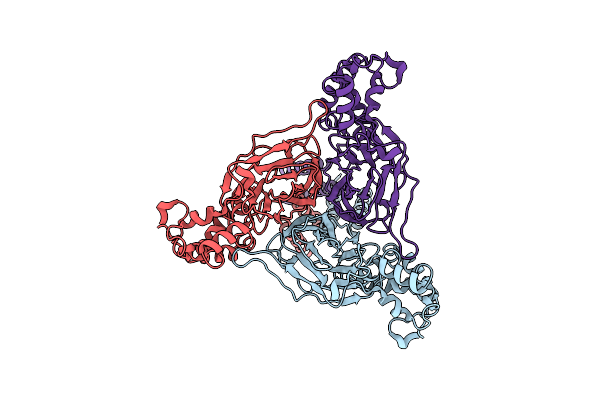
The Structure Of Brtnac1 At 4 Degrees Celsius
Organism: Scolopendra mutilans
Method: ELECTRON MICROSCOPY Release Date: 2025-03-05 Classification: MEMBRANE PROTEIN |
Organism: Scolopendra mutilans
Method: ELECTRON MICROSCOPY
Release Date: 2025-03-05
 |
The Structure Of Brtnac1 At 40 Degree Centigrade
Organism: Scolopendra mutilans
Method: ELECTRON MICROSCOPY Release Date: 2025-03-05 Classification: MEMBRANE PROTEIN |
Organism: Scolopendra mutilans
Method: ELECTRON MICROSCOPY
Release Date: 2025-03-05
 |
Structure Of The Human Glun1-N2B Nmda Receptors In The Mg2+ Bound State
Organism: Homo sapiens
Method: ELECTRON MICROSCOPY Resolution:3.27 Å Release Date: 2025-03-05 Classification: MEMBRANE PROTEIN Ligands: GLY, NAG, 7RC, MG |
Organism: Homo sapiens
Method: ELECTRON MICROSCOPY
Release Date: 2025-03-05
Ligands: GLY, NAG, 7RC, MG