

Search Count: 33
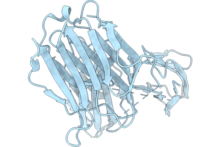 |
Crystal Structure Of Galectin-3 Bound To Fn3Con-9 And Fn3Con-41, A Cooperative Binder That Recognises The Galectin-3-Fn3Con-9 Interface
Organism: Synthetic construct, Homo sapiens
Method: X-RAY DIFFRACTION Resolution:1.96 Å Release Date: 2026-05-20 Classification: CARBOHYDRATE |
Organism: Synthetic construct, Homo sapiens
Method: X-RAY DIFFRACTION
Release Date: 2026-05-20
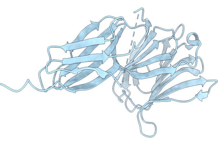 |
Crystal Structure Of The Galectin-3 Complex With Fn3Con-7, An Mrna Display-Derived Binding Protein
Organism: Homo sapiens, Synthetic construct
Method: X-RAY DIFFRACTION Resolution:1.92 Å Release Date: 2026-05-20 Classification: CARBOHYDRATE |
Organism: Homo sapiens, Synthetic construct
Method: X-RAY DIFFRACTION
Release Date: 2026-05-20
 |
Crystal Structure Of Galectin-3 In Complex With Fn3
Organism: Homo sapiens
Method: X-RAY DIFFRACTION Resolution:2.04 Å Release Date: 2026-02-18 Classification: PROTEIN BINDING Ligands: SO4 |
Organism: Homo sapiens
Method: X-RAY DIFFRACTION
Release Date: 2026-02-18
Ligands: SO4
 |
Crystal Structure Of Human Soluble Epoxide Hydrolase C-Terminal Domain In Complex With A Benzohomoadamantane-Based Urea Inhibitor
Organism: Homo sapiens
Method: X-RAY DIFFRACTION Resolution:2.80 Å Release Date: 2025-04-30 Classification: HYDROLASE Ligands: A1H8Y |
Organism: Homo sapiens
Method: X-RAY DIFFRACTION
Release Date: 2025-04-30
Ligands: A1H8Y
 |
Cryoem Structure Of Enterococcus Italicus Csm-Crrna-Ctr (3.2) Complex
Organism: Enterococcus italicus dsm 15952, Severe acute respiratory syndrome coronavirus 2
Method: ELECTRON MICROSCOPY Release Date: 2025-02-05 Classification: RNA BINDING PROTEIN |
 |
Cryoem Structure Of Enterococcus Italicus Csm-Crrna-Ctr (4.3) Complex
Organism: Enterococcus italicus dsm 15952, Severe acute respiratory syndrome coronavirus 2
Method: ELECTRON MICROSCOPY Release Date: 2025-02-05 Classification: RNA BINDING PROTEIN |
 |
Cryoem Structure Of Enterococcus Italicus Csm-Crrna (3.2 Complex)
Organism: Enterococcus italicus dsm 15952
Method: ELECTRON MICROSCOPY Release Date: 2025-01-29 Classification: RNA BINDING PROTEIN |
Organism: Enterococcus italicus dsm 15952
Method: ELECTRON MICROSCOPY
Release Date: 2025-01-29
 |
Cryoem Structure Of Enterococcus Italicus Csm-Crrna (4.3) Complex
Organism: Enterococcus italicus dsm 15952
Method: ELECTRON MICROSCOPY Release Date: 2025-01-29 Classification: RNA BINDING PROTEIN |
Organism: Enterococcus italicus dsm 15952
Method: ELECTRON MICROSCOPY
Release Date: 2025-01-29
 |
Cryoem Structure Of Enterococcus Italicus Csm-Crrna Complex Bound To Ampnpp
Organism: Enterococcus italicus dsm 15952
Method: ELECTRON MICROSCOPY Release Date: 2025-01-29 Classification: RNA BINDING PROTEIN Ligands: ZAN, MG |
Organism: Enterococcus italicus dsm 15952
Method: ELECTRON MICROSCOPY
Release Date: 2025-01-29
Ligands: ZAN, MG
 |
Cryoem Structure Of Enterococcus Italicus Csm-Crrna-Ctr Complex Bound To Ampnpp
Organism: Enterococcus italicus dsm 15952, Severe acute respiratory syndrome coronavirus 2
Method: ELECTRON MICROSCOPY Release Date: 2025-01-29 Classification: RNA BINDING PROTEIN Ligands: ZAN, MG |
Organism: Enterococcus italicus dsm 15952, Severe acute respiratory syndrome coronavirus 2
Method: ELECTRON MICROSCOPY
Release Date: 2025-01-29
Ligands: ZAN, MG
 |
Cryoem Structure Of Enterococcus Italicus Csm-Crrna-Ctr1 Complex (4.3) Bound To Ampnpp
Organism: Enterococcus italicus dsm 15952, Severe acute respiratory syndrome coronavirus 2
Method: ELECTRON MICROSCOPY Release Date: 2025-01-29 Classification: RNA BINDING PROTEIN Ligands: ZAN, MG |
Organism: Enterococcus italicus dsm 15952, Severe acute respiratory syndrome coronavirus 2
Method: ELECTRON MICROSCOPY
Release Date: 2025-01-29
Ligands: ZAN, MG
 |
Cryoem Structure Of Enterococcus Italicus Csm-Crrna-Ctr1 Complex Bound To Pnppa3 And Ampnpp
Organism: Enterococcus italicus dsm 15952, Severe acute respiratory syndrome coronavirus 2
Method: ELECTRON MICROSCOPY Release Date: 2025-01-29 Classification: RNA BINDING PROTEIN Ligands: A1II9, ANP, MG |
Organism: Enterococcus italicus dsm 15952, Severe acute respiratory syndrome coronavirus 2
Method: ELECTRON MICROSCOPY
Release Date: 2025-01-29
Ligands: A1II9, ANP, MG
 |
Cryoem Structure Of Enterococcus Italicus Csm-Crrna-Ctr2 Complex Bound To Pnppa3 And Ampnpp
Organism: Enterococcus italicus dsm 15952, Severe acute respiratory syndrome coronavirus 2
Method: ELECTRON MICROSCOPY Release Date: 2025-01-29 Classification: RNA BINDING PROTEIN Ligands: A1II9, ANP, MG |
Organism: Enterococcus italicus dsm 15952, Severe acute respiratory syndrome coronavirus 2
Method: ELECTRON MICROSCOPY
Release Date: 2025-01-29
Ligands: A1II9, ANP, MG
 |
Cryoem Structure Of Enterococcus Italicus Csm-Crrna Complex Bound To Pnppa3 And Ampnpp
Organism: Enterococcus italicus dsm 15952
Method: ELECTRON MICROSCOPY Release Date: 2025-01-29 Classification: RNA BINDING PROTEIN Ligands: A1II9, ANP, MN |
Organism: Enterococcus italicus dsm 15952
Method: ELECTRON MICROSCOPY
Release Date: 2025-01-29
Ligands: A1II9, ANP, MN
 |
Cryoem Structure Of Enterococcus Italicus Csm-Crrna-Ctr2 Complex (4.3) Bound To Ampnpp
Organism: Enterococcus italicus dsm 15952, Severe acute respiratory syndrome coronavirus 2
Method: ELECTRON MICROSCOPY Release Date: 2025-01-29 Classification: RNA BINDING PROTEIN Ligands: ZAN, MG |
Organism: Enterococcus italicus dsm 15952, Severe acute respiratory syndrome coronavirus 2
Method: ELECTRON MICROSCOPY
Release Date: 2025-01-29
Ligands: ZAN, MG
 |
Serratia Nucc Bound To Ca3
Organism: Serratia
Method: X-RAY DIFFRACTION Resolution:1.48 Å Release Date: 2022-10-26 Classification: RNA BINDING PROTEIN Ligands: CA, ACT |
Organism: Serratia
Method: X-RAY DIFFRACTION
Release Date: 2022-10-26
Ligands: CA, ACT
 |
Serratia Nucc Apo Form
Organism: Serratia
Method: X-RAY DIFFRACTION Resolution:1.83 Å Release Date: 2022-10-26 Classification: RNA BINDING PROTEIN |
Organism: Serratia
Method: X-RAY DIFFRACTION
Release Date: 2022-10-26
 |
Enterococcus Italicus Csm6 Bound To Cyclic Hexa-2'-Fluoro-Hexa-Damp
Organism: Enterococcus italicus (strain dsm 15952 / lmg 22039 / tp 1.5)
Method: X-RAY DIFFRACTION Resolution:2.42 Å Release Date: 2020-02-26 Classification: RNA BINDING PROTEIN Ligands: AF2, SO4 |
Organism: Enterococcus italicus (strain dsm 15952 / lmg 22039 / tp 1.5)
Method: X-RAY DIFFRACTION
Release Date: 2020-02-26
Ligands: AF2, SO4
 |
Structure Of The Carboxy-Terminal Domain Of The Bacteriophage T5 L- Shaped Tail Fibre
Organism: Escherichia phage t5
Method: X-RAY DIFFRACTION Resolution:2.30 Å Release Date: 2015-12-16 Classification: VIRAL PROTEIN |
Organism: Escherichia phage t5
Method: X-RAY DIFFRACTION
Release Date: 2015-12-16
 |
Structure Of The Carboxy-Terminal Domain Of The Bacteriophage T5 L- Shaped Tail Fiber Without Its Intra-Molecular Chaperone Domain
Organism: Enterobacteria phage t5
Method: X-RAY DIFFRACTION Resolution:2.52 Å Release Date: 2015-08-05 Classification: VIRAL PROTEIN Ligands: GOL Modified Residue(s): MSE |
Organism: Enterobacteria phage t5
Method: X-RAY DIFFRACTION
Release Date: 2015-08-05
Ligands: GOL