

Search Count: 21
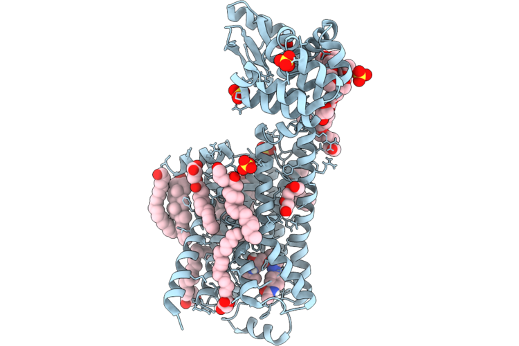 |
Dark Structure Of Beta-2 Adrenergic Receptor With Photoazolol In Dark State Recorded At Swissfel
Organism: Homo sapiens, Enterobacteria phage t4
Method: X-RAY DIFFRACTION Resolution:2.45 Å Release Date: 2026-04-22 Classification: MEMBRANE PROTEIN Ligands: SO4, CLR, 12P, OLC, 1PE, EDO, GOL, A1JHU, PLM |
Organism: Homo sapiens, Enterobacteria phage t4
Method: X-RAY DIFFRACTION
Release Date: 2026-04-22
Ligands: SO4, CLR, 12P, OLC, 1PE, EDO, GOL, A1JHU, PLM
 |
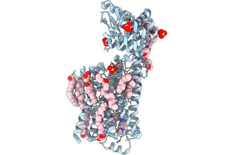
Mixed Model Refinement Of Beta-2 Adrenergic Receptor With Photoazolol In Dark State And Light State, 10 Seconds After Light Activation, Recorded At Swissfel
Organism: Homo sapiens, Tequatrovirus t4
Method: X-RAY DIFFRACTION Resolution:2.45 Å Release Date: 2026-04-22 Classification: MEMBRANE PROTEIN Ligands: SO4, ACM, CLR, PLM, 12P, OLC, 1PE, EDO, GOL, A1JHU |
Organism: Homo sapiens, Tequatrovirus t4
Method: X-RAY DIFFRACTION
Release Date: 2026-04-22
Ligands: SO4, ACM, CLR, PLM, 12P, OLC, 1PE, EDO, GOL, A1JHU
 |
Dark Structure Of Beta-2 Adrenergic Receptor With Photoazolol In Dark State Recorded At Lcls
Organism: Homo sapiens, Enterobacteria phage t4
Method: X-RAY DIFFRACTION Resolution:2.50 Å Release Date: 2026-04-22 Classification: MEMBRANE PROTEIN Ligands: SO4, CLR, PLM, 12P, A1JHU, OLC, EDO, GOL |
Organism: Homo sapiens, Enterobacteria phage t4
Method: X-RAY DIFFRACTION
Release Date: 2026-04-22
Ligands: SO4, CLR, PLM, 12P, A1JHU, OLC, EDO, GOL
 |
Mixed Model Refinement Of Beta-2 Adrenergic Receptor With Photoazolol In Dark State And Light State, 17 Nanoseconds After Light Activation, Recorded At Lcls
Organism: Homo sapiens, Tequatrovirus t4
Method: X-RAY DIFFRACTION Resolution:2.60 Å Release Date: 2026-04-22 Classification: MEMBRANE PROTEIN Ligands: SO4, ACM, CLR, PLM, 12P, OLC, EDO, GOL, 1PE, A1JHU |
Organism: Homo sapiens, Tequatrovirus t4
Method: X-RAY DIFFRACTION
Release Date: 2026-04-22
Ligands: SO4, ACM, CLR, PLM, 12P, OLC, EDO, GOL, 1PE, A1JHU
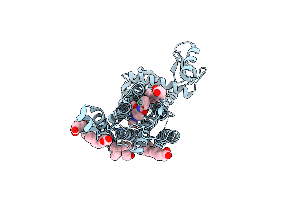 |
Dark Structure Of The Human Metabotropic Glutamate Receptor 5 Transmembrane Domain Bound To Photoswitchable Ligand Alloswitch-1
Organism: Homo sapiens, Enterobacteria phage t4
Method: X-RAY DIFFRACTION Resolution:2.33 Å Release Date: 2025-06-25 Classification: SIGNALING PROTEIN Ligands: OLA, 4YI |
Organism: Homo sapiens, Enterobacteria phage t4
Method: X-RAY DIFFRACTION
Release Date: 2025-06-25
Ligands: OLA, 4YI
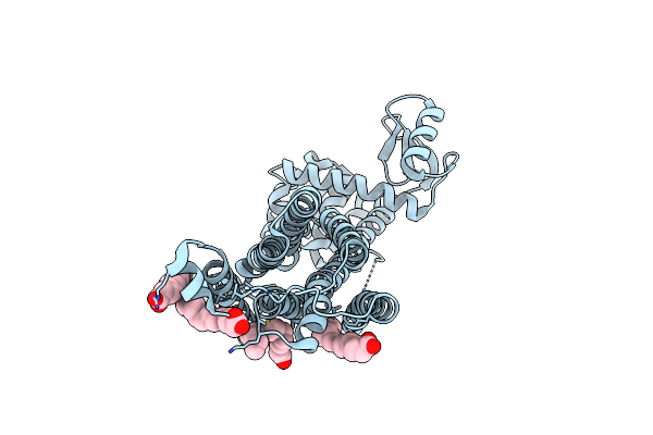 |
Apo-State Structure Of The Human Metabotropic Glutamate Receptor 5 Transmembrane Domain Freeze-Trapped After Light Activation Of Photoswitchable Ligand Alloswitch-1
Organism: Homo sapiens, Enterobacteria phage t4
Method: X-RAY DIFFRACTION Resolution:2.90 Å Release Date: 2025-06-25 Classification: SIGNALING PROTEIN Ligands: OLA |
Organism: Homo sapiens, Enterobacteria phage t4
Method: X-RAY DIFFRACTION
Release Date: 2025-06-25
Ligands: OLA
 |
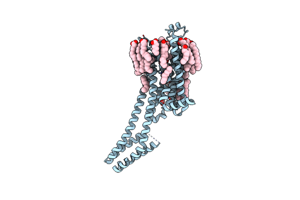
Dark Structure Of The Human Adenosine A2A Receptor Bound To Synthetic Photoswitch "Stilswitch3" Determined By Serial Synchrotron Crystallography
Organism: Homo sapiens
Method: X-RAY DIFFRACTION Resolution:2.65 Å Release Date: 2025-01-08 Classification: MEMBRANE PROTEIN Ligands: OLA, A1H3H, CLR, NA |
Organism: Homo sapiens
Method: X-RAY DIFFRACTION
Release Date: 2025-01-08
Ligands: OLA, A1H3H, CLR, NA
 |
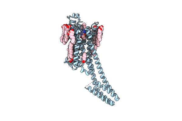
Crystal Structure Of The Adenosine A2A Receptor In Complex With Istradefylline
Organism: Homo sapiens
Method: X-RAY DIFFRACTION Resolution:1.94 Å Release Date: 2025-01-08 Classification: MEMBRANE PROTEIN Ligands: JQ9, CLR, OLA, NA |
Organism: Homo sapiens
Method: X-RAY DIFFRACTION
Release Date: 2025-01-08
Ligands: JQ9, CLR, OLA, NA
 |
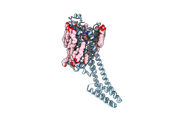
Crystal Structure Of The Adenosine A2A Receptor In Complex With The Synthetic Photoswitch 'Azoswitch2
Organism: Homo sapiens
Method: X-RAY DIFFRACTION Resolution:2.20 Å Release Date: 2025-01-08 Classification: MEMBRANE PROTEIN Ligands: A1H3L, CLR, OLA, NA |
Organism: Homo sapiens
Method: X-RAY DIFFRACTION
Release Date: 2025-01-08
Ligands: A1H3L, CLR, OLA, NA
 |
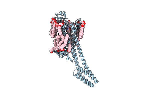
Crystal Structure Of The Adenosine A2A Receptor In Complex With The Synthetic Photoswitch 'Stilswitch1
Organism: Homo sapiens
Method: X-RAY DIFFRACTION Resolution:2.25 Å Release Date: 2025-01-08 Classification: MEMBRANE PROTEIN Ligands: A1H3J, CLR, OLA, OLC, OLB, NA |
Organism: Homo sapiens
Method: X-RAY DIFFRACTION
Release Date: 2025-01-08
Ligands: A1H3J, CLR, OLA, OLC, OLB, NA
 |
Crystal Structure Of The Adenosine A2A Receptor In Complex With The Synthetic Photoswitch 'Stilswitch2
Organism: Homo sapiens
Method: X-RAY DIFFRACTION Resolution:2.31 Å Release Date: 2025-01-08 Classification: MEMBRANE PROTEIN Ligands: A1H3I, CLR, OLC, OLA, NA |
Organism: Homo sapiens
Method: X-RAY DIFFRACTION
Release Date: 2025-01-08
Ligands: A1H3I, CLR, OLC, OLA, NA
 |
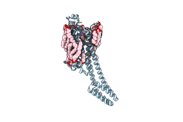
Crystal Structure Of The Adenosine A2A Receptor In Complex With The Synthetic Photoswitch 'Stilswitch3
Organism: Homo sapiens
Method: X-RAY DIFFRACTION Resolution:2.05 Å Release Date: 2025-01-08 Classification: MEMBRANE PROTEIN Ligands: A1H3H, CLR, OLA, NA |
Organism: Homo sapiens
Method: X-RAY DIFFRACTION
Release Date: 2025-01-08
Ligands: A1H3H, CLR, OLA, NA
 |
Crystal Structure Of The Adenosine A2A Receptor In Complex With The Synthetic Photoswitch 'Stilswitch4
Organism: Homo sapiens
Method: X-RAY DIFFRACTION Resolution:2.20 Å Release Date: 2025-01-08 Classification: MEMBRANE PROTEIN Ligands: A1H3K, CLR, OLA, OLC, NA |
Organism: Homo sapiens
Method: X-RAY DIFFRACTION
Release Date: 2025-01-08
Ligands: A1H3K, CLR, OLA, OLC, NA
 |
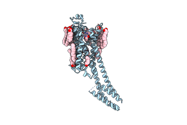
Dark Structure Of The Human Adenosine A2A Receptor Bound To Synthetic Photoswitch 'Stilswitch2' Determined By Serial Synchrotron Crystallography
Organism: Homo sapiens
Method: X-RAY DIFFRACTION Resolution:2.45 Å Release Date: 2025-01-08 Classification: MEMBRANE PROTEIN Ligands: A1H3I, CLR, OLA, OLB, OLC, NA |
Organism: Homo sapiens
Method: X-RAY DIFFRACTION
Release Date: 2025-01-08
Ligands: A1H3I, CLR, OLA, OLB, OLC, NA
 |
Steady State Structure Of The Human Adenosine A2A Receptor Bound To Synthetic Photoswitch 'Stilswitch2' Determined By Serial Synchrotron Crystallography
Organism: Homo sapiens
Method: X-RAY DIFFRACTION Resolution:2.80 Å Release Date: 2025-01-08 Classification: MEMBRANE PROTEIN Ligands: A1H3I, CLR, OLA, NA |
Organism: Homo sapiens
Method: X-RAY DIFFRACTION
Release Date: 2025-01-08
Ligands: A1H3I, CLR, OLA, NA
 |
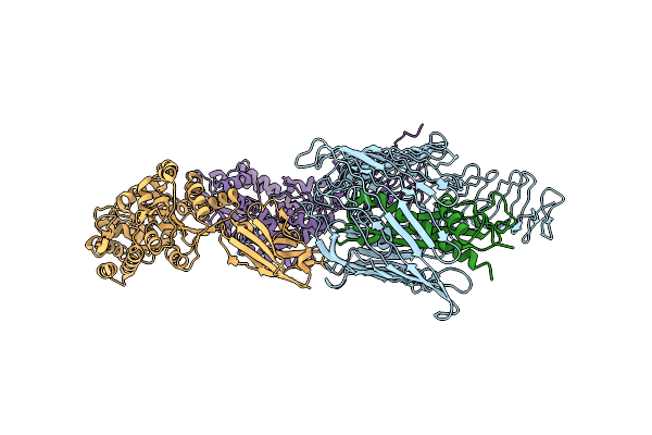
Steady State Structure Of The Human Adenosine A2A Receptor Bound To Synthetic Photoswitch 'Stilswitch3' Determined By Serial Synchrotron Crystallography
Organism: Homo sapiens, Escherichia coli
Method: X-RAY DIFFRACTION Resolution:3.05 Å Release Date: 2025-01-08 Classification: MEMBRANE PROTEIN Ligands: A1H3H, OLA, NA |
Organism: Homo sapiens, Escherichia coli
Method: X-RAY DIFFRACTION
Release Date: 2025-01-08
Ligands: A1H3H, OLA, NA
 |
Cryo-Em Structure Of N. Gonorhoeae Lptde In Complex With Promacrobodies (Mbps Have Not Been Built De Novo)
Organism: Neisseria gonorrhoeae, Synthetic construct, Methanosarcina mazei
Method: ELECTRON MICROSCOPY Release Date: 2022-05-04 Classification: TRANSPORT PROTEIN |
Organism: Neisseria gonorrhoeae, Synthetic construct, Methanosarcina mazei
Method: ELECTRON MICROSCOPY
Release Date: 2022-05-04
 |
Crystal Structure Of Promacrobody 21 With Bound Maltose
Organism: Synthetic construct
Method: X-RAY DIFFRACTION Resolution:2.00 Å Release Date: 2022-05-04 Classification: IMMUNE SYSTEM Ligands: P6G, MG |
Organism: Synthetic construct
Method: X-RAY DIFFRACTION
Release Date: 2022-05-04
Ligands: P6G, MG
 |
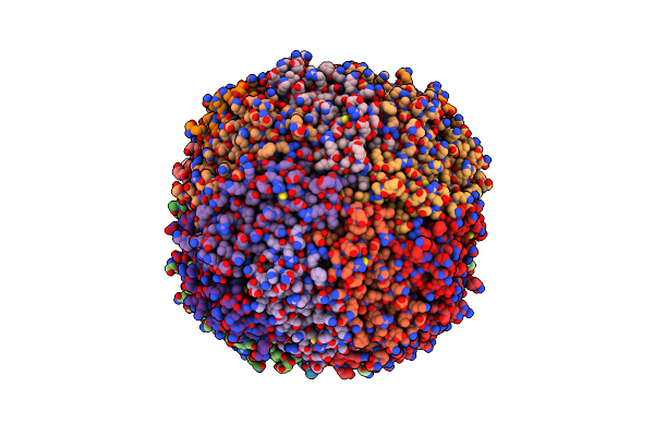
Structure Of A Modified Mouse H-Chain Ferritin With A Lanthanide Binding Motif
Organism: Mus musculus
Method: X-RAY DIFFRACTION Resolution:2.85 Å Release Date: 2018-07-25 Classification: METAL TRANSPORT Ligands: FE |
Organism: Mus musculus
Method: X-RAY DIFFRACTION
Release Date: 2018-07-25
Ligands: FE
 |
Structure Of A Modified Mouse H Chain Ferritin With A Lanthanide Binding Motif In Complex With Terbium
Organism: Mus musculus
Method: X-RAY DIFFRACTION Resolution:2.65 Å Release Date: 2018-07-25 Classification: METAL TRANSPORT Ligands: TB |
Organism: Mus musculus
Method: X-RAY DIFFRACTION
Release Date: 2018-07-25
Ligands: TB