

Search Count: 33
 |
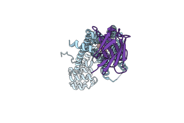
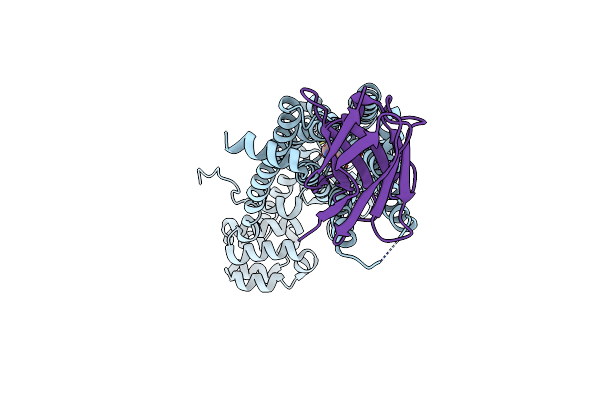
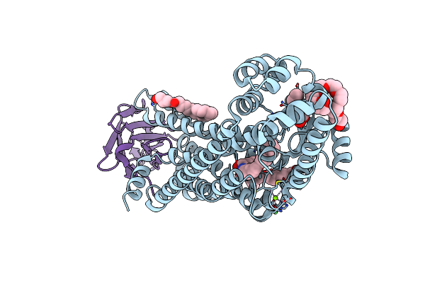
Structure Of The Lm189-Bound Beta2Ar-Gi Complex
Organism: Homo sapiens, Mus musculus
Method: ELECTRON MICROSCOPY Release Date: 2025-05-28 Classification: SIGNALING PROTEIN/IMMUNE SYSTEM Ligands: CLR, A1ASM, KBY |
Organism: Homo sapiens, Mus musculus
Method: ELECTRON MICROSCOPY
Release Date: 2025-05-28
Ligands: CLR, A1ASM, KBY
 |
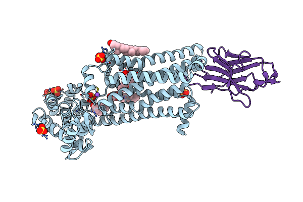
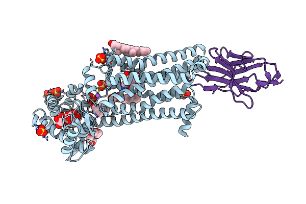
Structure Of Human Beta2 Adrenergic Receptor Bound To Constrained Isoproterenol
Organism: enterobacteria phage t4, Homo sapiens
Method: X-RAY DIFFRACTION Resolution:3.40 Å Release Date: 2023-04-26 Classification: MEMBRANE PROTEIN Ligands: GJ6, NA |
Organism: enterobacteria phage t4, Homo sapiens
Method: X-RAY DIFFRACTION
Release Date: 2023-04-26
Ligands: GJ6, NA
 |
Structure Of Human Beta2 Adrenergic Receptor Bound To Constrained Epinephrine
Organism: enterobacteria phage t4, Homo sapiens
Method: X-RAY DIFFRACTION Resolution:3.10 Å Release Date: 2023-04-26 Classification: MEMBRANE PROTEIN Ligands: G1I, NA |
Organism: enterobacteria phage t4, Homo sapiens
Method: X-RAY DIFFRACTION
Release Date: 2023-04-26
Ligands: G1I, NA
 |
Structure Of Human Beta1 Adrenergic Receptor Bound To Epinephrine And Nanobody 6B9
Organism: Enterobacteria phage t4, Homo sapiens, Vicugna pacos
Method: X-RAY DIFFRACTION Resolution:3.13 Å Release Date: 2020-12-02 Classification: MEMBRANE PROTEIN Ligands: ALE, SO4, NA, EPE, CLR, 1WV |
Organism: Enterobacteria phage t4, Homo sapiens, Vicugna pacos
Method: X-RAY DIFFRACTION
Release Date: 2020-12-02
Ligands: ALE, SO4, NA, EPE, CLR, 1WV
 |
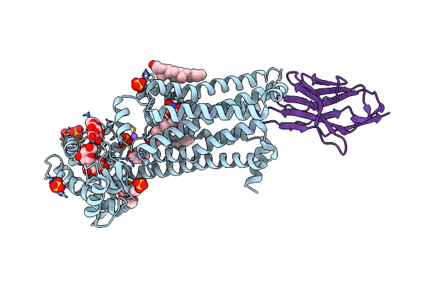
Structure Of Human Beta1 Adrenergic Receptor Bound To Norepinephrine And Nanobody 6B9
Organism: Enterobacteria phage t4, Homo sapiens, Camelidae mixed library
Method: X-RAY DIFFRACTION Resolution:2.70 Å Release Date: 2020-12-02 Classification: MEMBRANE PROTEIN Ligands: E5E, SO4, NA, EPE, 480, CLR, PG4 |
Organism: Enterobacteria phage t4, Homo sapiens, Camelidae mixed library
Method: X-RAY DIFFRACTION
Release Date: 2020-12-02
Ligands: E5E, SO4, NA, EPE, 480, CLR, PG4
 |
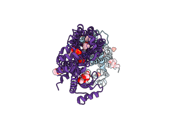
Structure Of Human Beta1 Adrenergic Receptor Bound To Bi-167107 And Nanobody 6B9
Organism: Enterobacteria phage t4, Homo sapiens, Camelidae mixed library
Method: X-RAY DIFFRACTION Resolution:2.60 Å Release Date: 2020-12-02 Classification: MEMBRANE PROTEIN Ligands: SO4, CLR, 1WV, NA, EPE, P0G, P6G |
Organism: Enterobacteria phage t4, Homo sapiens, Camelidae mixed library
Method: X-RAY DIFFRACTION
Release Date: 2020-12-02
Ligands: SO4, CLR, 1WV, NA, EPE, P0G, P6G
 |
Structure Of Human Beta1 Adrenergic Receptor Bound To Carazolol
Organism: Enterobacteria phage t4, Homo sapiens
Method: X-RAY DIFFRACTION Resolution:2.50 Å Release Date: 2020-12-02 Classification: MEMBRANE PROTEIN Ligands: CLR, CAU, SO4, 1WV, NA, CIT, PG4 |
Organism: Enterobacteria phage t4, Homo sapiens
Method: X-RAY DIFFRACTION
Release Date: 2020-12-02
Ligands: CLR, CAU, SO4, 1WV, NA, CIT, PG4
 |
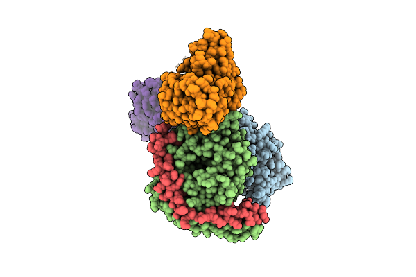
Muscarinic Acetylcholine Receptor 1 - Muscarinic Toxin 7 Complex
Organism: Homo sapiens, Enterobacteria phage t4, Dendroaspis angusticeps
Method: X-RAY DIFFRACTION Resolution:2.55 Å Release Date: 2020-07-08 Classification: MEMBRANE PROTEIN Ligands: ACM, Y01, OIN |
Organism: Homo sapiens, Enterobacteria phage t4, Dendroaspis angusticeps
Method: X-RAY DIFFRACTION
Release Date: 2020-07-08
Ligands: ACM, Y01, OIN
 |
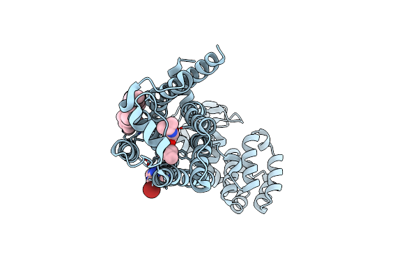
Structure Of A D2 Dopamine Receptor-G-Protein Complex In A Lipid Membrane
Organism: Rattus norvegicus, Homo sapiens, Enterobacteria phage t4
Method: ELECTRON MICROSCOPY Release Date: 2020-06-17 Classification: SIGNALING PROTEIN Ligands: 08Y |
Organism: Rattus norvegicus, Homo sapiens, Enterobacteria phage t4
Method: ELECTRON MICROSCOPY
Release Date: 2020-06-17
Ligands: 08Y
 |
The Beta2 Adrenergic Receptor Bound To A Negative Allosteric Modulator
Organism: Homo sapiens, Enterobacteria phage t4
Method: X-RAY DIFFRACTION Resolution:3.10 Å Release Date: 2020-03-25 Classification: SIGNALING PROTEIN Ligands: CLR, JTZ, M3J |
Organism: Homo sapiens, Enterobacteria phage t4
Method: X-RAY DIFFRACTION
Release Date: 2020-03-25
Ligands: CLR, JTZ, M3J
 |
M3 Muscarinic Acetylcholine Receptor In Complex With A Selective Antagonist
Organism: Rattus norvegicus, Enterobacteria phage t4
Method: X-RAY DIFFRACTION Resolution:3.10 Å Release Date: 2018-11-28 Classification: MEMBRANE PROTEIN Ligands: PG4, 9EC, P6G, CIT |
Organism: Rattus norvegicus, Enterobacteria phage t4
Method: X-RAY DIFFRACTION
Release Date: 2018-11-28
Ligands: PG4, 9EC, P6G, CIT
 |
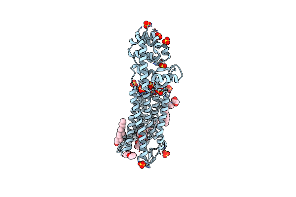
Crystal Structure Of Human Beta2 Adrenergic Receptor Bound To Salmeterol And Nb71
Organism: Enterobacteria phage t4, Homo sapiens, Lama glama
Method: X-RAY DIFFRACTION Resolution:2.96 Å Release Date: 2018-11-14 Classification: SIGNALING PROTEIN/HORMONE Ligands: K5Y, MG, NI, OLC, HTO, OLA, P33 |
Organism: Enterobacteria phage t4, Homo sapiens, Lama glama
Method: X-RAY DIFFRACTION
Release Date: 2018-11-14
Ligands: K5Y, MG, NI, OLC, HTO, OLA, P33
 |
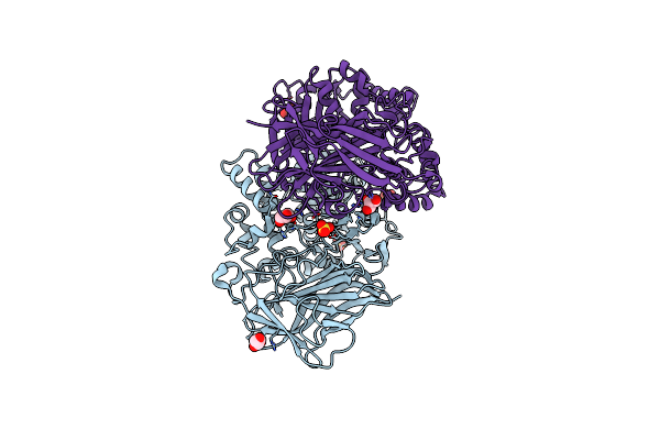
Crystal Structure Of The Mu-Opioid Receptor Bound To A Morphinan Antagonist
Organism: Mus musculus, Enterobacteria phage t4
Method: X-RAY DIFFRACTION Resolution:2.80 Å Release Date: 2012-03-21 Classification: SIGNALING PROTEIN/ANTAGONIST Ligands: BF0, SO4, CLR, MPG, 1PE, CL |
Organism: Mus musculus, Enterobacteria phage t4
Method: X-RAY DIFFRACTION
Release Date: 2012-03-21
Ligands: BF0, SO4, CLR, MPG, 1PE, CL
 |
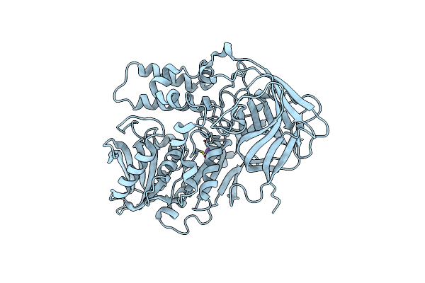
Cocaine Esterase, Wild-Type Biologically Active Dimer
Organism: Rhodococcus sp.
Method: X-RAY DIFFRACTION Resolution:2.30 Å Release Date: 2011-09-21 Classification: HYDROLASE Ligands: GOL, SO4 |
Organism: Rhodococcus sp.
Method: X-RAY DIFFRACTION
Release Date: 2011-09-21
Ligands: GOL, SO4
 |
Cocaine Esterase With Mutations G4C, S10C
Organism: Rhodococcus sp.
Method: X-RAY DIFFRACTION Resolution:1.53 Å Release Date: 2011-09-21 Classification: HYDROLASE Ligands: CL, NA |
Organism: Rhodococcus sp.
Method: X-RAY DIFFRACTION
Release Date: 2011-09-21
Ligands: CL, NA
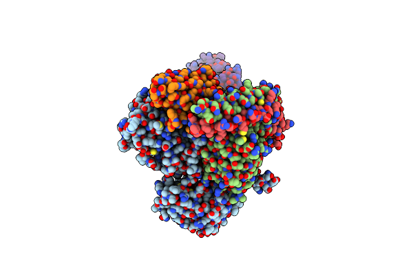 |
Crystal Structure Of The Beta2 Adrenergic Receptor-Gs Protein Complex
Organism: Bos taurus, Rattus norvegicus, Enterobacteria phage t4, Homo sapiens, Lama glama
Method: X-RAY DIFFRACTION Resolution:3.20 Å Release Date: 2011-07-20 Classification: SIGNALING PROTEIN/Hydrolase Ligands: P0G |
Organism: Bos taurus, Rattus norvegicus, Enterobacteria phage t4, Homo sapiens, Lama glama
Method: X-RAY DIFFRACTION
Release Date: 2011-07-20
Ligands: P0G
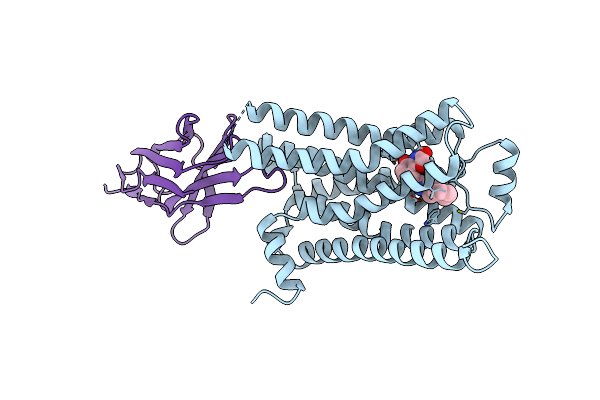 |
Structure Of A Nanobody-Stabilized Active State Of The Beta2 Adrenoceptor
Organism: Homo sapiens, Enterobacteria phage t4, Lama glama
Method: X-RAY DIFFRACTION Resolution:3.50 Å Release Date: 2011-01-19 Classification: SIGNALING PROTEIN, Hydrolase Ligands: P0G |
Organism: Homo sapiens, Enterobacteria phage t4, Lama glama
Method: X-RAY DIFFRACTION
Release Date: 2011-01-19
Ligands: P0G
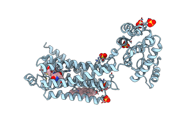 |
Irreversible Agonist-Beta2 Adrenoceptor Complex
Organism: Homo sapiens, Enterobacteria phage t4
Method: X-RAY DIFFRACTION Resolution:3.50 Å Release Date: 2011-01-12 Classification: Membrane Protein/Hydrolase Ligands: ERC, CLR, SO4 |
Organism: Homo sapiens, Enterobacteria phage t4
Method: X-RAY DIFFRACTION
Release Date: 2011-01-12
Ligands: ERC, CLR, SO4
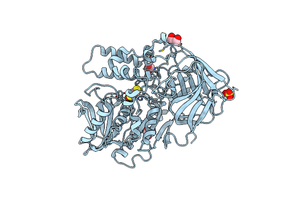 |
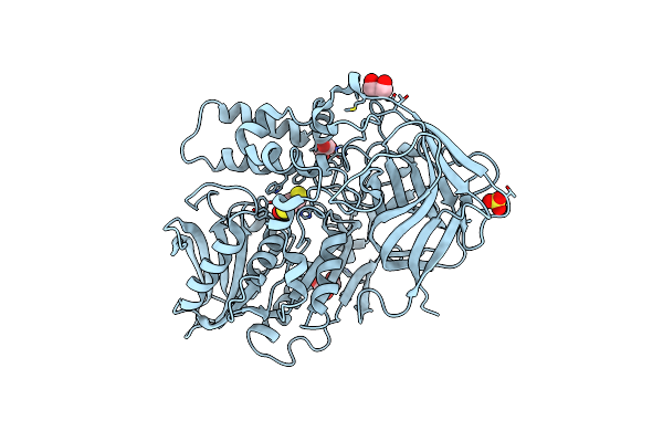
Cocaine Esterase With Mutations T172R / G173Q, Bound To Dtt Adduct
Organism: Rhodococcus sp. mb1 'bresler 1999'
Method: X-RAY DIFFRACTION Resolution:2.50 Å Release Date: 2010-06-16 Classification: HYDROLASE Ligands: CL, DBC, GOL, SO4 |
Organism: Rhodococcus sp. mb1 'bresler 1999'
Method: X-RAY DIFFRACTION
Release Date: 2010-06-16
Ligands: CL, DBC, GOL, SO4
 |
Cocaine Esterase With Mutation G173Q, Bound To Dtt Adduct
Organism: Rhodococcus sp. mb1 'bresler 1999'
Method: X-RAY DIFFRACTION Resolution:2.50 Å Release Date: 2010-06-16 Classification: HYDROLASE Ligands: CL, DBC, GOL, SO4 |
Organism: Rhodococcus sp. mb1 'bresler 1999'
Method: X-RAY DIFFRACTION
Release Date: 2010-06-16
Ligands: CL, DBC, GOL, SO4