

Search Count: 217
 |
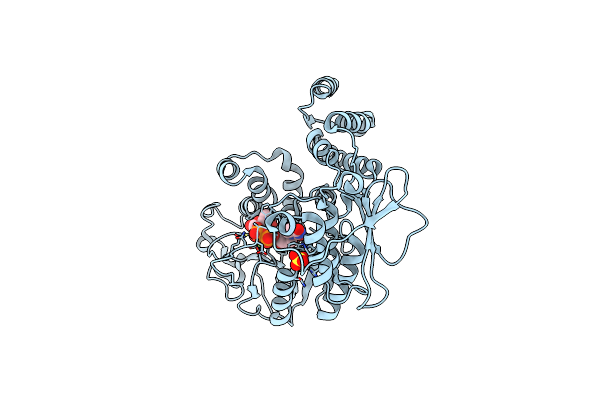
Crystal Structure Of The Glycosyltransferase Domain Of Legionella Seta (Unbound)
Organism: Legionella pneumophila
Method: X-RAY DIFFRACTION Resolution:2.54 Å Release Date: 2025-02-19 Classification: TRANSFERASE Ligands: MG |
Organism: Legionella pneumophila
Method: X-RAY DIFFRACTION
Release Date: 2025-02-19
Ligands: MG
 |
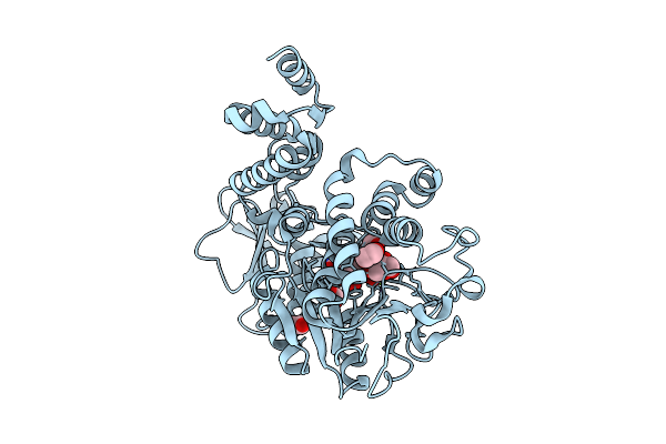
Crystal Structure Of The Glycosyltransferase Domain Of Legionella Seta In Complex With Updglc
Organism: Legionella pneumophila
Method: X-RAY DIFFRACTION Resolution:1.90 Å Release Date: 2025-02-19 Classification: TRANSFERASE Ligands: UDP, MG, BGC, SO4 |
Organism: Legionella pneumophila
Method: X-RAY DIFFRACTION
Release Date: 2025-02-19
Ligands: UDP, MG, BGC, SO4
 |
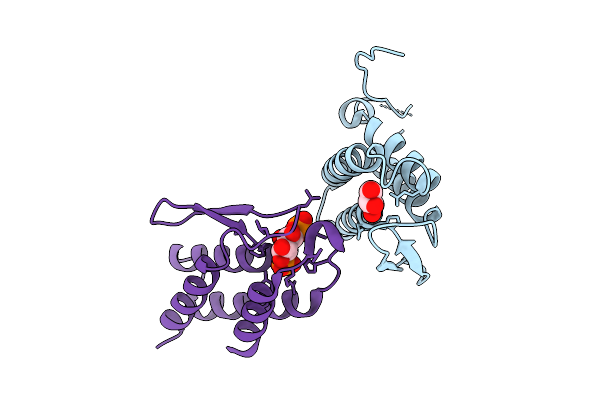
Crystal Structure Of The Glycosyltransferase Domain Of Legionella Seta In Complex With Upd
Organism: Legionella pneumophila
Method: X-RAY DIFFRACTION Resolution:1.79 Å Release Date: 2025-02-19 Classification: TRANSFERASE Ligands: UDP, MG, EDO, GOL |
Organism: Legionella pneumophila
Method: X-RAY DIFFRACTION
Release Date: 2025-02-19
Ligands: UDP, MG, EDO, GOL
 |
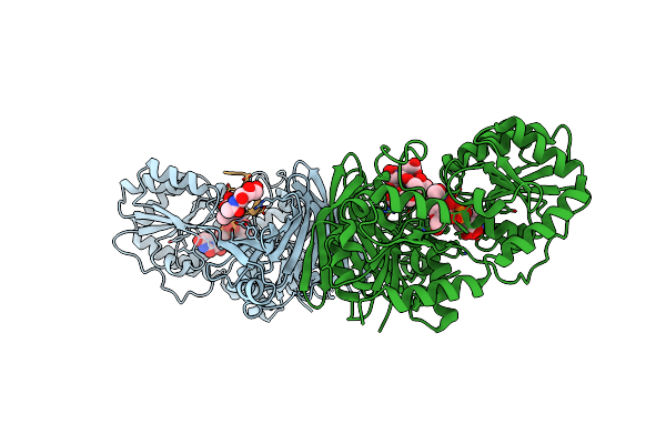
Crystal Structure Of The Pi3P-Binding Domain Of Legionella Seta In Complex With Inositol 1,3-Bisphosphate
Organism: Legionella pneumophila
Method: X-RAY DIFFRACTION Resolution:2.46 Å Release Date: 2025-02-19 Classification: TRANSFERASE Ligands: GOL, ITP |
Organism: Legionella pneumophila
Method: X-RAY DIFFRACTION
Release Date: 2025-02-19
Ligands: GOL, ITP
 |
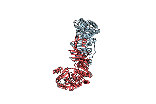
Crystal Structure Of Sdgb (Complexed With Udp, Glcnac, And Glycosylated Peptide)
Organism: Staphylococcus aureus (strain usa300), Synthetic construct
Method: X-RAY DIFFRACTION Resolution:2.50 Å Release Date: 2022-03-16 Classification: TRANSFERASE Ligands: UDP, NAG |
Organism: Staphylococcus aureus (strain usa300), Synthetic construct
Method: X-RAY DIFFRACTION
Release Date: 2022-03-16
Ligands: UDP, NAG
 |
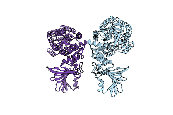
Crystal Structure Of Sdgb (Complexed With Peptides)
Organism: Staphylococcus aureus subsp. aureus usa300, Synthetic construct
Method: X-RAY DIFFRACTION Resolution:1.90 Å Release Date: 2022-03-16 Classification: TRANSFERASE |
Organism: Staphylococcus aureus subsp. aureus usa300, Synthetic construct
Method: X-RAY DIFFRACTION
Release Date: 2022-03-16
 |
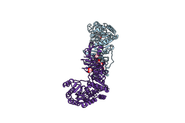
Crystal Structure Of Sdgb (Complexed With Phosphate Ions)
Organism: Staphylococcus aureus (strain usa300)
Method: X-RAY DIFFRACTION Resolution:3.20 Å Release Date: 2022-03-16 Classification: TRANSFERASE Ligands: PO4 |
Organism: Staphylococcus aureus (strain usa300)
Method: X-RAY DIFFRACTION
Release Date: 2022-03-16
Ligands: PO4
 |
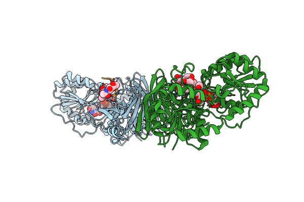
Crystal Structure Of Sdgb (Ligand-Free Form)
Organism: Staphylococcus aureus subsp. aureus usa300
Method: X-RAY DIFFRACTION Resolution:1.84 Å Release Date: 2021-11-24 Classification: TRANSFERASE Ligands: GOL, PGE |
Organism: Staphylococcus aureus subsp. aureus usa300
Method: X-RAY DIFFRACTION
Release Date: 2021-11-24
Ligands: GOL, PGE
 |
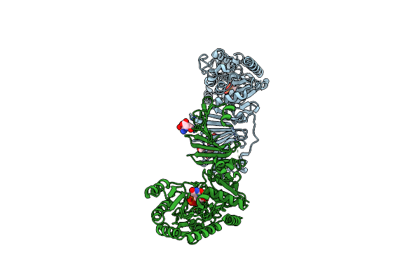
Crystal Structure Of Sdgb (Udp, Nag, And O-Glycosylated Sd Peptide-Binding Form)
Organism: Staphylococcus aureus (strain usa300), Synthetic construct
Method: X-RAY DIFFRACTION Resolution:2.45 Å Release Date: 2021-11-24 Classification: TRANSFERASE Ligands: UDP, NAG |
Organism: Staphylococcus aureus (strain usa300), Synthetic construct
Method: X-RAY DIFFRACTION
Release Date: 2021-11-24
Ligands: UDP, NAG
 |
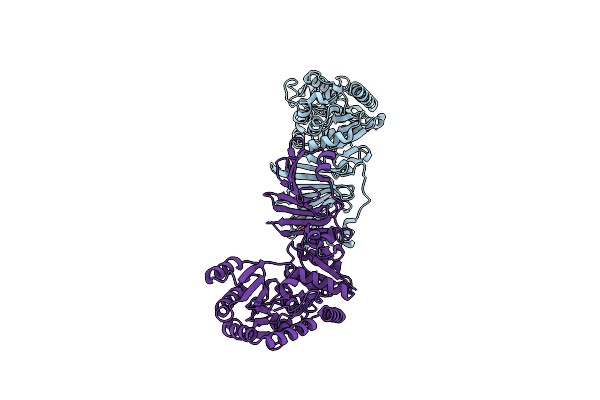
Crystal Structure Of Sdgb (Udp And Sd Peptide-Binding Form)
Organism: Staphylococcus aureus subsp. aureus usa300, Synthetic construct
Method: X-RAY DIFFRACTION Resolution:2.28 Å Release Date: 2021-11-24 Classification: TRANSFERASE Ligands: UDP |
Organism: Staphylococcus aureus subsp. aureus usa300, Synthetic construct
Method: X-RAY DIFFRACTION
Release Date: 2021-11-24
Ligands: UDP
 |
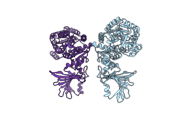
Crystal Structure Of Sdgb (Sd Peptide-Binding Form)
Organism: Staphylococcus aureus subsp. aureus usa300, Synthetic construct
Method: X-RAY DIFFRACTION Resolution:1.90 Å Release Date: 2021-11-24 Classification: TRANSFERASE |
Organism: Staphylococcus aureus subsp. aureus usa300, Synthetic construct
Method: X-RAY DIFFRACTION
Release Date: 2021-11-24
 |
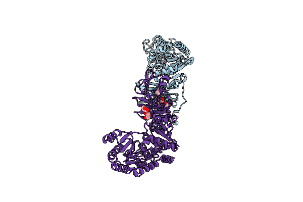
Crystal Structure Of Sdgb (Phosphate-Binding Form)
Organism: Staphylococcus aureus subsp. aureus usa300
Method: X-RAY DIFFRACTION Resolution:3.20 Å Release Date: 2021-11-24 Classification: TRANSFERASE Ligands: PO4 |
Organism: Staphylococcus aureus subsp. aureus usa300
Method: X-RAY DIFFRACTION
Release Date: 2021-11-24
Ligands: PO4
 |
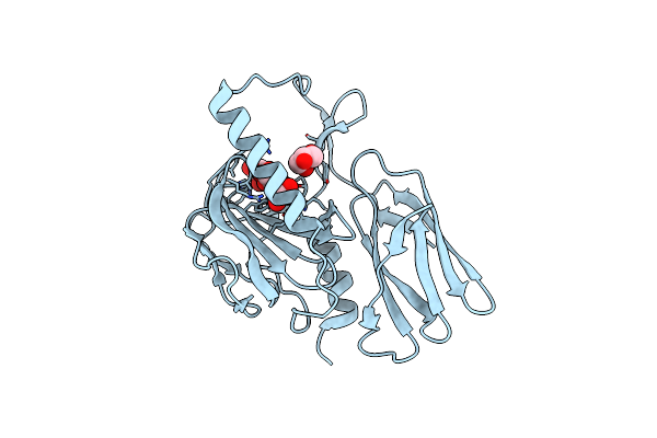
Crystal Structure Of Sdgb (Ligand-Free Form)
Organism: Staphylococcus aureus subsp. aureus usa300
Method: X-RAY DIFFRACTION Resolution:1.85 Å Release Date: 2021-03-24 Classification: TRANSFERASE Ligands: GOL, PGE |
Organism: Staphylococcus aureus subsp. aureus usa300
Method: X-RAY DIFFRACTION
Release Date: 2021-03-24
Ligands: GOL, PGE
 |
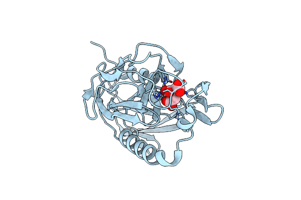
Structure Of Open Form Of Peptidoglycan Peptidase
Organism: Campylobacter jejuni
Method: X-RAY DIFFRACTION Resolution:1.86 Å Release Date: 2020-01-15 Classification: HYDROLASE Ligands: ZN, TAR, GOL |
Organism: Campylobacter jejuni
Method: X-RAY DIFFRACTION
Release Date: 2020-01-15
Ligands: ZN, TAR, GOL
 |
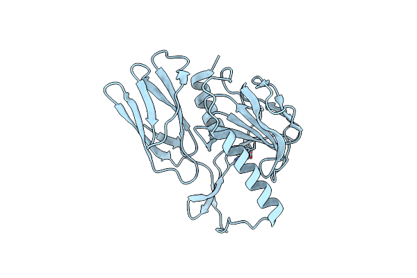
Structure Of Wild Type Closed Form Of Peptidoglycan Peptidase
Organism: Campylobacter jejuni
Method: X-RAY DIFFRACTION Resolution:1.66 Å Release Date: 2020-01-15 Classification: HYDROLASE Ligands: ZN, CIT |
Organism: Campylobacter jejuni
Method: X-RAY DIFFRACTION
Release Date: 2020-01-15
Ligands: ZN, CIT
 |
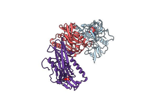
Structure Of H247A Mutant Open Form Peptidoglycan Peptidase
Organism: Campylobacter jejuni
Method: X-RAY DIFFRACTION Resolution:1.92 Å Release Date: 2020-01-15 Classification: HYDROLASE Ligands: ZN |
Organism: Campylobacter jejuni
Method: X-RAY DIFFRACTION
Release Date: 2020-01-15
Ligands: ZN
 |
Structure Of H247A Mutant Peptidoglycan Peptidase Complex With Tetra-Tri Peptide
Organism: Campylobacter jejuni, Synthetic construct
Method: X-RAY DIFFRACTION Resolution:2.16 Å Release Date: 2020-01-15 Classification: HYDROLASE Ligands: ZN |
Organism: Campylobacter jejuni, Synthetic construct
Method: X-RAY DIFFRACTION
Release Date: 2020-01-15
Ligands: ZN
 |
Structure Of H247A Mutant Peptidoglycan Peptidase Complex With Penta Peptide
Organism: Campylobacter jejuni, Synthetic construct
Method: X-RAY DIFFRACTION Resolution:2.38 Å Release Date: 2020-01-15 Classification: HYDROLASE Ligands: ZN |
Organism: Campylobacter jejuni, Synthetic construct
Method: X-RAY DIFFRACTION
Release Date: 2020-01-15
Ligands: ZN
 |
Structure Of H216A Mutant Closed Form Peptidoglycan Peptidase
Organism: Campylobacter jejuni
Method: X-RAY DIFFRACTION Resolution:2.04 Å Release Date: 2020-01-15 Classification: HYDROLASE Ligands: ZN, TAR |
Organism: Campylobacter jejuni
Method: X-RAY DIFFRACTION
Release Date: 2020-01-15
Ligands: ZN, TAR
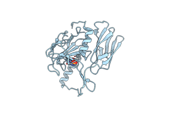 |
Structure Of H216A Mutant Open Form Peptidoglycan Peptidase
Organism: Campylobacter jejuni
Method: X-RAY DIFFRACTION Resolution:2.11 Å Release Date: 2020-01-15 Classification: HYDROLASE Ligands: ZN, SO4 |
Organism: Campylobacter jejuni
Method: X-RAY DIFFRACTION
Release Date: 2020-01-15
Ligands: ZN, SO4