
Search Count: 341
All
Selected
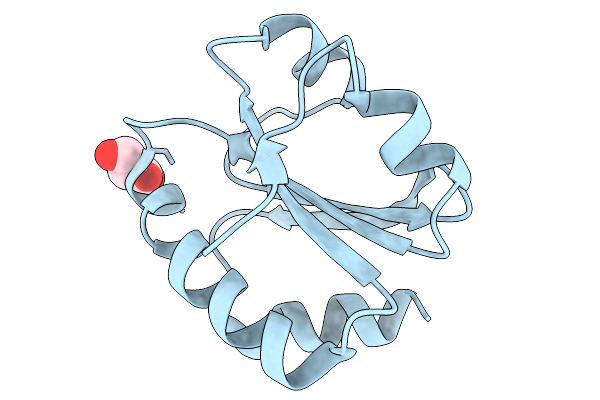 |
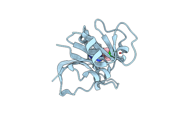
Organism: Treponema pallidum subsp. pallidum str. nichols
Method: X-RAY DIFFRACTION Resolution:2.64 Å Release Date: 2026-02-11 Classification: STRUCTURAL PROTEIN Ligands: 3OH |
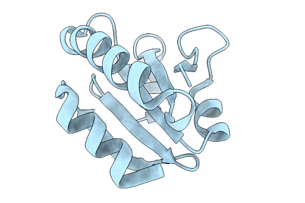 |
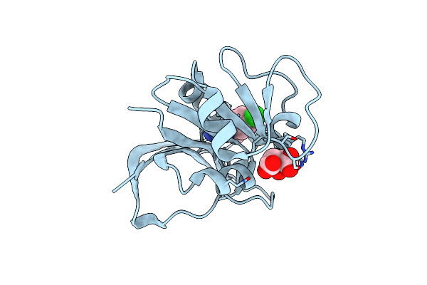
Organism: Treponema pallidum subsp. pallidum str. nichols
Method: X-RAY DIFFRACTION Resolution:1.70 Å Release Date: 2026-02-11 Classification: STRUCTURAL PROTEIN |
 |
Structure Of Methanogen Mtxx (Methanogen Marker Protein Mmp4) From Methanothermobacter Thermautotrophicus
Organism: Methanothermobacter thermautotrophicus str. delta h
Method: X-RAY DIFFRACTION Resolution:1.60 Å Release Date: 2026-02-11 Classification: UNKNOWN FUNCTION Ligands: EDO |
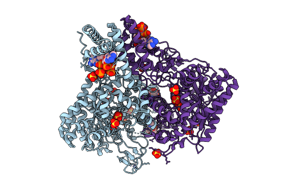 |
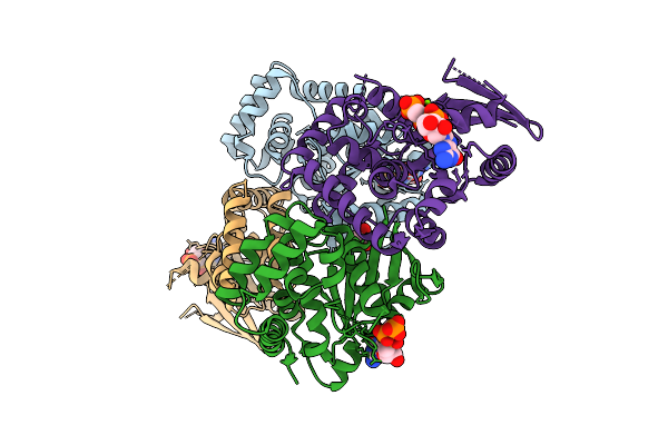
Crystal Structure Of S. Thermophilus Class Iii Ribonucleotide Reductase Bound To Datp
Organism: Streptococcus thermophilus
Method: X-RAY DIFFRACTION Resolution:2.60 Å Release Date: 2025-12-17 Classification: OXIDOREDUCTASE Ligands: MG, DTP, ZN, SO4 |
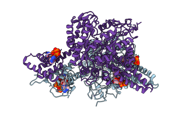 |
Organism: Streptococcus thermophilus
Method: ELECTRON MICROSCOPY Release Date: 2025-12-17 Classification: OXIDOREDUCTASE Ligands: TTP, DTP, MG, ZN |
 |
Organism: Streptococcus thermophilus
Method: ELECTRON MICROSCOPY Release Date: 2025-12-17 Classification: OXIDOREDUCTASE Ligands: ATP, MG, TTP, ZN |
 |
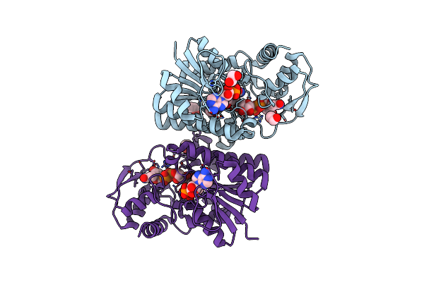
High-Resolution Crystal Structure Of Vibrio Cholerae Nfeob In The Gdp-Bound Form
Organism: Vibrio cholerae
Method: X-RAY DIFFRACTION Resolution:1.82 Å Release Date: 2025-09-03 Classification: TRANSPORT PROTEIN Ligands: GDP, GOL |
 |
Organism: Vibrio cholerae
Method: X-RAY DIFFRACTION Resolution:2.11 Å Release Date: 2025-09-03 Classification: TRANSPORT PROTEIN Ligands: GCP, GOL, MG |
 |
Crystal Structure Of Lysyl-Trna Synthetase From Mycobacterium Tuberculosis Complexed With L-Lysine And Inhibitor Ddd01991231
Organism: Mycobacterium tuberculosis
Method: X-RAY DIFFRACTION Resolution:2.40 Å Release Date: 2025-08-13 Classification: LIGASE Ligands: LYS, A1I5H |
 |
Crystal Structure Of Lysyl-Trna Synthetase From Mycobacterium Tuberculosis Complexed With L-Lysine And Inhibitor Ddd01993593
Organism: Mycobacterium tuberculosis
Method: X-RAY DIFFRACTION Resolution:2.28 Å Release Date: 2025-08-13 Classification: LIGASE Ligands: LYS, A1I50 |
 |
Crystal Structure Of Lysyl-Trna Synthetase From Mycobacterium Tuberculosis Complexed With L-Lysine And Inhibitor Ddd01869767
Organism: Mycobacterium tuberculosis
Method: X-RAY DIFFRACTION Resolution:2.60 Å Release Date: 2025-08-13 Classification: LIGASE Ligands: LYS, A1I5Z |
 |
Crystal Structure Of Lysyl-Trna Synthetase From Mycobacterium Tuberculosis Complexed With L-Lysine And Inhibitor Ddd018870911
Organism: Mycobacterium tuberculosis
Method: X-RAY DIFFRACTION Resolution:2.60 Å Release Date: 2025-08-13 Classification: LIGASE Ligands: LYS, A1I60 |
 |
Crystal Structure Of Lysyl-Trna Synthetase From Mycobacterium Tuberculosis Complexed With L-Lysine And Inhibitor Ddd01839469
Organism: Mycobacterium tuberculosis
Method: X-RAY DIFFRACTION Resolution:2.30 Å Release Date: 2025-08-13 Classification: LIGASE Ligands: LYS, A1I61 |
 |
Crystal Structure Of Lysyl-Trna Synthetase From Mycobacterium Tuberculosis Complexed With L-Lysine And Inhibitor Ddd01866774
Organism: Mycobacterium tuberculosis
Method: X-RAY DIFFRACTION Resolution:2.40 Å Release Date: 2025-08-13 Classification: LIGASE Ligands: LYS, A1I62 |
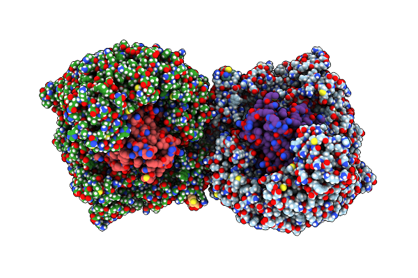 |
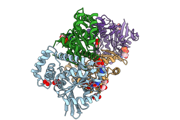
Organism: Homo sapiens, Human immunodeficiency virus 1
Method: ELECTRON MICROSCOPY Release Date: 2025-04-30 Classification: RNA BINDING PROTEIN/RNA |
 |
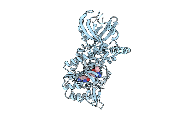
Structure Of Epimerase Mth373 From The Thermophilic Pseudomurein-Containing Methanogen Methanothermobacter Thermautotrophicus
Organism: Methanothermobacter thermautotrophicus str. delta h
Method: X-RAY DIFFRACTION Resolution:2.00 Å Release Date: 2025-02-26 Classification: SUGAR BINDING PROTEIN Ligands: UDP, NAD, EDO, MG, CL |
 |
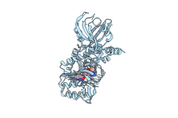
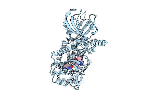
Structure Of E. Coli Dihydrofolate Reductase (Dhfr) In An Occluded Conformation And In Complex With A Cycloguanil Derivative
Organism: Escherichia coli
Method: X-RAY DIFFRACTION Resolution:2.35 Å Release Date: 2025-02-26 Classification: OXIDOREDUCTASE Ligands: A1A3E |
 |
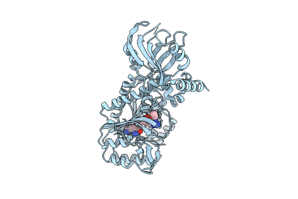
Structure Of E. Coli Dihydrofolate Reductase (Dhfr) In An Occluded Conformation And In Complex With Cycloguanil
Organism: Escherichia coli
Method: X-RAY DIFFRACTION Resolution:2.17 Å Release Date: 2025-02-26 Classification: OXIDOREDUCTASE Ligands: 1CY, FLC |
 |
Organism: Methanothermobacter thermautotrophicus str. delta h
Method: X-RAY DIFFRACTION Resolution:1.97 Å Release Date: 2025-01-29 Classification: SUGAR BINDING PROTEIN Ligands: NAD, EDO, UDX, UDP, GOL |
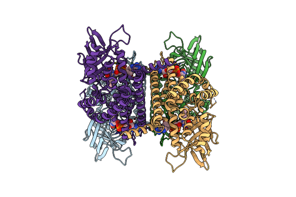 |
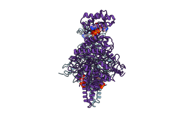
Organism: Mus musculus
Method: ELECTRON MICROSCOPY Release Date: 2024-12-11 Classification: OXIDOREDUCTASE Ligands: FAD |