

Search Count: 151
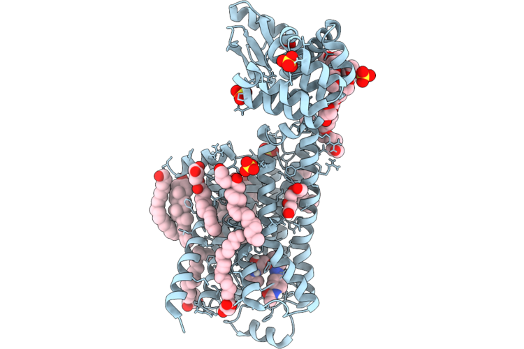 |
Dark Structure Of Beta-2 Adrenergic Receptor With Photoazolol In Dark State Recorded At Swissfel
Organism: Homo sapiens, Enterobacteria phage t4
Method: X-RAY DIFFRACTION Resolution:2.45 Å Release Date: 2026-04-22 Classification: MEMBRANE PROTEIN Ligands: SO4, CLR, 12P, OLC, 1PE, EDO, GOL, A1JHU, PLM |
Organism: Homo sapiens, Enterobacteria phage t4
Method: X-RAY DIFFRACTION
Release Date: 2026-04-22
Ligands: SO4, CLR, 12P, OLC, 1PE, EDO, GOL, A1JHU, PLM
 |
Mixed Model Refinement Of Beta-2 Adrenergic Receptor With Photoazolol In Dark State And Light State, 10 Seconds After Light Activation, Recorded At Swissfel
Organism: Homo sapiens, Tequatrovirus t4
Method: X-RAY DIFFRACTION Resolution:2.45 Å Release Date: 2026-04-22 Classification: MEMBRANE PROTEIN Ligands: SO4, ACM, CLR, PLM, 12P, OLC, 1PE, EDO, GOL, A1JHU |
Organism: Homo sapiens, Tequatrovirus t4
Method: X-RAY DIFFRACTION
Release Date: 2026-04-22
Ligands: SO4, ACM, CLR, PLM, 12P, OLC, 1PE, EDO, GOL, A1JHU
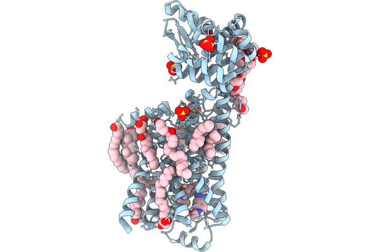 |
Dark Structure Of Beta-2 Adrenergic Receptor With Photoazolol In Dark State Recorded At Lcls
Organism: Homo sapiens, Enterobacteria phage t4
Method: X-RAY DIFFRACTION Resolution:2.50 Å Release Date: 2026-04-22 Classification: MEMBRANE PROTEIN Ligands: SO4, CLR, PLM, 12P, A1JHU, OLC, EDO, GOL |
Organism: Homo sapiens, Enterobacteria phage t4
Method: X-RAY DIFFRACTION
Release Date: 2026-04-22
Ligands: SO4, CLR, PLM, 12P, A1JHU, OLC, EDO, GOL
 |
Mixed Model Refinement Of Beta-2 Adrenergic Receptor With Photoazolol In Dark State And Light State, 17 Nanoseconds After Light Activation, Recorded At Lcls
Organism: Homo sapiens, Tequatrovirus t4
Method: X-RAY DIFFRACTION Resolution:2.60 Å Release Date: 2026-04-22 Classification: MEMBRANE PROTEIN Ligands: SO4, ACM, CLR, PLM, 12P, OLC, EDO, GOL, 1PE, A1JHU |
Organism: Homo sapiens, Tequatrovirus t4
Method: X-RAY DIFFRACTION
Release Date: 2026-04-22
Ligands: SO4, ACM, CLR, PLM, 12P, OLC, EDO, GOL, 1PE, A1JHU
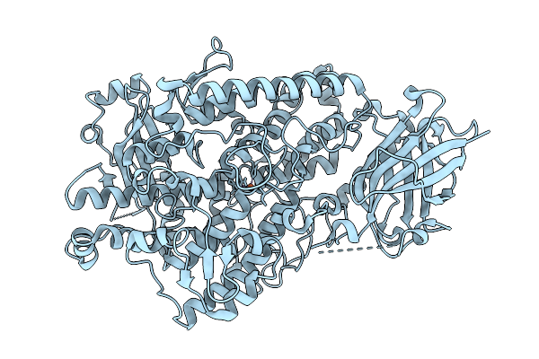 |
Rt Xfel Structure Of Soybean Lipoxygenase-1 In Small Unit-Cell
Organism: Glycine max
Method: X-RAY DIFFRACTION Resolution:1.95 Å Release Date: 2026-04-08 Classification: OXIDOREDUCTASE Ligands: FE |
Organism: Glycine max
Method: X-RAY DIFFRACTION
Release Date: 2026-04-08
Ligands: FE
 |
Rt Xfel Structure Of Soybean Lipoxygenase-1 In Large Unit-Cell
Organism: Glycine max
Method: X-RAY DIFFRACTION Resolution:1.95 Å Release Date: 2026-04-08 Classification: OXIDOREDUCTASE Ligands: FE |
Organism: Glycine max
Method: X-RAY DIFFRACTION
Release Date: 2026-04-08
Ligands: FE
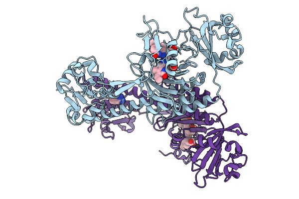 |
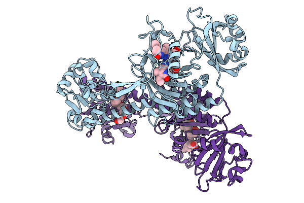
Photoactivation In Bacteriophytochromes, Reference (Dark) Structure For The 3 Ps Time Point
Organism: Stigmatella aurantiaca
Method: X-RAY DIFFRACTION Resolution:2.30 Å Release Date: 2025-10-08 Classification: SIGNALING PROTEIN Ligands: 3Q8, BEN |
Organism: Stigmatella aurantiaca
Method: X-RAY DIFFRACTION
Release Date: 2025-10-08
Ligands: 3Q8, BEN
 |
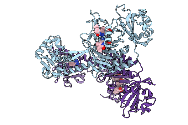
Photoactivation In Bacteriophytochrome, High Resolution Cryo Structure In The Dark.
Organism: Stigmatella aurantiaca
Method: X-RAY DIFFRACTION Resolution:1.40 Å Release Date: 2025-10-08 Classification: SIGNALING PROTEIN Ligands: EL5, P33 |
Organism: Stigmatella aurantiaca
Method: X-RAY DIFFRACTION
Release Date: 2025-10-08
Ligands: EL5, P33
 |
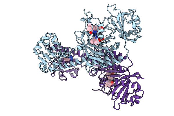
Photoactivation In Bacteriophytochromes, Reference (Dark) Structure For The 100 Ps Time Point
Organism: Stigmatella aurantiaca
Method: X-RAY DIFFRACTION Resolution:1.93 Å Release Date: 2025-10-08 Classification: SIGNALING PROTEIN Ligands: EL5, BEN |
Organism: Stigmatella aurantiaca
Method: X-RAY DIFFRACTION
Release Date: 2025-10-08
Ligands: EL5, BEN
 |
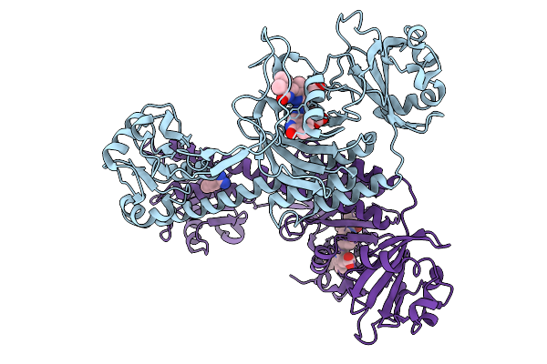
Photoactivation In Bacteriophytochromes; 3Ps Tr-Sfx Structure
Organism: Stigmatella aurantiaca
Method: X-RAY DIFFRACTION Resolution:2.30 Å Release Date: 2025-10-08 Classification: SIGNALING PROTEIN Ligands: BLA, BEN |
Organism: Stigmatella aurantiaca
Method: X-RAY DIFFRACTION
Release Date: 2025-10-08
Ligands: BLA, BEN
 |
Photoactivation In Bacteriophytochrome, 100 Ps Structure
Organism: Stigmatella aurantiaca
Method: X-RAY DIFFRACTION Resolution:2.30 Å Release Date: 2025-10-08 Classification: SIGNALING PROTEIN Ligands: BLA, BEN |
Organism: Stigmatella aurantiaca
Method: X-RAY DIFFRACTION
Release Date: 2025-10-08
Ligands: BLA, BEN
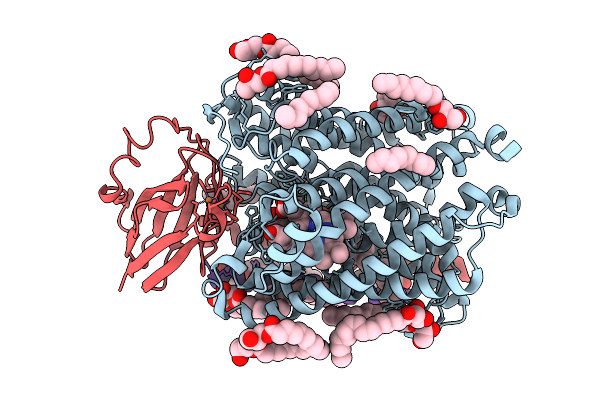 |
Chloride Bound Structure Of Oxidized Ba3-Type Cytochrome C Oxidase Confirmed By Single-Wavelength Anomalous Diffraction
Organism: Thermus thermophilus
Method: X-RAY DIFFRACTION Resolution:2.28 Å Release Date: 2025-08-13 Classification: OXIDOREDUCTASE Ligands: CU, HEM, HAS, OLC, CL, CUA |
Organism: Thermus thermophilus
Method: X-RAY DIFFRACTION
Release Date: 2025-08-13
Ligands: CU, HEM, HAS, OLC, CL, CUA
 |
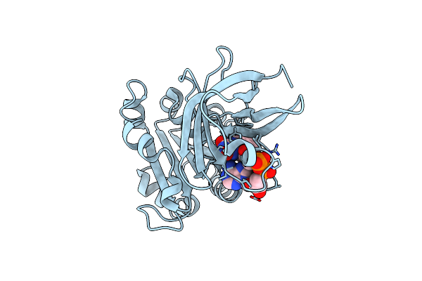
Room Temperature Structure Of Fad-Containing Ferrodoxin-Nadp Reductase From Brucella Ovis At Euxfel
Organism: Brucella ovis atcc 25840
Method: X-RAY DIFFRACTION Resolution:1.90 Å Release Date: 2024-11-27 Classification: OXIDOREDUCTASE Ligands: FAD |
Organism: Brucella ovis atcc 25840
Method: X-RAY DIFFRACTION
Release Date: 2024-11-27
Ligands: FAD
 |
Room Temperature Structure Of Fad-Containing Ferrodoxin-Nadp Reductase From Brucella Ovis At Lcls
Organism: Brucella ovis atcc 25840
Method: X-RAY DIFFRACTION Resolution:2.20 Å Release Date: 2024-11-27 Classification: OXIDOREDUCTASE Ligands: FAD |
Organism: Brucella ovis atcc 25840
Method: X-RAY DIFFRACTION
Release Date: 2024-11-27
Ligands: FAD
 |
Pseudomonas Fluorescens G150T Isocyanide Hydratase At 298 K Xfel Data, Free Enzyme
Organism: Pseudomonas fluorescens
Method: X-RAY DIFFRACTION Resolution:1.30 Å Release Date: 2024-02-21 Classification: LYASE Ligands: CL |
Organism: Pseudomonas fluorescens
Method: X-RAY DIFFRACTION
Release Date: 2024-02-21
Ligands: CL
 |
Pseudomonas Fluorescens G150T Isocyanide Hydratase At 298 K Xfel Data, Thioimidate Intermediate
Organism: Pseudomonas fluorescens
Method: X-RAY DIFFRACTION Resolution:1.30 Å Release Date: 2024-02-21 Classification: LYASE Ligands: QCV, CL |
Organism: Pseudomonas fluorescens
Method: X-RAY DIFFRACTION
Release Date: 2024-02-21
Ligands: QCV, CL
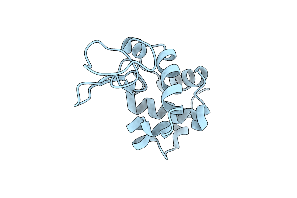 |
Lysozyme Crystallized In Cyclic Olefin Copolymer-Based Microfluidic Chips
Organism: Gallus gallus
Method: X-RAY DIFFRACTION Resolution:1.45 Å Release Date: 2023-10-25 Classification: HYDROLASE |
Organism: Gallus gallus
Method: X-RAY DIFFRACTION
Release Date: 2023-10-25
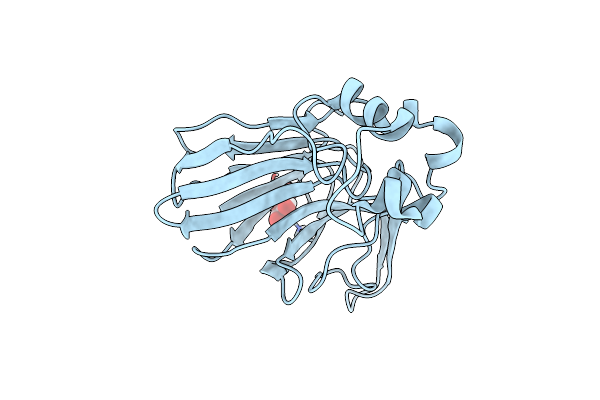 |
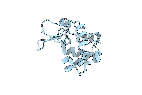
Thaumatin Crystallized In Cyclic Olefin Copolymer-Based Microfluidic Chips
Organism: Thaumatococcus daniellii
Method: X-RAY DIFFRACTION Resolution:1.48 Å Release Date: 2023-10-18 Classification: PLANT PROTEIN Ligands: TLA |
Organism: Thaumatococcus daniellii
Method: X-RAY DIFFRACTION
Release Date: 2023-10-18
Ligands: TLA
 |
Lysozyme Crystallized In Cyclic Olefin Copolymer-Based Microfluidic Chips
Organism: Gallus gallus
Method: X-RAY DIFFRACTION Resolution:1.70 Å Release Date: 2023-10-18 Classification: HYDROLASE |
Organism: Gallus gallus
Method: X-RAY DIFFRACTION
Release Date: 2023-10-18
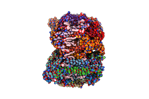 |
Sfx Structure Of Oxidized Cytochrome C Oxidase At 2.38 Angstrom Resolution
Organism: Bos taurus
Method: X-RAY DIFFRACTION Resolution:2.38 Å Release Date: 2023-09-27 Classification: MEMBRANE PROTEIN Ligands: CU, MG, NA, PGV, HEA, OH, CUA, TGL, PSC, CHD, PEK, CDL, DMU, ZN, SAC |