

Search Count: 54
 |
Neutron Structure Of Cellulase Cel6A From Phanerochaete Chrysosporium At Room Temperature
Organism: Phanerodontia chrysosporium
Method: X-RAY DIFFRACTION, NEUTRON DIFFRACTION Resolution:1.36 Å, 1.86 Å Release Date: 2025-03-12 Classification: HYDROLASE |
Organism: Phanerodontia chrysosporium
Method: X-RAY DIFFRACTION, NEUTRON DIFFRACTION
Release Date: 2025-03-12
 |
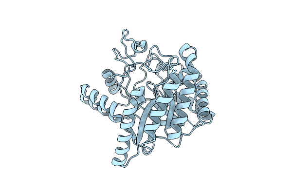
Neutron Structure Of Cellulase Cel6A From Phanerochaete Chrysosporium At Room Temperature, Enzyme-Product Complex
Organism: Phanerodontia chrysosporium
Method: X-RAY DIFFRACTION, NEUTRON DIFFRACTION Resolution:1.40 Å, 1.8 Å Release Date: 2025-03-12 Classification: HYDROLASE Ligands: BGC, NA |
Organism: Phanerodontia chrysosporium
Method: X-RAY DIFFRACTION, NEUTRON DIFFRACTION
Release Date: 2025-03-12
Ligands: BGC, NA
 |
Neutron Structure Of Cellulase Cel6A From Phanerochaete Chrysosporium At Room Temperature, Low-D2O-Solvent
Organism: Phanerodontia chrysosporium
Method: X-RAY DIFFRACTION, NEUTRON DIFFRACTION Resolution:1.40 Å, 2.15 Å Release Date: 2025-03-12 Classification: HYDROLASE |
Organism: Phanerodontia chrysosporium
Method: X-RAY DIFFRACTION, NEUTRON DIFFRACTION
Release Date: 2025-03-12
 |
Neutron Structure Of Cellulase Cel6A From Phanerochaete Chrysosporium At Room Temperature, Enzyme-Product Complex, H2O Solvent
Organism: Phanerodontia chrysosporium
Method: X-RAY DIFFRACTION, NEUTRON DIFFRACTION Resolution:1.80 Å, 2.59 Å Release Date: 2025-03-12 Classification: HYDROLASE Ligands: BGC, NA |
Organism: Phanerodontia chrysosporium
Method: X-RAY DIFFRACTION, NEUTRON DIFFRACTION
Release Date: 2025-03-12
Ligands: BGC, NA
 |
High-Resolution X-Ray Structure Of Cellulase Cel6A From Phanerochaete Chrysosporium At Cryogenic Temperature
Organism: Phanerodontia chrysosporium
Method: X-RAY DIFFRACTION Resolution:0.80 Å Release Date: 2025-03-12 Classification: HYDROLASE Ligands: MPD, ACT |
Organism: Phanerodontia chrysosporium
Method: X-RAY DIFFRACTION
Release Date: 2025-03-12
Ligands: MPD, ACT
 |
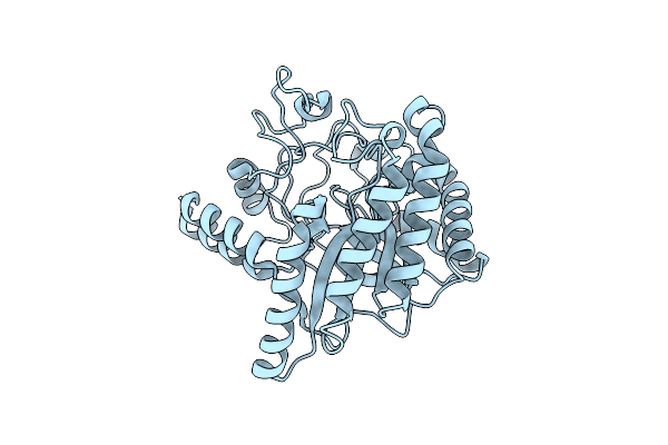
High-Resolution X-Ray Structure Of Cellulase Cel6A From Phanerochaete Chrysosporium At Cryogenic Temperature, Enzyme-Product Complex
Organism: Phanerodontia chrysosporium
Method: X-RAY DIFFRACTION Resolution:0.85 Å Release Date: 2025-03-12 Classification: HYDROLASE Ligands: BGC, NA, CL, PEG |
Organism: Phanerodontia chrysosporium
Method: X-RAY DIFFRACTION
Release Date: 2025-03-12
Ligands: BGC, NA, CL, PEG
 |
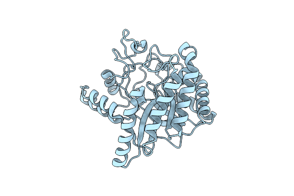
X-Ray Crystal Structure Of Glycoside Hydrolase Family 6 Cellobiohydrolase From Phanerochaete Chrysosporium Pccel6A Wild-Type
Organism: Phanerodontia chrysosporium
Method: X-RAY DIFFRACTION Resolution:0.99 Å Release Date: 2024-09-04 Classification: HYDROLASE |
Organism: Phanerodontia chrysosporium
Method: X-RAY DIFFRACTION
Release Date: 2024-09-04
 |
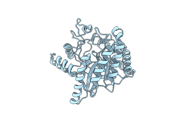
X-Ray Crystal Structure Of Glycoside Hydrolase Family 6 Cellobiohydrolase From Phanerochaete Chrysosporium Pccel6A C240S
Organism: Phanerodontia chrysosporium
Method: X-RAY DIFFRACTION Resolution:1.01 Å Release Date: 2024-09-04 Classification: HYDROLASE |
Organism: Phanerodontia chrysosporium
Method: X-RAY DIFFRACTION
Release Date: 2024-09-04
 |
X-Ray Crystal Structure Of Glycoside Hydrolase Family 6 Cellobiohydrolase From Phanerochaete Chrysosporium Pccel6A C393S
Organism: Phanerodontia chrysosporium
Method: X-RAY DIFFRACTION Resolution:1.00 Å Release Date: 2024-09-04 Classification: HYDROLASE |
Organism: Phanerodontia chrysosporium
Method: X-RAY DIFFRACTION
Release Date: 2024-09-04
 |
X-Ray Crystal Structure Of Glycoside Hydrolase Family 6 Cellobiohydrolase From Phanerochaete Chrysosporium Pccel6A C240S/C393S
Organism: Phanerodontia chrysosporium
Method: X-RAY DIFFRACTION Resolution:0.99 Å Release Date: 2024-09-04 Classification: HYDROLASE |
Organism: Phanerodontia chrysosporium
Method: X-RAY DIFFRACTION
Release Date: 2024-09-04
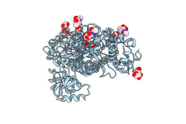 |
The Structure Of Beta-Xylosidase From Phanerochaete Chrysosporium(Pcbxl3)
Organism: Phanerochaete chrysosporium
Method: X-RAY DIFFRACTION Resolution:2.54 Å Release Date: 2022-02-09 Classification: HYDROLASE Ligands: NAG, GOL |
Organism: Phanerochaete chrysosporium
Method: X-RAY DIFFRACTION
Release Date: 2022-02-09
Ligands: NAG, GOL
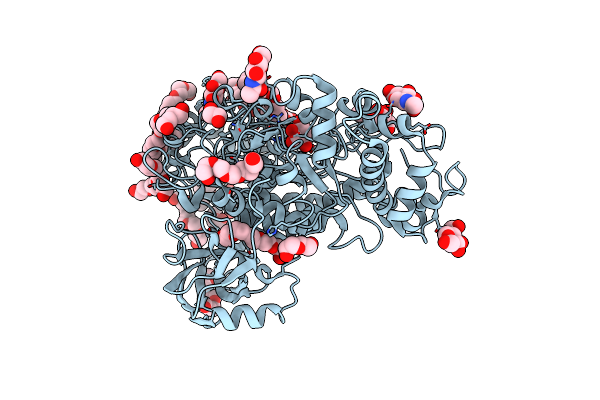 |
The Structure Of Beta-Xylosidase From Phanerochaete Chrysosporium(Pcbxl3)
Organism: Phanerochaete chrysosporium
Method: X-RAY DIFFRACTION Resolution:3.08 Å Release Date: 2022-02-09 Classification: HYDROLASE Ligands: 1PE, PEG, EDO, NAG, XYP |
Organism: Phanerochaete chrysosporium
Method: X-RAY DIFFRACTION
Release Date: 2022-02-09
Ligands: 1PE, PEG, EDO, NAG, XYP
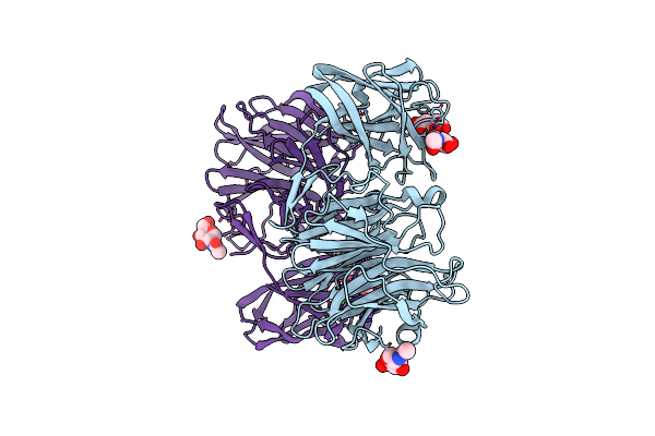 |
Crystal Structure Of Exo-Beta-1,3-Galactanase From Phanerochaete Chrysosporium Pc1,3Gal43A Apo Form
Organism: Phanerochaete chrysosporium
Method: X-RAY DIFFRACTION Resolution:1.40 Å Release Date: 2020-11-04 Classification: HYDROLASE Ligands: NAG, CA, CIT |
Organism: Phanerochaete chrysosporium
Method: X-RAY DIFFRACTION
Release Date: 2020-11-04
Ligands: NAG, CA, CIT
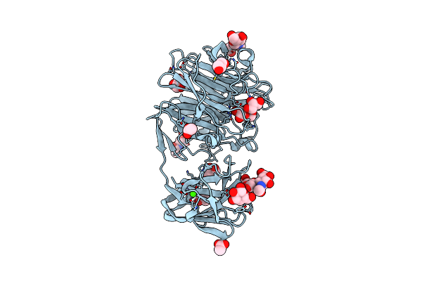 |
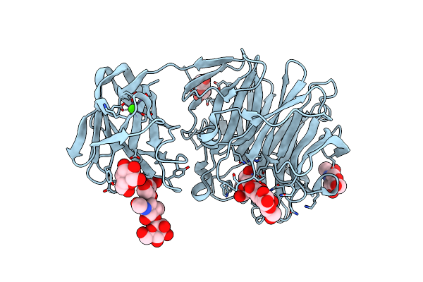
Crystal Structure Of Exo-Beta-1,3-Galactanase From Phanerochaete Chrysosporium Pc1,3Gal43A With Galactose
Organism: Phanerochaete chrysosporium
Method: X-RAY DIFFRACTION Resolution:1.50 Å Release Date: 2020-11-04 Classification: HYDROLASE Ligands: NAG, CA, GLA, GAL, ACY, ACT, GOL |
Organism: Phanerochaete chrysosporium
Method: X-RAY DIFFRACTION
Release Date: 2020-11-04
Ligands: NAG, CA, GLA, GAL, ACY, ACT, GOL
 |
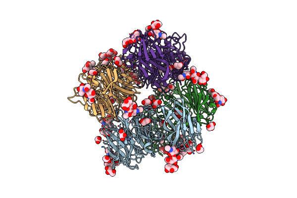
Crystal Structure Of Exo-Beta-1,3-Galactanase From Phanerochaete Chrysosporium Pc1,3Gal43A E208Q With Beta-1,3-Galactotriose
Organism: Phanerochaete chrysosporium
Method: X-RAY DIFFRACTION Resolution:2.50 Å Release Date: 2020-11-04 Classification: HYDROLASE Ligands: NAG, CA, GAL |
Organism: Phanerochaete chrysosporium
Method: X-RAY DIFFRACTION
Release Date: 2020-11-04
Ligands: NAG, CA, GAL
 |
Crystal Structure Of Exo-Beta-1,3-Galactanase From Phanerochaete Chrysosporium Pc1,3Gal43A E208A With Beta-1,3-Galactotriose
Organism: Phanerochaete chrysosporium
Method: X-RAY DIFFRACTION Resolution:2.30 Å Release Date: 2020-11-04 Classification: HYDROLASE Ligands: CA, ACT, GOL, NAG |
Organism: Phanerochaete chrysosporium
Method: X-RAY DIFFRACTION
Release Date: 2020-11-04
Ligands: CA, ACT, GOL, NAG
 |
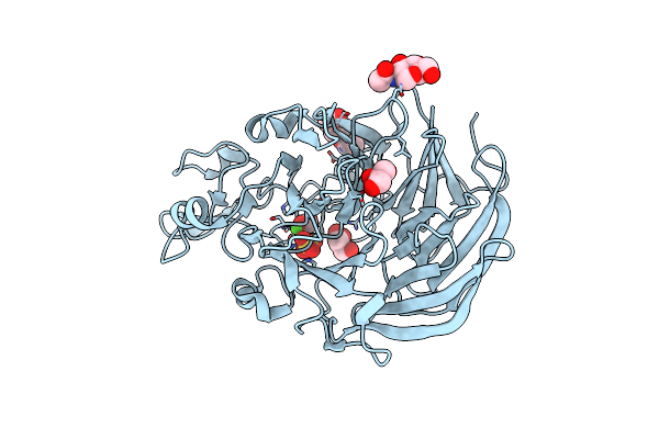
Crystal Structure Of Cytochrome B Domain Of Pyranose Dehydrogenase From Coprinopsis Cinerea
Organism: Coprinopsis cinerea
Method: X-RAY DIFFRACTION Resolution:2.00 Å Release Date: 2019-11-13 Classification: OXIDOREDUCTASE Ligands: NAG, HEM, MPD, ACT |
Organism: Coprinopsis cinerea
Method: X-RAY DIFFRACTION
Release Date: 2019-11-13
Ligands: NAG, HEM, MPD, ACT
 |
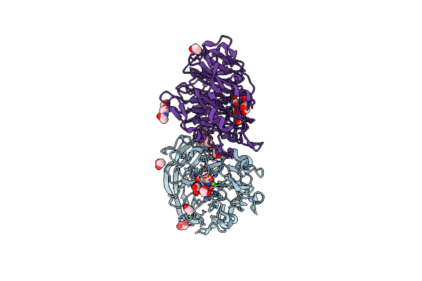
Crystal Structure Of Pqq Doamin Of Pyranose Dehydrogenase From Coprinopsis Cinerea: Apo-From
Organism: Coprinopsis cinerea
Method: X-RAY DIFFRACTION Resolution:1.50 Å Release Date: 2019-11-06 Classification: OXIDOREDUCTASE Ligands: NAG, CA, SO4, FMT, PGE, GOL |
Organism: Coprinopsis cinerea
Method: X-RAY DIFFRACTION
Release Date: 2019-11-06
Ligands: NAG, CA, SO4, FMT, PGE, GOL
 |
Holo Form Of Pyranose Dehydrogenase Pqq Domain From Coprinopsis Cinerea
Organism: Coprinopsis cinerea
Method: X-RAY DIFFRACTION Resolution:1.30 Å Release Date: 2019-11-06 Classification: OXIDOREDUCTASE Ligands: CA, PQQ, EDO, ACT, NAG |
Organism: Coprinopsis cinerea
Method: X-RAY DIFFRACTION
Release Date: 2019-11-06
Ligands: CA, PQQ, EDO, ACT, NAG
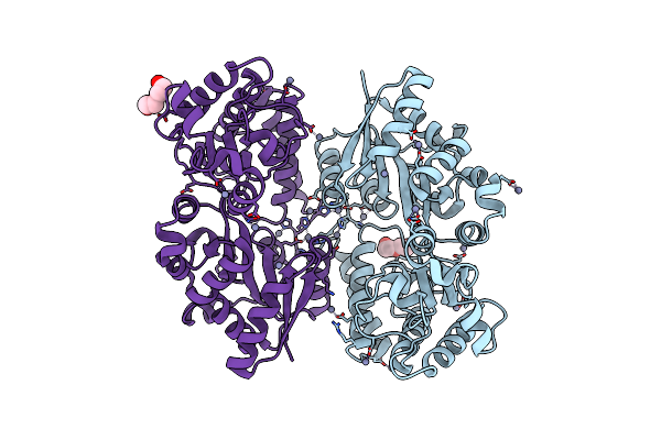 |
Crystal Structure Of Beta-1,2-Glucooligosaccharide Binding Protein In Ligand-Free Form
Organism: Listeria innocua serovar 6a (strain atcc baa-680 / clip 11262)
Method: X-RAY DIFFRACTION Resolution:2.20 Å Release Date: 2018-05-02 Classification: SUGAR BINDING PROTEIN Ligands: ZN, PEG |
Organism: Listeria innocua serovar 6a (strain atcc baa-680 / clip 11262)
Method: X-RAY DIFFRACTION
Release Date: 2018-05-02
Ligands: ZN, PEG