

Search Count: 43
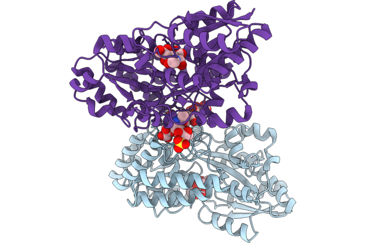 |
Crystal Structure Of A Sialic Acid Binding Protein, R113K:G213Q:Q216N Mutant, From Streptococcus Pneumoniae Bound To Neu5Ac
Organism: Streptococcus pneumoniae tigr4
Method: X-RAY DIFFRACTION Resolution:2.00 Å Release Date: 2026-07-29 Classification: SUGAR BINDING PROTEIN Ligands: SIA, SLB, SO4, CIT |
Organism: Streptococcus pneumoniae tigr4
Method: X-RAY DIFFRACTION
Release Date: 2026-07-29
Ligands: SIA, SLB, SO4, CIT
 |
Structural Characterization Of A N-Acetyl-Beta-Neuraminic Acid-Binding Site In Human Rnase2
Organism: Homo sapiens
Method: X-RAY DIFFRACTION Resolution:1.01 Å Release Date: 2026-03-18 Classification: PROTEIN BINDING Ligands: SLB, B3P, ACT |
Organism: Homo sapiens
Method: X-RAY DIFFRACTION
Release Date: 2026-03-18
Ligands: SLB, B3P, ACT
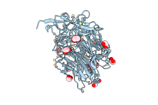 |
Crystal Structure Of Treponema Denticola Sialidase (Tde_0471) Bound To Neu5Ac (Nana)
Organism: Treponema denticola
Method: X-RAY DIFFRACTION Resolution:1.56 Å Release Date: 2026-02-25 Classification: HYDROLASE Ligands: CD, SLB, EDO, PEG, PGE |
Organism: Treponema denticola
Method: X-RAY DIFFRACTION
Release Date: 2026-02-25
Ligands: CD, SLB, EDO, PEG, PGE
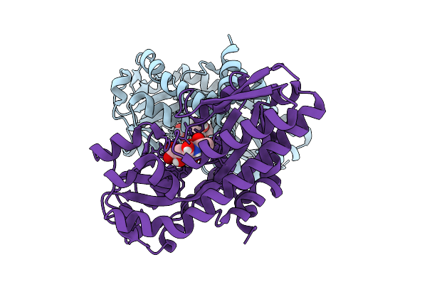 |
Structure Of Apo Aggregatibacter Actinomycetemcomitans Siap Protein
Organism: Aggregatibacter actinomycetemcomitans
Method: X-RAY DIFFRACTION Resolution:1.90 Å Release Date: 2025-10-08 Classification: MEMBRANE PROTEIN Ligands: SLB |
Organism: Aggregatibacter actinomycetemcomitans
Method: X-RAY DIFFRACTION
Release Date: 2025-10-08
Ligands: SLB
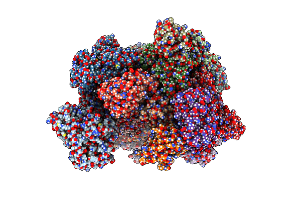 |
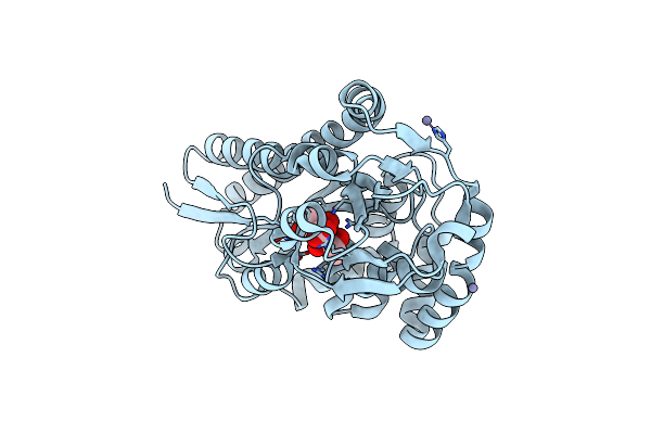
Crystal Structure Of Vcsiap W73A Mutant In Complex With Sialic Acid And A Vhh Antibody (Vhh_Vcp#2)
Organism: Vicugna pacos
Method: X-RAY DIFFRACTION Resolution:2.81 Å Release Date: 2024-11-06 Classification: TRANSPORT PROTEIN Ligands: SLB, GOL |
Organism: Vicugna pacos
Method: X-RAY DIFFRACTION
Release Date: 2024-11-06
Ligands: SLB, GOL
 |
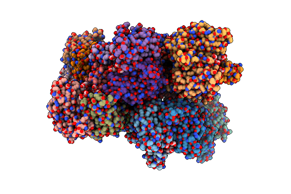
Structure Of The Disulfide-Locked Substrate Binding Protein Hisiap.
Organism: Haemophilus influenzae
Method: X-RAY DIFFRACTION Resolution:1.90 Å Release Date: 2023-12-27 Classification: SUGAR BINDING PROTEIN Ligands: SLB, ZN |
Organism: Haemophilus influenzae
Method: X-RAY DIFFRACTION
Release Date: 2023-12-27
Ligands: SLB, ZN
 |
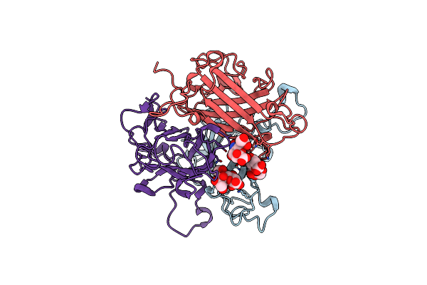
Human Adenovirus Type 25 Fiber-Knob Protein Complexed With Sialic Acid
Organism: Human adenovirus 25
Method: X-RAY DIFFRACTION Resolution:2.51 Å Release Date: 2023-09-20 Classification: VIRAL PROTEIN Ligands: SLB |
Organism: Human adenovirus 25
Method: X-RAY DIFFRACTION
Release Date: 2023-09-20
Ligands: SLB
 |
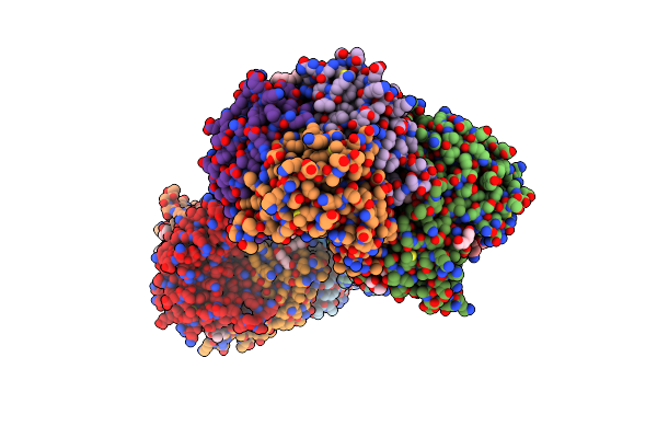
Human Adenovirus Type 30 Fiber-Knob Protein Complexed With Sialic Acid
Organism: Human adenovirus 30
Method: X-RAY DIFFRACTION Resolution:2.56 Å Release Date: 2023-09-20 Classification: VIRAL PROTEIN Ligands: SLB |
Organism: Human adenovirus 30
Method: X-RAY DIFFRACTION
Release Date: 2023-09-20
Ligands: SLB
 |
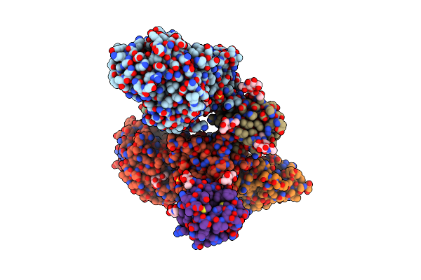
Human Adenovirus Type 53 Fiber-Knob Protein Complexed With Sialic Acid
Organism: Human adenovirus 53
Method: X-RAY DIFFRACTION Resolution:1.77 Å Release Date: 2023-09-20 Classification: VIRAL PROTEIN Ligands: PEG, EDO, P4G, SLB, SIA |
Organism: Human adenovirus 53
Method: X-RAY DIFFRACTION
Release Date: 2023-09-20
Ligands: PEG, EDO, P4G, SLB, SIA
 |
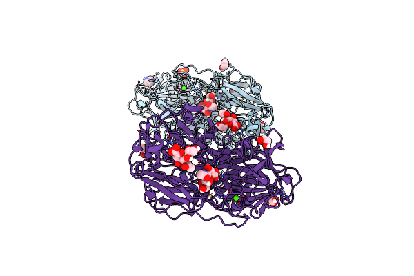
Crystal Structure Of Bont/E Receptor Binding Domain In Complex With Sv2, Vhh, And Sialic Acid
Organism: Vicugna pacos, Homo sapiens, Clostridium botulinum
Method: X-RAY DIFFRACTION Resolution:2.77 Å Release Date: 2023-04-05 Classification: TOXIN Ligands: NAG, SO4, PG4, SLB |
Organism: Vicugna pacos, Homo sapiens, Clostridium botulinum
Method: X-RAY DIFFRACTION
Release Date: 2023-04-05
Ligands: NAG, SO4, PG4, SLB
 |
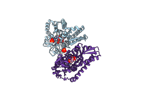
Sialidases And Fucosidases Of Akkermansia Muciniphila Are Key For Rapid Growth On Colonic Mucin And Nutrient Sharing Amongst Mucin-Associated Human Gut Microbiota
Organism: Akkermansia muciniphila
Method: X-RAY DIFFRACTION Resolution:1.30 Å Release Date: 2023-03-01 Classification: HYDROLASE Ligands: SLB, IMD, EDO, CL, CA, SO4, DAN |
Organism: Akkermansia muciniphila
Method: X-RAY DIFFRACTION
Release Date: 2023-03-01
Ligands: SLB, IMD, EDO, CL, CA, SO4, DAN
 |
Structure Of The Sialic Acid Bound Tripartite Atp-Independent Periplasmic (Trap) Periplasmic Component Siap From Photobacterium Profundum
Organism: Photobacterium profundum
Method: X-RAY DIFFRACTION Resolution:1.04 Å Release Date: 2022-12-14 Classification: TRANSPORT PROTEIN Ligands: SLB, SO4 |
Organism: Photobacterium profundum
Method: X-RAY DIFFRACTION
Release Date: 2022-12-14
Ligands: SLB, SO4
 |
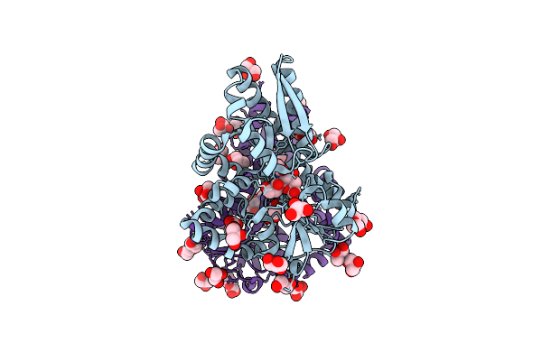
Crystal Structure Of Tetrameric Fibrinogen-Like Recognition Domain Of Fibcd1 With Neu5Ac Ligand Bound
Organism: Homo sapiens
Method: X-RAY DIFFRACTION Resolution:1.85 Å Release Date: 2021-07-21 Classification: SUGAR BINDING PROTEIN Ligands: CA, SO4, ACY, SLB |
Organism: Homo sapiens
Method: X-RAY DIFFRACTION
Release Date: 2021-07-21
Ligands: CA, SO4, ACY, SLB
 |
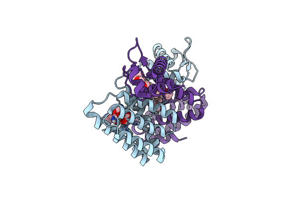
Crystal Structure Of Vcsiap Bound To Sialic Acid
Organism: Vibrio cholerae
Method: X-RAY DIFFRACTION Resolution:1.68 Å Release Date: 2020-12-30 Classification: TRANSPORT PROTEIN Ligands: SLB, GOL, PGE, BGC |
Organism: Vibrio cholerae
Method: X-RAY DIFFRACTION
Release Date: 2020-12-30
Ligands: SLB, GOL, PGE, BGC
 |
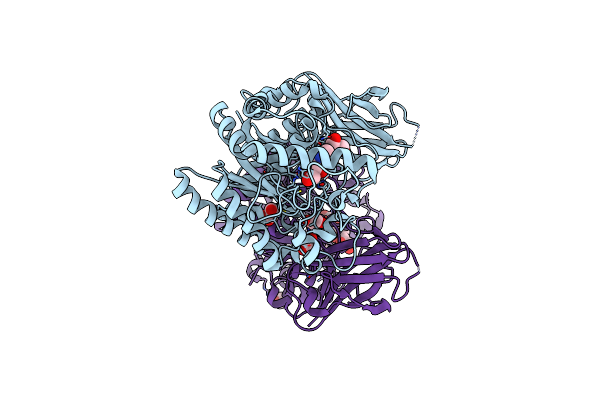
Crystal Structure Of The Gntr-Type Sialoregulator Nanr From Escherichia Coli, In Complex With Sialic Acid
Organism: Escherichia coli k-12
Method: X-RAY DIFFRACTION Resolution:2.10 Å Release Date: 2020-07-08 Classification: DNA BINDING PROTEIN Ligands: SLB, ZN, P4K |
Organism: Escherichia coli k-12
Method: X-RAY DIFFRACTION
Release Date: 2020-07-08
Ligands: SLB, ZN, P4K
 |
Crystal Structure Of An Inverting Family Gh156 Exosialidase From Uncultured Bacterium Pg7 In Complex With N-Acetylneuraminic Acid
Organism: Uncultured bacterium pg7
Method: X-RAY DIFFRACTION Resolution:2.00 Å Release Date: 2019-11-06 Classification: HYDROLASE Ligands: GOL, SLB, PG4 |
Organism: Uncultured bacterium pg7
Method: X-RAY DIFFRACTION
Release Date: 2019-11-06
Ligands: GOL, SLB, PG4
 |
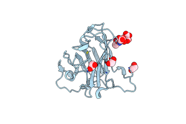
Adenovirus Serotype 26 (Ad26) In Complex With Sialic Acid, Ph4.0
Organism: Human adenovirus 26
Method: X-RAY DIFFRACTION Resolution:1.03 Å Release Date: 2019-09-18 Classification: VIRAL PROTEIN Ligands: SIA, SLB, SO4, EDO, PEG |
Organism: Human adenovirus 26
Method: X-RAY DIFFRACTION
Release Date: 2019-09-18
Ligands: SIA, SLB, SO4, EDO, PEG
 |
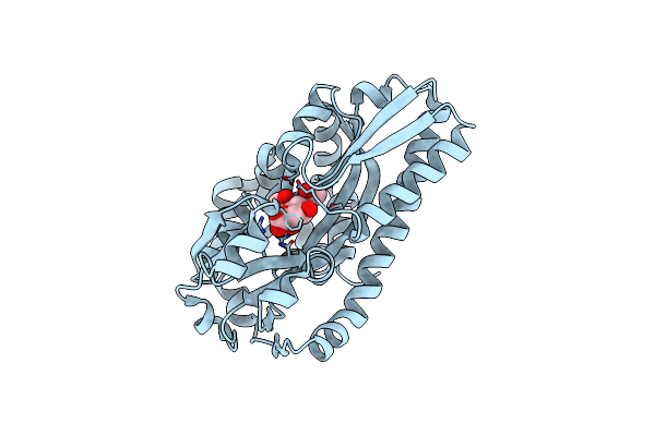
Adenovirus Serotype 26 (Ad26) In Complex With Sialic Acid, Ph8.0
Organism: Human adenovirus 26
Method: X-RAY DIFFRACTION Resolution:1.19 Å Release Date: 2019-09-18 Classification: VIRAL PROTEIN Ligands: SIA, SLB, EDO |
Organism: Human adenovirus 26
Method: X-RAY DIFFRACTION
Release Date: 2019-09-18
Ligands: SIA, SLB, EDO
 |
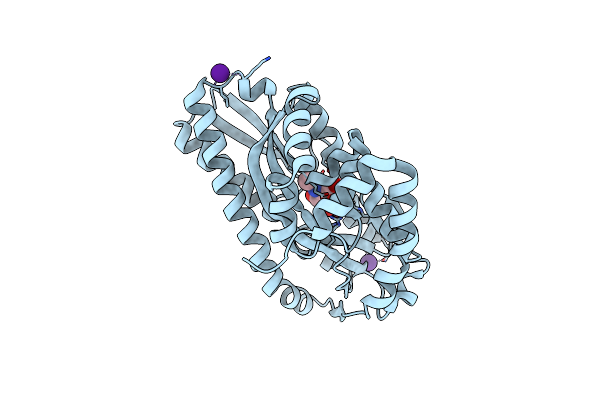
Siap A11N In Complex With Neu5Ac (Rt)
Organism: Haemophilus influenzae rd kw20
Method: X-RAY DIFFRACTION Resolution:1.45 Å Release Date: 2019-08-14 Classification: TRANSPORT PROTEIN Ligands: SLB |
Organism: Haemophilus influenzae rd kw20
Method: X-RAY DIFFRACTION
Release Date: 2019-08-14
Ligands: SLB
 |
Siap In Complex With Neu5Ac (Rt)
Organism: Haemophilus influenzae rd kw20
Method: X-RAY DIFFRACTION Resolution:1.50 Å Release Date: 2019-08-14 Classification: TRANSPORT PROTEIN Ligands: SLB, CL, CS |
Organism: Haemophilus influenzae rd kw20
Method: X-RAY DIFFRACTION
Release Date: 2019-08-14
Ligands: SLB, CL, CS