
Search Count: 1,04,516
All
Selected
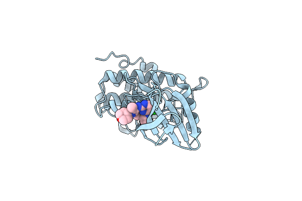 |
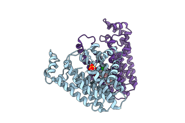
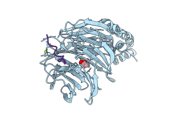
Structure Of Chk1 10-Pt. Mutant Complex With Macrocyclic Lrrk2 Inhibitor Compound 1 ((11R)-8-Chloro-3,11-Dimethyl-2-(Oxan-4-Yl)-2,4,10,11,12,13-Hexahydro-9,5-(Azeno)Pyrazolo[3,4-B][1,4,6,10]Oxatriazacyclotridecine)
Organism: Homo sapiens
Method: X-RAY DIFFRACTION Resolution:2.03 Å Release Date: 2026-04-08 Classification: TRANSFERASE/INHIBITOR Ligands: A1C5F |
 |
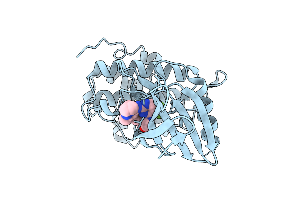
Structure Of Chk1 10-Pt. Mutant Complex With Macrocyclic Lrrk2 Inhibitor Compound 7 ((10As,13As)-3-Cyclobutyl-1-Methyl-8-(Trifluoromethyl)-3,4,10A,11,13A,14-Hexahydro-10H,13H-9,5-(Azeno)Furo[3,4-K]Pyrazolo[4,3-B][1,4,6,10]Oxatriazacyclotridecine)
Organism: Homo sapiens
Method: X-RAY DIFFRACTION Resolution:1.67 Å Release Date: 2026-04-08 Classification: TRANSFERASE/INHIBITOR Ligands: A1C5G |
 |
Structure Of Chk1 10-Pt. Mutant Complex With Macrocyclic Lrrk2 Inhibitor Compound 12 ((10As,13As)-3-Cyclopropyl-1-Methyl-8-(Trifluoromethyl)-3,4,10A,11,13A,14-Hexahydro-10H,13H-9,5-(Azeno)Furo[3,4-K]Pyrazolo[4,3-B][1,4,6,10]Oxatriazacyclotridecine)
Organism: Homo sapiens
Method: X-RAY DIFFRACTION Resolution:1.74 Å Release Date: 2026-04-08 Classification: TRANSFERASE/INHIBITOR Ligands: A1C5H |
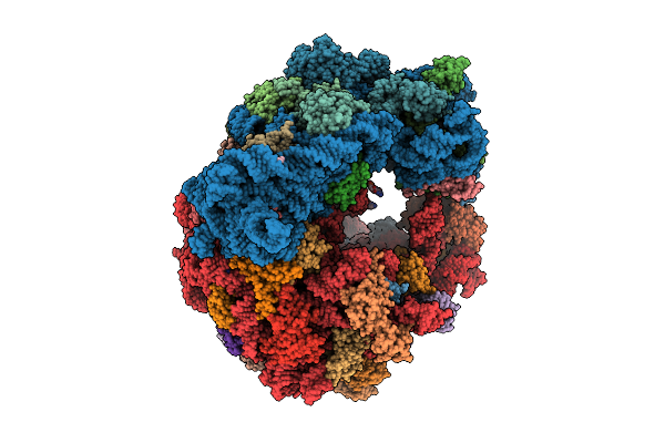 |
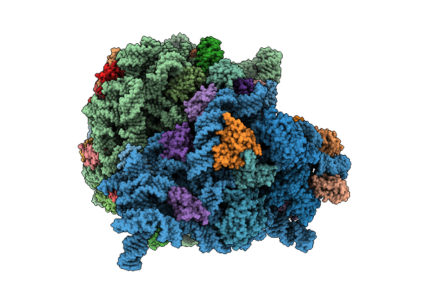
Organism: Escherichia coli
Method: ELECTRON MICROSCOPY Release Date: 2026-04-08 Classification: RIBOSOME Ligands: ZN, MG, SPD, SPM, C, G |
 |
Chimeric Escherichia Coli 70S Ribosome Containing An Evolved Vibrio Cholerae 16S Rrna (Vc-S4.4)
Organism: Escherichia coli
Method: ELECTRON MICROSCOPY Release Date: 2026-04-08 Classification: RIBOSOME Ligands: ZN, MG, SPD, SPM |
 |
Chimeric Escherichia Coli 70S Ribosome Containing An Evolved 16S Rrna From Pseudomonas Aeruginosa (Pa-S3.3)
Organism: Escherichia coli
Method: ELECTRON MICROSCOPY Release Date: 2026-04-08 Classification: RIBOSOME Ligands: MG |
 |
Chimeric Escherichia Coli 70S Ribosome Containing An Evolved 16S Rrna From Pseudomonas Aeruginosa (Pa-St)
Organism: Escherichia coli
Method: ELECTRON MICROSCOPY Release Date: 2026-04-08 Classification: RIBOSOME Ligands: MG |
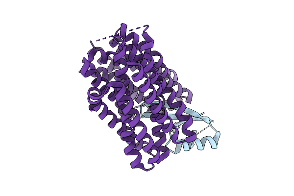 |
Organism: Synthetic construct, Staphylococcus aureus, Escherichia coli
Method: ELECTRON MICROSCOPY Release Date: 2026-04-08 Classification: TRANSPORT PROTEIN |
 |
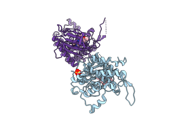
Organism: Homo sapiens
Method: X-RAY DIFFRACTION Resolution:2.10 Å Release Date: 2026-04-08 Classification: TRANSFERASE Ligands: EDO, A1J3A, IOD |
 |
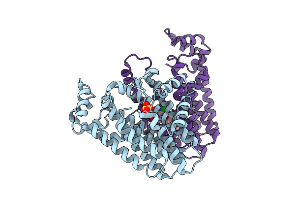
Organism: Homo sapiens
Method: X-RAY DIFFRACTION Resolution:2.50 Å Release Date: 2026-04-08 Classification: TRANSFERASE Ligands: SO4, A1J3B |
 |
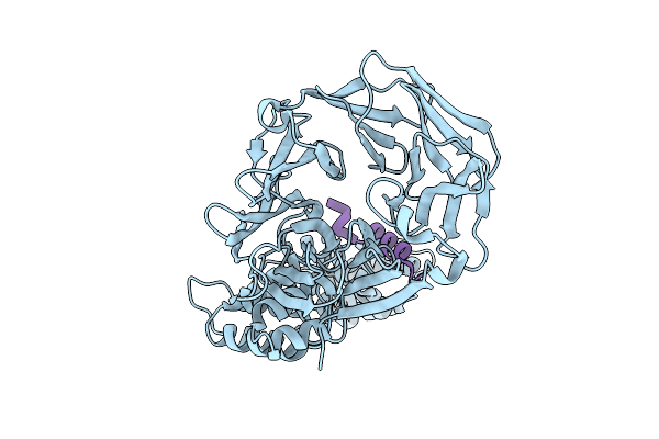
Organism: Severe acute respiratory syndrome coronavirus 2
Method: X-RAY DIFFRACTION Resolution:1.03 Å Release Date: 2026-04-08 Classification: VIRAL PROTEIN,HYDROLASE/INHIBITOR Ligands: A1A5Z, CL |
 |
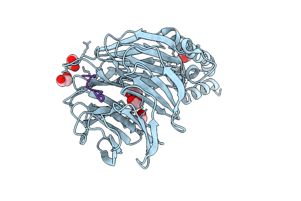
Organism: Severe acute respiratory syndrome coronavirus 2
Method: X-RAY DIFFRACTION Resolution:1.02 Å Release Date: 2026-04-08 Classification: VIRAL PROTEIN,HYDROLASE/INHIBITOR Ligands: A1A54, CL |
 |
Organism: Oryza sativa japonica group
Method: X-RAY DIFFRACTION Resolution:1.83 Å Release Date: 2026-04-08 Classification: TRANSFERASE Ligands: SO4, A1EC1 |
 |
Organism: Oryza sativa japonica group
Method: X-RAY DIFFRACTION Resolution:1.99 Å Release Date: 2026-04-08 Classification: TRANSFERASE Ligands: SO4, A1EC2 |
 |
Organism: Rattus norvegicus, Sus scrofa
Method: X-RAY DIFFRACTION Resolution:2.51 Å Release Date: 2026-04-08 Classification: CELL CYCLE Ligands: GTP, GDP, A1B97, SO4 |
 |
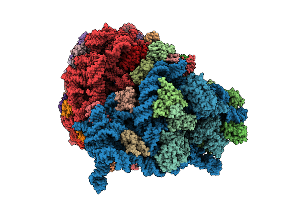
Organism: Mus musculus
Method: ELECTRON MICROSCOPY Release Date: 2026-04-08 Classification: RIBOSOME Ligands: MG, ZN |
 |
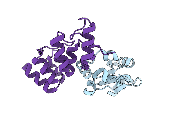
Crystal Structure Of Clathrin Heavy Chain In Complex With A Peptidomimetic Inhibitor Of The Tacc3 Interaction
Organism: Rattus norvegicus, Homo sapiens
Method: X-RAY DIFFRACTION Resolution:2.15 Å Release Date: 2026-04-08 Classification: CELL CYCLE |
 |
Crystal Structure Of The Acl1 Ankyrin Repeat Domain In Complex With The Second Rpl1 Domain
Organism: Saccharomyces cerevisiae
Method: X-RAY DIFFRACTION Resolution:2.55 Å Release Date: 2026-04-08 Classification: RIBOSOMAL PROTEIN |
 |
Crystal Structure Of Beta-Trcp Bound By Diphosphorylated I-Kappa-B-Alpha Degron Peptide
Organism: Homo sapiens
Method: X-RAY DIFFRACTION Resolution:1.16 Å Release Date: 2026-04-08 Classification: TRANSFERASE Ligands: EDO |
 |
Crystal Structure Of Beta-Trcp Bound By Diphosphorylated Pdcd4 Degron Peptide
Organism: Homo sapiens
Method: X-RAY DIFFRACTION Resolution:1.22 Å Release Date: 2026-04-08 Classification: TRANSFERASE Ligands: MG, EDO |