

Search Count: 23
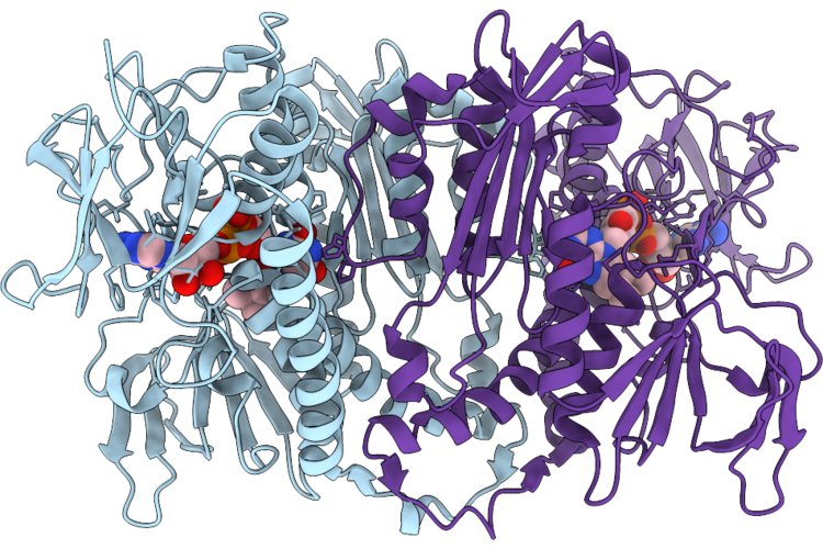 |
Structure Of Trypanothione Reductase From Leishmania Donovani
Organism: Leishmania donovani
Method: X-RAY DIFFRACTION Resolution:2.38 Å Release Date: 2026-07-08 Classification: OXIDOREDUCTASE Ligands: FAD |
Organism: Leishmania donovani
Method: X-RAY DIFFRACTION
Release Date: 2026-07-08
Ligands: FAD
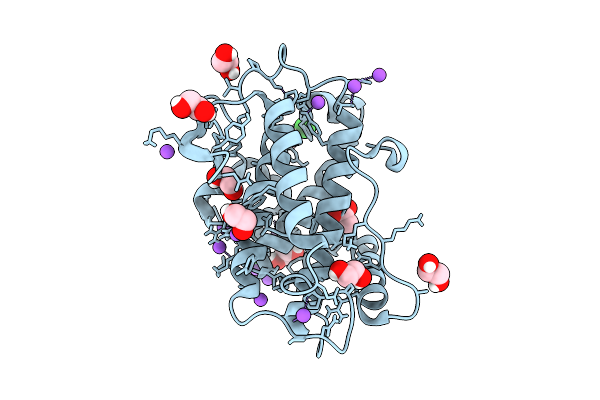 |
L. Donovani Cell Cycle Associated Protein Mob1
Organism: Leishmania donovani
Method: X-RAY DIFFRACTION Resolution:1.30 Å Release Date: 2026-04-01 Classification: CELL CYCLE Ligands: ZN, ACT, NA, CL, EDO |
Organism: Leishmania donovani
Method: X-RAY DIFFRACTION
Release Date: 2026-04-01
Ligands: ZN, ACT, NA, CL, EDO
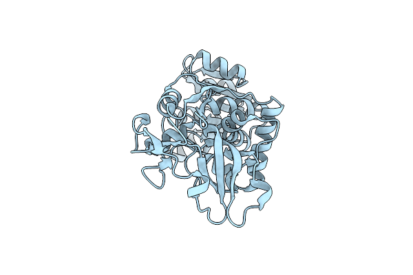 |
Structure Insights Into The Nadph Quinone Oxidoreductase From Leishmania Donovani
Organism: Leishmania donovani
Method: X-RAY DIFFRACTION Resolution:2.05 Å Release Date: 2023-11-22 Classification: OXIDOREDUCTASE |
Organism: Leishmania donovani
Method: X-RAY DIFFRACTION
Release Date: 2023-11-22
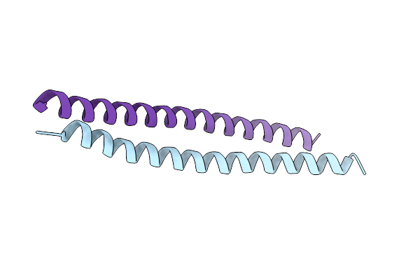 |
Structure Of Coiled Coil Domain Of Trypanosoma Brucei Coronin
Organism: Trypanosoma brucei
Method: X-RAY DIFFRACTION Resolution:2.06 Å Release Date: 2021-11-10 Classification: CONTRACTILE PROTEIN |
Organism: Trypanosoma brucei
Method: X-RAY DIFFRACTION
Release Date: 2021-11-10
 |
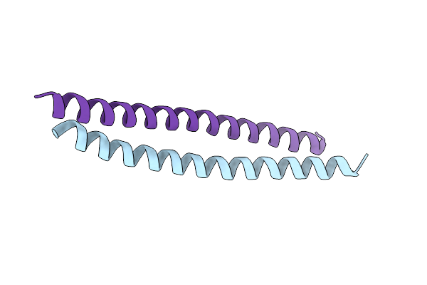
Double Mutant- V493I-V507M Coiled Coil Domain Of Trypanosoma Brucei Coronin
Organism: Trypanosoma brucei
Method: X-RAY DIFFRACTION Resolution:2.41 Å Release Date: 2021-11-10 Classification: CONTRACTILE PROTEIN |
Organism: Trypanosoma brucei
Method: X-RAY DIFFRACTION
Release Date: 2021-11-10
 |
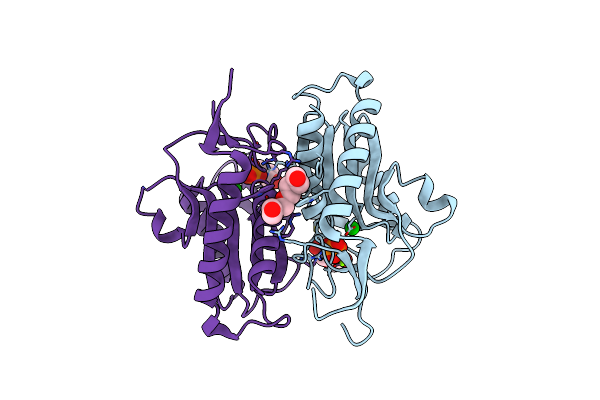
Mutant V507M Coiled Coil Domain Of Trypanosoma Brucei Coronin
Organism: Trypanosoma brucei
Method: X-RAY DIFFRACTION Resolution:3.02 Å Release Date: 2021-11-10 Classification: CONTRACTILE PROTEIN |
Organism: Trypanosoma brucei
Method: X-RAY DIFFRACTION
Release Date: 2021-11-10
 |
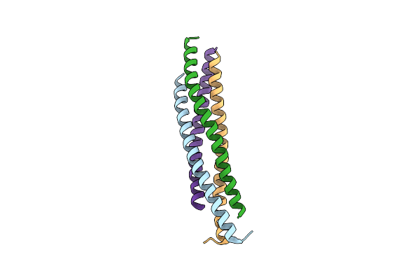
Crystal Structure Of L.Donovani Hypoxanthine-Guanine Phosphoribosyl Transferase (Hgprt)
Organism: Leishmania donovani (strain bpk282a1)
Method: X-RAY DIFFRACTION Resolution:2.76 Å Release Date: 2021-08-04 Classification: TRANSFERASE Ligands: MG, BA, PO4, PEG, PG4, CL |
Organism: Leishmania donovani (strain bpk282a1)
Method: X-RAY DIFFRACTION
Release Date: 2021-08-04
Ligands: MG, BA, PO4, PEG, PG4, CL
 |
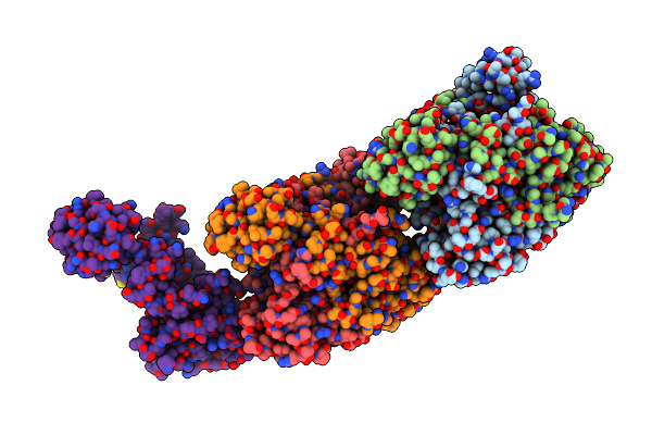
Ldcorocc Mutant-I486A
Organism: Leishmania donovani
Method: X-RAY DIFFRACTION Resolution:2.50 Å Release Date: 2019-10-09 Classification: STRUCTURAL PROTEIN |
Organism: Leishmania donovani
Method: X-RAY DIFFRACTION
Release Date: 2019-10-09
 |
Crystal Structure Of Hypothetical Protein (Rv3272) From Mycobacterium Tuberculosis
Organism: Mycobacterium tuberculosis (strain atcc 25618 / h37rv)
Method: X-RAY DIFFRACTION Resolution:2.79 Å Release Date: 2018-11-07 Classification: TRANSFERASE |
Organism: Mycobacterium tuberculosis (strain atcc 25618 / h37rv)
Method: X-RAY DIFFRACTION
Release Date: 2018-11-07
 |
Crystal Structure Of D175A Mutant Of Rv3272 From Mycobacterium Tuberculosis
Organism: Mycobacterium tuberculosis (strain atcc 25618 / h37rv)
Method: X-RAY DIFFRACTION Resolution:2.50 Å Release Date: 2018-11-07 Classification: TRANSFERASE Ligands: CL |
Organism: Mycobacterium tuberculosis (strain atcc 25618 / h37rv)
Method: X-RAY DIFFRACTION
Release Date: 2018-11-07
Ligands: CL
 |
Crystal Structure Of Rv3272 From M. Tuberculosis Orthorhombic Form
Organism: Mycobacterium tuberculosis (strain atcc 25618 / h37rv)
Method: X-RAY DIFFRACTION Resolution:2.20 Å Release Date: 2018-11-07 Classification: TRANSFERASE Ligands: BEN, GOL |
Organism: Mycobacterium tuberculosis (strain atcc 25618 / h37rv)
Method: X-RAY DIFFRACTION
Release Date: 2018-11-07
Ligands: BEN, GOL
 |
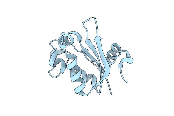
Nucleoside Diphosphate Kinase From Vibrio Cholerae Is A Thermolabile Type Ii Tetramer
Organism: Vibrio cholerae serotype o1 (strain m66-2)
Method: X-RAY DIFFRACTION Resolution:3.06 Å Release Date: 2018-01-31 Classification: TRANSFERASE |
Organism: Vibrio cholerae serotype o1 (strain m66-2)
Method: X-RAY DIFFRACTION
Release Date: 2018-01-31
 |
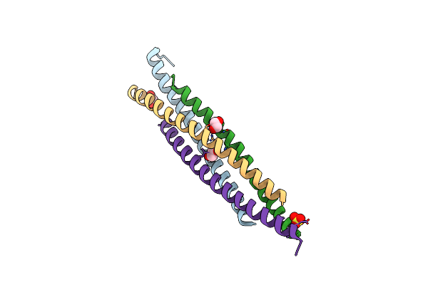
Structural, Functional Characterization And Discovery Of Novel Inhibitors Of Leishmania Amazonensis Nucleoside Diphosphatase Kinase (Ndk)
Organism: Leishmania amazonensis
Method: X-RAY DIFFRACTION Resolution:2.50 Å Release Date: 2017-07-26 Classification: TRANSFERASE |
Organism: Leishmania amazonensis
Method: X-RAY DIFFRACTION
Release Date: 2017-07-26
 |
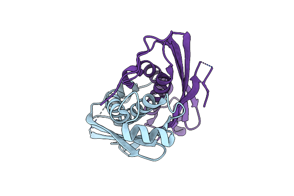
Structure Of Coiled Coil Domain Of Leishmania Donovani Coronin
Organism: Leishmania donovani
Method: X-RAY DIFFRACTION Resolution:2.21 Å Release Date: 2016-07-27 Classification: STRUCTURAL PROTEIN Ligands: EDO, SO4 |
Organism: Leishmania donovani
Method: X-RAY DIFFRACTION
Release Date: 2016-07-27
Ligands: EDO, SO4
 |
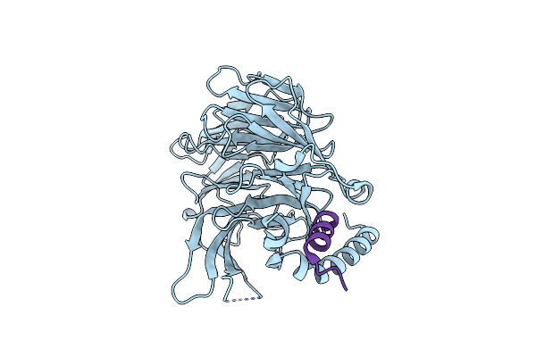
Structural Insights Into Putative Molybdenum Cofactor Biosynthesis Protein C (Moac2) From Mycobacterium Tuberculosis H37Rv
Organism: Mycobacterium tuberculosis
Method: X-RAY DIFFRACTION Resolution:2.20 Å Release Date: 2013-04-10 Classification: BIOSYNTHETIC PROTEIN |
Organism: Mycobacterium tuberculosis
Method: X-RAY DIFFRACTION
Release Date: 2013-04-10
 |
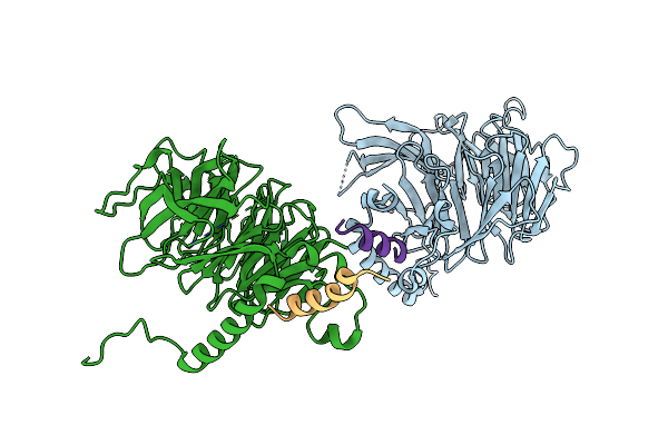
Structural Basis Of The Interaction Of Rbap46/Rbap48 With Histone H4
Organism: Homo sapiens
Method: X-RAY DIFFRACTION Resolution:2.40 Å Release Date: 2008-06-10 Classification: HISTONE/CHAPERONE Ligands: ARS |
Organism: Homo sapiens
Method: X-RAY DIFFRACTION
Release Date: 2008-06-10
Ligands: ARS
 |
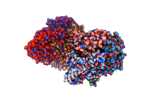
Structural Basis Of The Interaction Of Rbap46/Rbap48 With Histone H4
Organism: Homo sapiens
Method: X-RAY DIFFRACTION Resolution:2.60 Å Release Date: 2008-06-10 Classification: HISTONE/CHAPERONE Ligands: ARS |
Organism: Homo sapiens
Method: X-RAY DIFFRACTION
Release Date: 2008-06-10
Ligands: ARS
 |
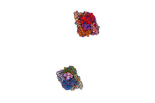
New Crystal Form Of The Pseudomonas Putida Branched-Chain Dehydrogenase (E1)
Organism: Pseudomonas putida
Method: X-RAY DIFFRACTION Resolution:2.90 Å Release Date: 2005-08-10 Classification: OXIDOREDUCTASE |
Organism: Pseudomonas putida
Method: X-RAY DIFFRACTION
Release Date: 2005-08-10
 |
The Crystal Structure Of Pyruvate Dehydrogenase E1 Bound To The Peripheral Subunit Binding Domain Of E2
Organism: Geobacillus stearothermophilus
Method: X-RAY DIFFRACTION Resolution:2.00 Å Release Date: 2004-11-02 Classification: OXIDOREDUCTASE Ligands: MG, PEG, TPP, K |
Organism: Geobacillus stearothermophilus
Method: X-RAY DIFFRACTION
Release Date: 2004-11-02
Ligands: MG, PEG, TPP, K
 |
The Crystal Structure Of Pyruvate Dehydrogenase E1(D180N,E183Q) Bound To The Peripheral Subunit Binding Domain Of E2
Organism: Geobacillus stearothermophilus
Method: X-RAY DIFFRACTION Resolution:2.30 Å Release Date: 2004-11-02 Classification: OXIDOREDUCTASE Ligands: MG, TPP |
Organism: Geobacillus stearothermophilus
Method: X-RAY DIFFRACTION
Release Date: 2004-11-02
Ligands: MG, TPP