

Search Count: 32
 |
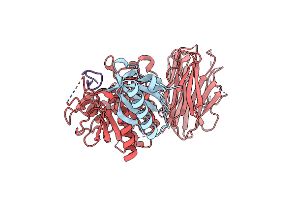
Pcsk9 In Complex With An Inhibitor
Organism: Homo sapiens, Synthetic construct
Method: X-RAY DIFFRACTION Resolution:3.00 Å Release Date: 2024-10-16 Classification: HYDROLASE/INHIBITOR Ligands: CA |
Organism: Homo sapiens, Synthetic construct
Method: X-RAY DIFFRACTION
Release Date: 2024-10-16
Ligands: CA
 |
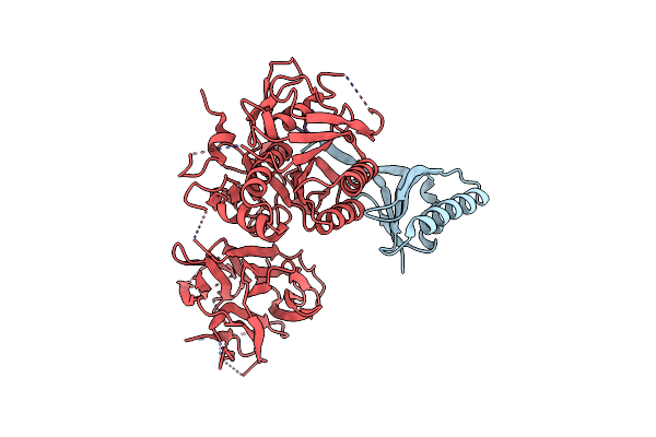
Pcsk9 In Complex With An Inhibitor
Organism: Homo sapiens, Synthetic construct
Method: X-RAY DIFFRACTION Resolution:2.50 Å Release Date: 2024-10-16 Classification: HYDROLASE/INHIBITOR Ligands: CA |
Organism: Homo sapiens, Synthetic construct
Method: X-RAY DIFFRACTION
Release Date: 2024-10-16
Ligands: CA
 |
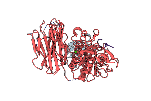
Pcsk9 In Complex With An Inhibitor
Organism: Homo sapiens, Synthetic construct
Method: X-RAY DIFFRACTION Resolution:1.96 Å Release Date: 2024-10-16 Classification: HYDROLASE/INHIBITOR |
Organism: Homo sapiens, Synthetic construct
Method: X-RAY DIFFRACTION
Release Date: 2024-10-16
 |
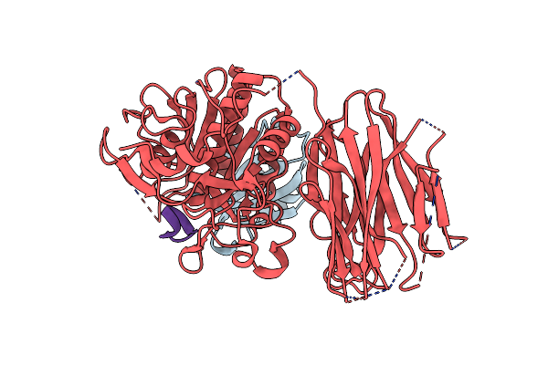
Pcsk9 In Complex With An Inhibitor
Organism: Homo sapiens, Synthetic construct
Method: X-RAY DIFFRACTION Resolution:2.85 Å Release Date: 2024-10-16 Classification: HYDROLASE/INHIBITOR Ligands: CA |
Organism: Homo sapiens, Synthetic construct
Method: X-RAY DIFFRACTION
Release Date: 2024-10-16
Ligands: CA
 |
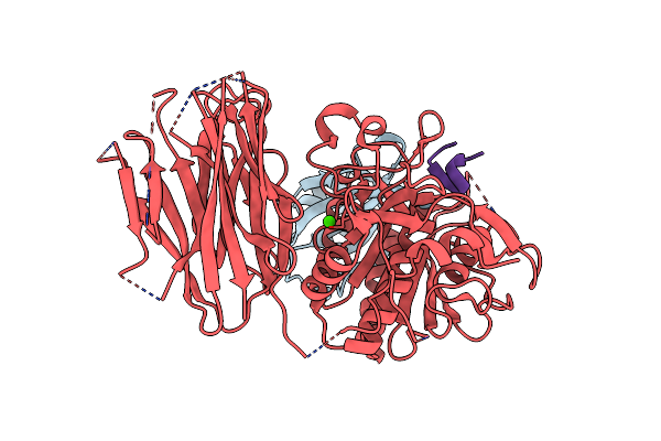
Pcsk9 In Complex With An Inhibitor
Organism: Homo sapiens, Synthetic construct
Method: X-RAY DIFFRACTION Resolution:2.71 Å Release Date: 2024-10-16 Classification: HYDROLASE/INHIBITOR |
Organism: Homo sapiens, Synthetic construct
Method: X-RAY DIFFRACTION
Release Date: 2024-10-16
 |
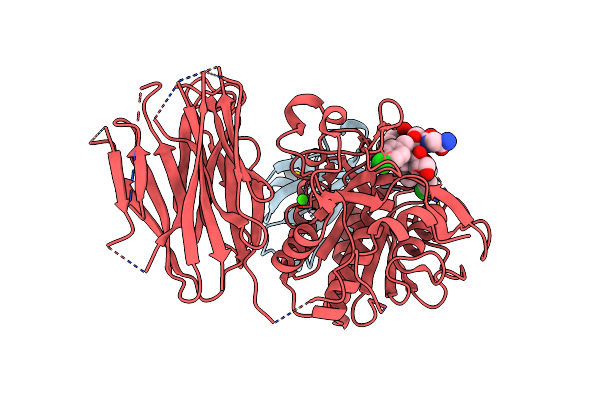
Pcsk9 In Complex With An Inhibitor
Organism: Homo sapiens, Synthetic construct
Method: X-RAY DIFFRACTION Resolution:2.65 Å Release Date: 2024-10-16 Classification: HYDROLASE/INHIBITOR Ligands: CA |
Organism: Homo sapiens, Synthetic construct
Method: X-RAY DIFFRACTION
Release Date: 2024-10-16
Ligands: CA
 |
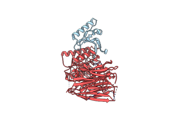
Pcsk9 In Complex With An Inhibitor
Organism: Homo sapiens, Synthetic construct
Method: X-RAY DIFFRACTION Resolution:2.60 Å Release Date: 2024-10-16 Classification: HYDROLASE/INHIBITOR Ligands: CA |
Organism: Homo sapiens, Synthetic construct
Method: X-RAY DIFFRACTION
Release Date: 2024-10-16
Ligands: CA
 |
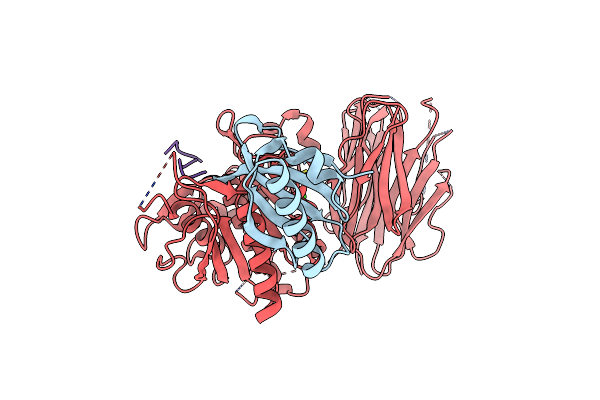
Pcsk9 In Complex With An Inhibitor
Organism: Homo sapiens, Synthetic construct
Method: X-RAY DIFFRACTION Resolution:2.57 Å Release Date: 2024-10-16 Classification: HYDROLASE/INHIBITOR |
Organism: Homo sapiens, Synthetic construct
Method: X-RAY DIFFRACTION
Release Date: 2024-10-16
 |
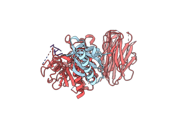
Pcsk9 In Complex With A Cyclic Peptide Ldlr Disruptor
Organism: Homo sapiens, Synthetic construct
Method: X-RAY DIFFRACTION Resolution:2.80 Å Release Date: 2021-11-24 Classification: LIPID TRANSPORT, HYDROLASE Ligands: CA |
Organism: Homo sapiens, Synthetic construct
Method: X-RAY DIFFRACTION
Release Date: 2021-11-24
Ligands: CA
 |
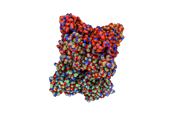
Pcsk9 In Complex With Pcsk9I A 13Mer Cyclic Peptide Ldlr Disruptor
Organism: Homo sapiens, Synthetic construct
Method: X-RAY DIFFRACTION Resolution:2.45 Å Release Date: 2021-11-10 Classification: LIPID TRANSPORT, HYDROLASE Ligands: CA |
Organism: Homo sapiens, Synthetic construct
Method: X-RAY DIFFRACTION
Release Date: 2021-11-10
Ligands: CA
 |
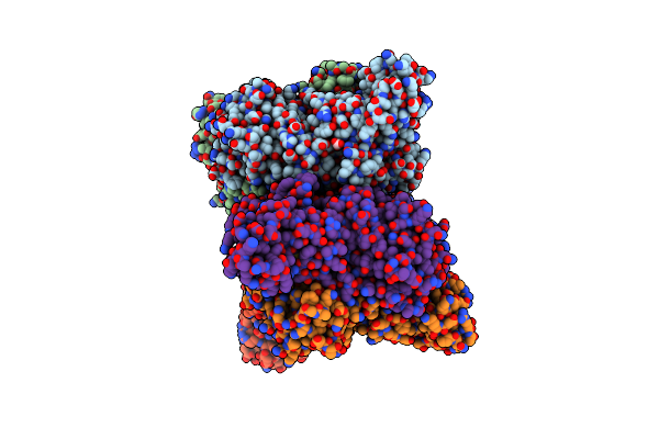
Hexameric Form Ii Rubisco From Rhodopseudomonas Palustris, Activated And Complexed With 2-Cabp
Organism: Rhodopseudomonas palustris
Method: X-RAY DIFFRACTION Resolution:2.38 Å Release Date: 2014-06-25 Classification: lyase, oxidoreductase Ligands: CAP, MG Modified Residue(s): KCX |
Organism: Rhodopseudomonas palustris
Method: X-RAY DIFFRACTION
Release Date: 2014-06-25
Ligands: CAP, MG
 |
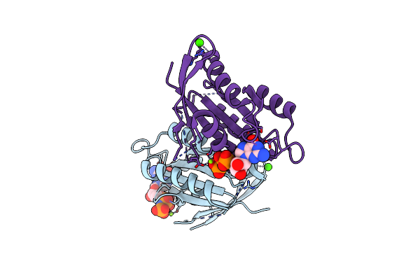
Hexameric Form Ii Rubisco From Rhodopseudomonas Palustris, Activated And Complexed With Sulfate And Magnesium
Organism: Rhodopseudomonas palustris
Method: X-RAY DIFFRACTION Resolution:2.38 Å Release Date: 2014-06-25 Classification: lyase, oxidoreductase Ligands: MG, SO4, CO3 Modified Residue(s): KCX |
Organism: Rhodopseudomonas palustris
Method: X-RAY DIFFRACTION
Release Date: 2014-06-25
Ligands: MG, SO4, CO3
 |
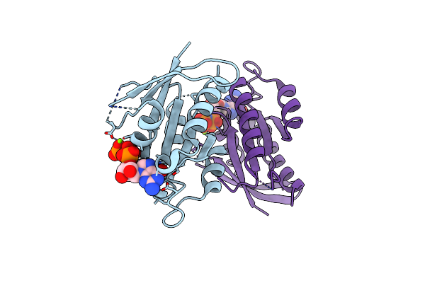
Crystal Structure Of Rad G-Domain-Gtp Analog Complex
Organism: Homo sapiens
Method: X-RAY DIFFRACTION Resolution:1.66 Å Release Date: 2011-09-21 Classification: SIGNALING PROTEIN Ligands: GNP, MG, CA |
Organism: Homo sapiens
Method: X-RAY DIFFRACTION
Release Date: 2011-09-21
Ligands: GNP, MG, CA
 |
Crystal Structure Of Rad G-Domain-Gtp Analog Complex
Organism: Homo sapiens
Method: X-RAY DIFFRACTION Resolution:2.50 Å Release Date: 2011-09-21 Classification: SIGNALING PROTEIN Ligands: GNP, MG |
Organism: Homo sapiens
Method: X-RAY DIFFRACTION
Release Date: 2011-09-21
Ligands: GNP, MG
 |
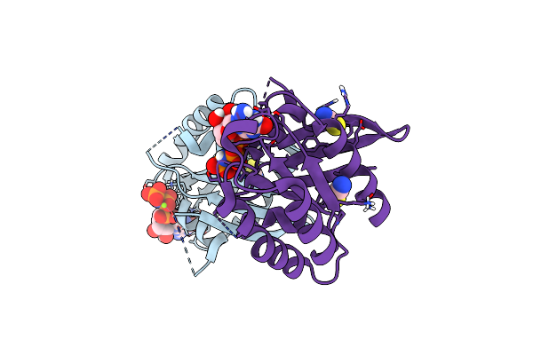
Crystal Structure Of Rad G-Domain Q148A-Gtp Analog Complex
Organism: Homo sapiens
Method: X-RAY DIFFRACTION Resolution:2.30 Å Release Date: 2011-09-21 Classification: SIGNALING PROTEIN Ligands: GNP, MG, CA |
Organism: Homo sapiens
Method: X-RAY DIFFRACTION
Release Date: 2011-09-21
Ligands: GNP, MG, CA
 |
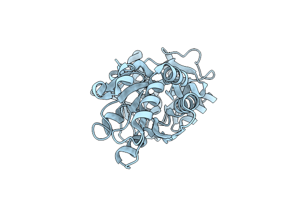
Crystal Structure Of Rem2 G-Domain -Gtp Analog Complex
Organism: Mus musculus
Method: X-RAY DIFFRACTION Resolution:1.76 Å Release Date: 2011-09-21 Classification: SIGNALING PROTEIN Ligands: GNP, MG, SCN |
Organism: Mus musculus
Method: X-RAY DIFFRACTION
Release Date: 2011-09-21
Ligands: GNP, MG, SCN
 |
M. Acetivorans Molybdate-Binding Protein (Moda) In Unliganded Open Form
Organism: Methanosarcina acetivorans
Method: X-RAY DIFFRACTION Resolution:1.95 Å Release Date: 2010-01-12 Classification: TRANSPORT PROTEIN |
Organism: Methanosarcina acetivorans
Method: X-RAY DIFFRACTION
Release Date: 2010-01-12
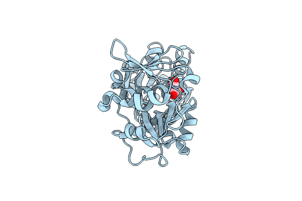 |
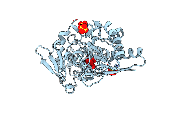
M. Acetivorans Molybdate-Binding Protein (Moda) In Citrate-Bound Open Form
Organism: Methanosarcina acetivorans
Method: X-RAY DIFFRACTION Resolution:1.69 Å Release Date: 2010-01-12 Classification: TRANSPORT PROTEIN Ligands: CIT |
Organism: Methanosarcina acetivorans
Method: X-RAY DIFFRACTION
Release Date: 2010-01-12
Ligands: CIT
 |
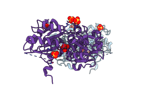
Apo And Ligand Bound Structures Of Moda From The Archaeon Methanosarcina Acetivorans
Organism: Methanosarcina acetivorans
Method: X-RAY DIFFRACTION Resolution:2.45 Å Release Date: 2010-01-12 Classification: TRANSPORT PROTEIN Ligands: MOO, SO4 |
Organism: Methanosarcina acetivorans
Method: X-RAY DIFFRACTION
Release Date: 2010-01-12
Ligands: MOO, SO4
 |
M. Acetivorans Molybdate-Binding Protein (Moda) In Molybdate-Bound Close Form With 2 Molecules In Asymmetric Unit Forming Beta Barrel
Organism: Methanosarcina acetivorans
Method: X-RAY DIFFRACTION Resolution:2.25 Å Release Date: 2010-01-12 Classification: TRANSPORT PROTEIN Ligands: MOO, SO4 |
Organism: Methanosarcina acetivorans
Method: X-RAY DIFFRACTION
Release Date: 2010-01-12
Ligands: MOO, SO4