

Search Count: 39
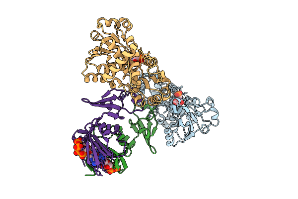 |
Crystal Structure Of E. Coli Aspartate Transcarbamoylase In The R-State Complexed With Pala, Atp, Gtp, And Mg2+
Organism: Escherichia coli
Method: X-RAY DIFFRACTION Resolution:2.19 Å Release Date: 2026-04-15 Classification: CYTOSOLIC PROTEIN Ligands: PAL, ZN, ATP, MG, GTP |
Organism: Escherichia coli
Method: X-RAY DIFFRACTION
Release Date: 2026-04-15
Ligands: PAL, ZN, ATP, MG, GTP
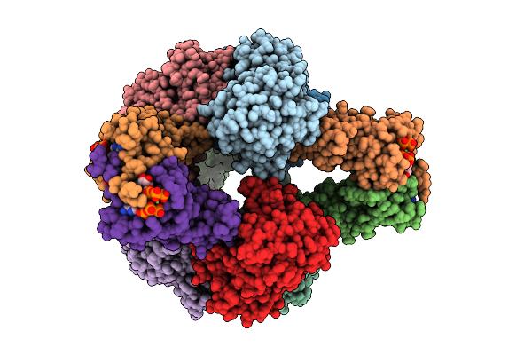 |
Crystal Structure Of E. Coli Aspartate Transcarbamoylase In The R-State Complexed With Cp, Succinate, Atp, And Mg2+
Organism: Escherichia coli
Method: X-RAY DIFFRACTION Resolution:3.00 Å Release Date: 2026-04-15 Classification: CYTOSOLIC PROTEIN Ligands: CP, SIN, ZN, ATP, MG |
Organism: Escherichia coli
Method: X-RAY DIFFRACTION
Release Date: 2026-04-15
Ligands: CP, SIN, ZN, ATP, MG
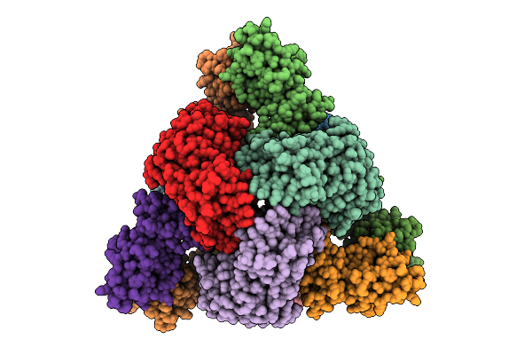 |
Cryo-Em Model Of E. Coli Aspartate Transcarbamoylase In The Ligand-Free T-State
Organism: Escherichia coli
Method: ELECTRON MICROSCOPY Release Date: 2026-04-15 Classification: CYTOSOLIC PROTEIN Ligands: ZN |
Organism: Escherichia coli
Method: ELECTRON MICROSCOPY
Release Date: 2026-04-15
Ligands: ZN
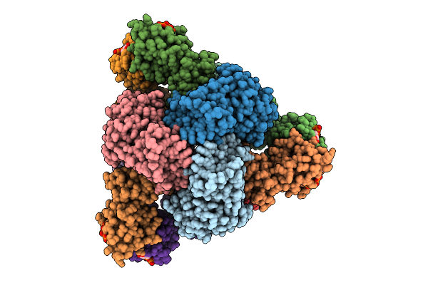 |
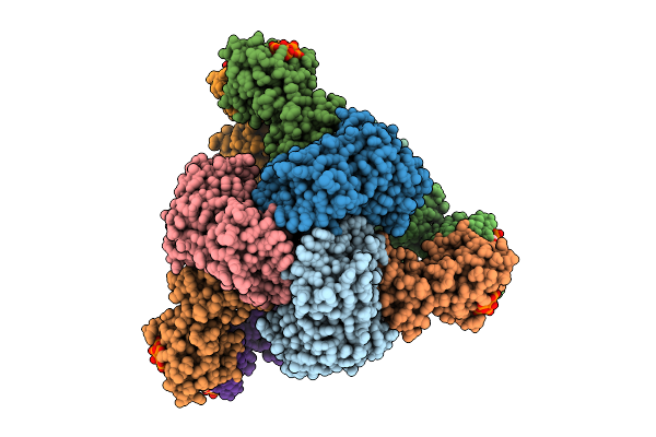
Cryo-Em Model Of E. Coli Aspartate Transcarbamoylase In The T-State Complexed With Cp, Ctp, Utp, And Mg2+
Organism: Escherichia coli
Method: ELECTRON MICROSCOPY Release Date: 2026-04-15 Classification: CYTOSOLIC PROTEIN Ligands: ZN, CTP, UTP, MG, CP |
Organism: Escherichia coli
Method: ELECTRON MICROSCOPY
Release Date: 2026-04-15
Ligands: ZN, CTP, UTP, MG, CP
 |
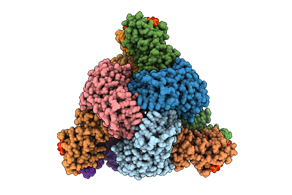
Cryo-Em Model Of E. Coli Aspartate Transcarbamoylase In The R-State Complexed With Cp, Succinate, Ctp, Utp, And Mg2+
Organism: Escherichia coli
Method: ELECTRON MICROSCOPY Release Date: 2026-04-15 Classification: CYTOSOLIC PROTEIN Ligands: CP, SIN, ZN, CTP, UTP, MG |
Organism: Escherichia coli
Method: ELECTRON MICROSCOPY
Release Date: 2026-04-15
Ligands: CP, SIN, ZN, CTP, UTP, MG
 |
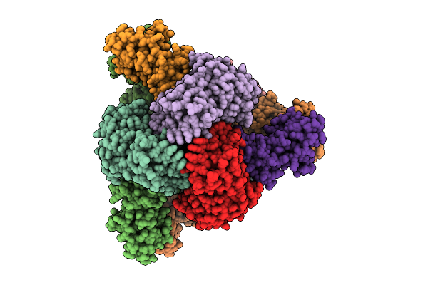
Cryo-Em Model Of E. Coli Aspartate Transcarbamoylase In The R-State Complexed With Cp, Succinate, Ctp, And Mg2+
Organism: Escherichia coli
Method: ELECTRON MICROSCOPY Release Date: 2026-04-15 Classification: CYTOSOLIC PROTEIN Ligands: CP, SIN, ZN, CTP, MG |
Organism: Escherichia coli
Method: ELECTRON MICROSCOPY
Release Date: 2026-04-15
Ligands: CP, SIN, ZN, CTP, MG
 |
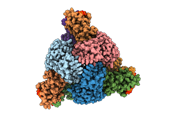
Cryo-Em Model Of E. Coli Aspartate Transcarbamoylase In The R-State Complexed With Cp And Succinate
Organism: Escherichia coli
Method: ELECTRON MICROSCOPY Release Date: 2026-04-15 Classification: CYTOSOLIC PROTEIN Ligands: CP, SIN, ZN |
Organism: Escherichia coli
Method: ELECTRON MICROSCOPY
Release Date: 2026-04-15
Ligands: CP, SIN, ZN
 |
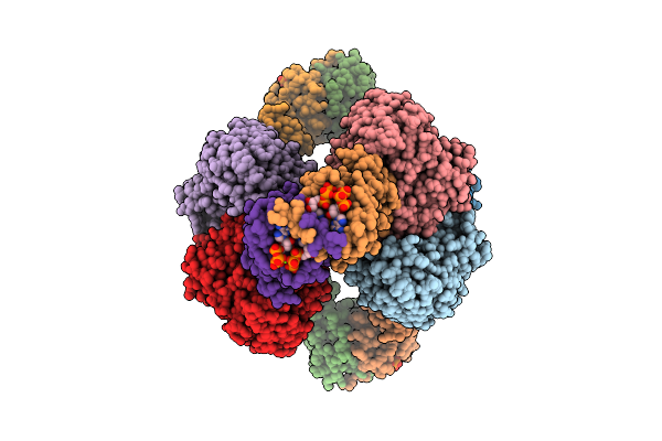
Cryo-Em Model Of E. Coli Aspartate Transcarbamoylase In The R-State Complexed With Cp, Succinate, Atp, And Mg2+
Organism: Escherichia coli
Method: ELECTRON MICROSCOPY Release Date: 2026-04-15 Classification: CYTOSOLIC PROTEIN Ligands: CP, SIN, ZN, ATP, MG |
Organism: Escherichia coli
Method: ELECTRON MICROSCOPY
Release Date: 2026-04-15
Ligands: CP, SIN, ZN, ATP, MG
 |
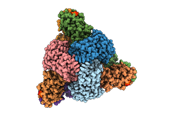
Cryo-Em Model Of E. Coli Aspartate Transcarbamoylase In The R-State Complexed With Cp, Succinate, Atp, Gtp, And Mg2+
Organism: Escherichia coli
Method: ELECTRON MICROSCOPY Release Date: 2026-04-15 Classification: CYTOSOLIC PROTEIN Ligands: CP, SIN, ZN, ATP, GTP, MG |
Organism: Escherichia coli
Method: ELECTRON MICROSCOPY
Release Date: 2026-04-15
Ligands: CP, SIN, ZN, ATP, GTP, MG
 |
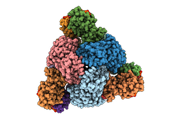
Cryo-Em Model Of E. Coli Aspartate Transcarbamoylase In An Expanded State Complexed With Cp, Atp, Gtp, And Mg2+
Organism: Escherichia coli
Method: ELECTRON MICROSCOPY Release Date: 2026-04-15 Classification: CYTOSOLIC PROTEIN Ligands: ZN, ATP, GTP, MG, CP |
Organism: Escherichia coli
Method: ELECTRON MICROSCOPY
Release Date: 2026-04-15
Ligands: ZN, ATP, GTP, MG, CP
 |
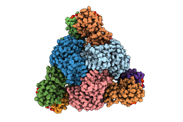
Cryo-Em Model Of E. Coli Aspartate Transcarbamoylase In The T-State Complexed With Cp, Atp, And Mg2+
Organism: Escherichia coli
Method: ELECTRON MICROSCOPY Release Date: 2026-04-15 Classification: CYTOSOLIC PROTEIN Ligands: ZN, ATP, MG, CP |
Organism: Escherichia coli
Method: ELECTRON MICROSCOPY
Release Date: 2026-04-15
Ligands: ZN, ATP, MG, CP
 |
Cryo-Em Model Of E. Coli Aspartate Transcarbamoylase In The T-State Complexed With Cp, Ctp, And Mg2+
Organism: Escherichia coli
Method: ELECTRON MICROSCOPY Release Date: 2026-04-01 Classification: CYTOSOLIC PROTEIN Ligands: ZN, CTP, MG, CP |
Organism: Escherichia coli
Method: ELECTRON MICROSCOPY
Release Date: 2026-04-01
Ligands: ZN, CTP, MG, CP
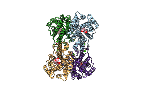 |
Crystal Structure Of Caryolan-1-Ol Synthase From S. Griseus With Peg Molecule In The Active Site
Organism: Streptomyces griseus
Method: X-RAY DIFFRACTION Resolution:2.33 Å Release Date: 2024-11-06 Classification: METAL BINDING PROTEIN Ligands: PG4, CA, 1PE |
Organism: Streptomyces griseus
Method: X-RAY DIFFRACTION
Release Date: 2024-11-06
Ligands: PG4, CA, 1PE
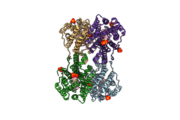 |
Crystal Structure Of Caryolan-1-Ol Synthase Complexed With 2-Fluorofarnesyl Diphosphate
Organism: Streptomyces griseus
Method: X-RAY DIFFRACTION Resolution:2.65 Å Release Date: 2024-11-06 Classification: METAL BINDING PROTEIN Ligands: FPF, PO4, MG, DPO |
Organism: Streptomyces griseus
Method: X-RAY DIFFRACTION
Release Date: 2024-11-06
Ligands: FPF, PO4, MG, DPO
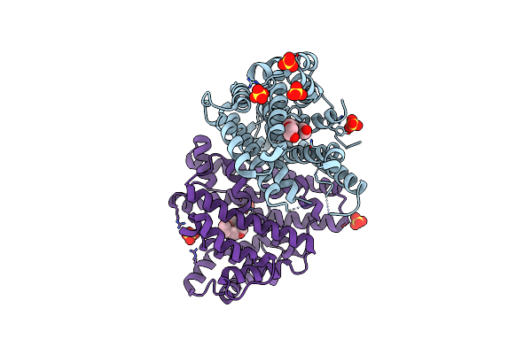 |
Crystal Structure Of Pentalenene Synthase Variant F76A With Peg Molecule In The Active Site
Organism: Streptomyces exfoliatus
Method: X-RAY DIFFRACTION Resolution:2.50 Å Release Date: 2024-11-06 Classification: METAL BINDING PROTEIN Ligands: PG4, SO4 |
Organism: Streptomyces exfoliatus
Method: X-RAY DIFFRACTION
Release Date: 2024-11-06
Ligands: PG4, SO4
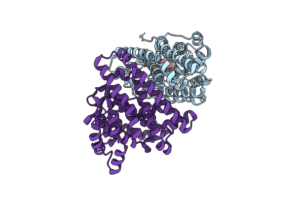 |
Crystal Structure Of Pentalenene Synthase Variant F76A Complexed With 2-Fluorofarnesyl Diphosphate
Organism: Streptomyces exfoliatus
Method: X-RAY DIFFRACTION Resolution:2.20 Å Release Date: 2024-11-06 Classification: METAL BINDING PROTEIN Ligands: FPF, MG |
Organism: Streptomyces exfoliatus
Method: X-RAY DIFFRACTION
Release Date: 2024-11-06
Ligands: FPF, MG
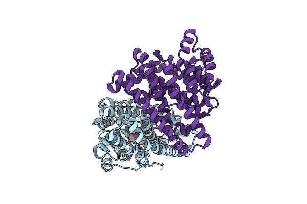 |
Crystal Structure Of Pentalenene Synthase Variant F76A Complexed With 12,13-Difluorofarnesyl Diphosphate
Organism: Streptomyces exfoliatus
Method: X-RAY DIFFRACTION Resolution:2.65 Å Release Date: 2024-11-06 Classification: METAL BINDING PROTEIN Ligands: FDF, MG |
Organism: Streptomyces exfoliatus
Method: X-RAY DIFFRACTION
Release Date: 2024-11-06
Ligands: FDF, MG
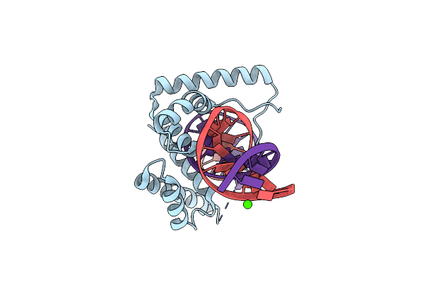 |
Crystal Structure Of The Hmg-C1 Domain Of Human Capicua Bound To Dna
Organism: Homo sapiens, Synthetic construct
Method: X-RAY DIFFRACTION Resolution:2.95 Å Release Date: 2022-04-06 Classification: DNA BINDING PROTEIN/DNA Ligands: CA |
Organism: Homo sapiens, Synthetic construct
Method: X-RAY DIFFRACTION
Release Date: 2022-04-06
Ligands: CA
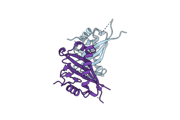 |
Human Muc16 Sea5 Domain
Organism: Homo sapiens
Method: X-RAY DIFFRACTION Resolution:1.69 Å Release Date: 2022-03-23 Classification: UNKNOWN FUNCTION |
Organism: Homo sapiens
Method: X-RAY DIFFRACTION
Release Date: 2022-03-23
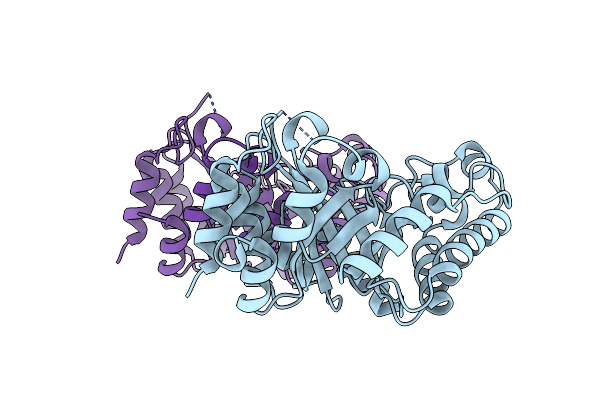 |
Crystal Structure Of Kemp Eliminase Hg3
Organism: Thermoascus aurantiacus
Method: X-RAY DIFFRACTION Resolution:1.08 Å Release Date: 2020-12-02 Classification: HYDROLASE |
Organism: Thermoascus aurantiacus
Method: X-RAY DIFFRACTION
Release Date: 2020-12-02