

Search Count: 19
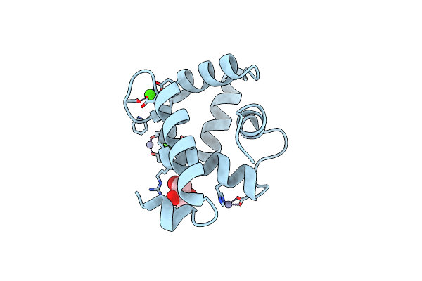 |
Crystal Structure Of The Efhd1/Swiprosin-2, A Mitochondrial Actin-Binding Protein
Organism: Mus musculus
Method: X-RAY DIFFRACTION Resolution:2.07 Å Release Date: 2021-01-06 Classification: METAL BINDING PROTEIN Ligands: CA, ZN, GOL |
Organism: Mus musculus
Method: X-RAY DIFFRACTION
Release Date: 2021-01-06
Ligands: CA, ZN, GOL
 |
Crystal Structure Of Ef-Hand Protein Mutant
Organism: Homo sapiens
Method: X-RAY DIFFRACTION Resolution:1.86 Å Release Date: 2017-09-13 Classification: METAL BINDING PROTEIN Ligands: CA |
Organism: Homo sapiens
Method: X-RAY DIFFRACTION
Release Date: 2017-09-13
Ligands: CA
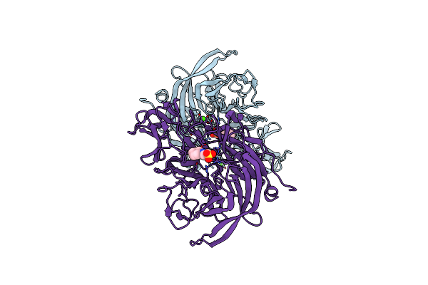 |
Crystal Structure Of The Catalytic Domain Of Clostridium Perfringens Neuraminidase (Nani) In Complex With A Ches
Organism: Clostridium perfringens atcc 13124
Method: X-RAY DIFFRACTION Resolution:1.24 Å Release Date: 2017-03-29 Classification: HYDROLASE Ligands: NHE, CA |
Organism: Clostridium perfringens atcc 13124
Method: X-RAY DIFFRACTION
Release Date: 2017-03-29
Ligands: NHE, CA
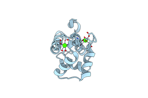 |
Structure Of Ef-Hand Containing Protein
Organism: Homo sapiens
Method: X-RAY DIFFRACTION Resolution:1.85 Å Release Date: 2016-12-28 Classification: METAL BINDING PROTEIN Ligands: CA |
Organism: Homo sapiens
Method: X-RAY DIFFRACTION
Release Date: 2016-12-28
Ligands: CA
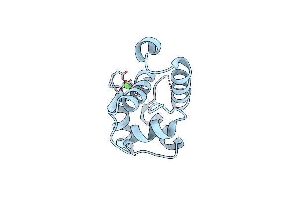 |
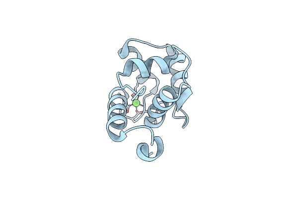
Structure Of Ef-Hand Containing Protein
Organism: Homo sapiens
Method: X-RAY DIFFRACTION Resolution:1.95 Å Release Date: 2016-12-28 Classification: METAL BINDING PROTEIN Ligands: CA |
Organism: Homo sapiens
Method: X-RAY DIFFRACTION
Release Date: 2016-12-28
Ligands: CA
 |
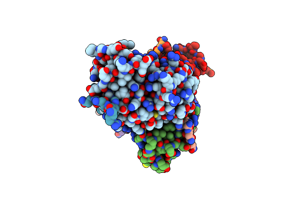
Structure Of Ef-Hand Containing Protein
Organism: Homo sapiens
Method: X-RAY DIFFRACTION Resolution:1.94 Å Release Date: 2016-12-28 Classification: METAL BINDING PROTEIN Ligands: CA |
Organism: Homo sapiens
Method: X-RAY DIFFRACTION
Release Date: 2016-12-28
Ligands: CA
 |
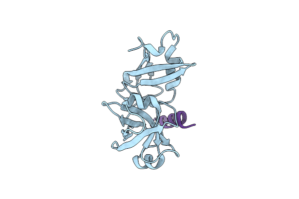
Structural Insights Into The Interaction Of P97 N-Terminal Domain And Shp Motif In Derlin-1 Rhomboid Pseudoprotease
Organism: Homo sapiens
Method: X-RAY DIFFRACTION Resolution:2.25 Å Release Date: 2016-11-09 Classification: HYDROLASE/TRANSPORT PROTEIN |
Organism: Homo sapiens
Method: X-RAY DIFFRACTION
Release Date: 2016-11-09
 |
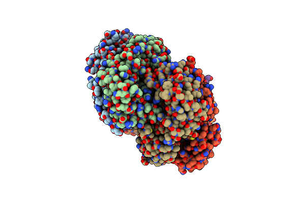
Structural Insights Into The Interaction Of P97 N-Terminus Domain And Vbm Motif In Rhomboid Protease, Rhbdl4
Organism: Homo sapiens
Method: X-RAY DIFFRACTION Resolution:1.88 Å Release Date: 2016-09-14 Classification: HYDROLASE |
Organism: Homo sapiens
Method: X-RAY DIFFRACTION
Release Date: 2016-09-14
 |
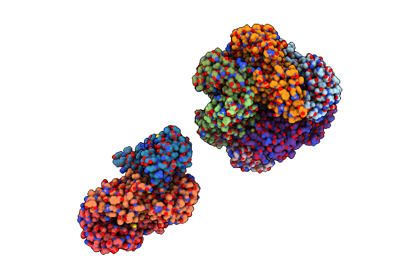
Crystal Structure Of Human Quinolinate Phosphoribosyltransferase
Organism: Homo sapiens
Method: X-RAY DIFFRACTION Resolution:2.80 Å Release Date: 2016-02-03 Classification: TRANSFERASE |
Organism: Homo sapiens
Method: X-RAY DIFFRACTION
Release Date: 2016-02-03
 |
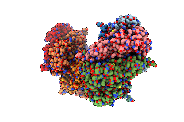
Crystal Structure Of Human Quinolinate Phosphoribosyltransferase In Complex With The Reactant Quinolinate
Organism: Homo sapiens
Method: X-RAY DIFFRACTION Resolution:3.09 Å Release Date: 2016-02-03 Classification: TRANSFERASE Ligands: NTM |
Organism: Homo sapiens
Method: X-RAY DIFFRACTION
Release Date: 2016-02-03
Ligands: NTM
 |
Crystal Structure Of Human Quinolinate Phosphoribosyltransferase In Complex With The Product Nicotinate Mononucleotide
Organism: Homo sapiens
Method: X-RAY DIFFRACTION Resolution:2.60 Å Release Date: 2016-02-03 Classification: TRANSFERASE Ligands: NCN |
Organism: Homo sapiens
Method: X-RAY DIFFRACTION
Release Date: 2016-02-03
Ligands: NCN
 |
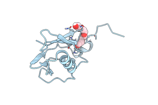
Crystal Structure Of The N-Terminal Domain Of The Human Mitochondrial Calcium Uniporter Fused With T4 Lysozyme
Organism: Enterobacteria phage t4, Homo sapiens
Method: X-RAY DIFFRACTION Resolution:1.80 Å Release Date: 2015-09-16 Classification: TRANSPORT PROTEIN Ligands: SO4 |
Organism: Enterobacteria phage t4, Homo sapiens
Method: X-RAY DIFFRACTION
Release Date: 2015-09-16
Ligands: SO4
 |
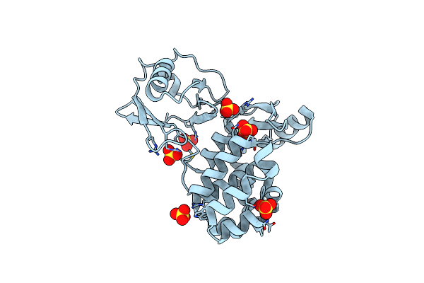
Crystal Structure Of The N-Terminal Domain Of The Human Mitochondrial Calcium Uniporter
Organism: Homo sapiens
Method: X-RAY DIFFRACTION Resolution:1.50 Å Release Date: 2015-09-16 Classification: TRANSPORT PROTEIN Ligands: PG4 |
Organism: Homo sapiens
Method: X-RAY DIFFRACTION
Release Date: 2015-09-16
Ligands: PG4
 |
Crystal Structure Of The N-Terminal Domain Single Mutant (S92A) Of The Human Mitochondrial Calcium Uniporter Fused With T4 Lysozyme
Organism: Enterobacteria phage t4, Homo sapiens
Method: X-RAY DIFFRACTION Resolution:2.75 Å Release Date: 2015-09-16 Classification: TRANSPORT PROTEIN Ligands: SO4 |
Organism: Enterobacteria phage t4, Homo sapiens
Method: X-RAY DIFFRACTION
Release Date: 2015-09-16
Ligands: SO4
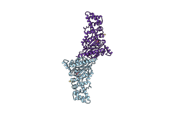 |
Crystal Structure Of Yeast Primase Catalytic Subunit
Organism: Saccharomyces cerevisiae
Method: X-RAY DIFFRACTION Resolution:1.60 Å Release Date: 2014-09-10 Classification: REPLICATION Ligands: CD, CIT |
Organism: Saccharomyces cerevisiae
Method: X-RAY DIFFRACTION
Release Date: 2014-09-10
Ligands: CD, CIT
 |
Crystal Structure Of Human Primase Catalytic Subunit
Organism: Homo sapiens
Method: X-RAY DIFFRACTION Resolution:2.20 Å Release Date: 2014-09-03 Classification: REPLICATION Ligands: ZN, CIT |
Organism: Homo sapiens
Method: X-RAY DIFFRACTION
Release Date: 2014-09-03
Ligands: ZN, CIT
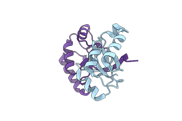 |
Crystal Structure Of Dimerized N-Terminal Domain Of Minc
Organism: Escherichia coli
Method: X-RAY DIFFRACTION Resolution:2.28 Å Release Date: 2013-10-23 Classification: PROTEIN BINDING |
Organism: Escherichia coli
Method: X-RAY DIFFRACTION
Release Date: 2013-10-23
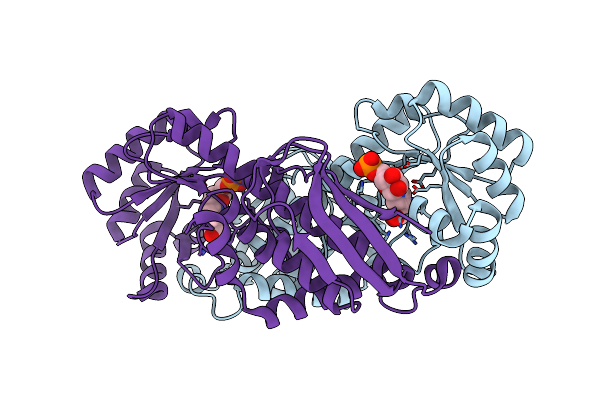 |
Crystal Structure Of Sus Scrofa Quinolinate Phosphoribosyltransferase In Complex With Nicotinate Mononucleotide
Organism: Sus scrofa
Method: X-RAY DIFFRACTION Resolution:2.10 Å Release Date: 2013-05-01 Classification: TRANSFERASE Ligands: NCN |
Organism: Sus scrofa
Method: X-RAY DIFFRACTION
Release Date: 2013-05-01
Ligands: NCN
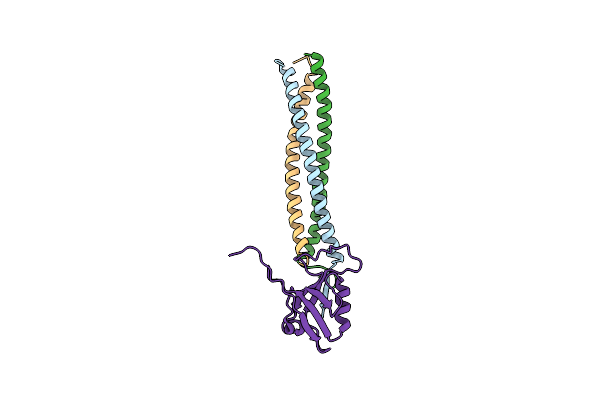 |
Crystal Structure Of Betapix Coiled-Coil Domain And Shank Pdz Complex
Organism: Rattus norvegicus
Method: X-RAY DIFFRACTION Resolution:2.80 Å Release Date: 2010-02-16 Classification: SIGNALING PROTEIN/PROTEIN BINDING |
Organism: Rattus norvegicus
Method: X-RAY DIFFRACTION
Release Date: 2010-02-16