

Search Count: 264
 |
Structure Of Pmhmgr Bound To Mevalonate, Coa And Nad 4 Minutes After Reaction Initiation At Ph 9
Organism: Pseudomonas sp. 'mevalonii'
Method: X-RAY DIFFRACTION Resolution:2.25 Å Release Date: 2026-11-04 Classification: OXIDOREDUCTASE/SUBSTRATE/INTERMEDIATE Ligands: ZKE, COA, A1AFY, NAI, MEV |
Organism: Pseudomonas sp. 'mevalonii'
Method: X-RAY DIFFRACTION
Release Date: 2026-11-04
Ligands: ZKE, COA, A1AFY, NAI, MEV
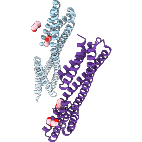 |
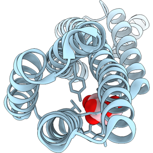
Structure Of The Ligand Binding Domain Of The Ancestral Reconstructed Pseudomonas Chemoreceptor Apcpi In Complex With Benzoate
Organism: Pseudomonas sp. sid14000
Method: X-RAY DIFFRACTION Resolution:2.20 Å Release Date: 2026-09-02 Classification: LIPID BINDING PROTEIN Ligands: BEZ, PEG |
Organism: Pseudomonas sp. sid14000
Method: X-RAY DIFFRACTION
Release Date: 2026-09-02
Ligands: BEZ, PEG
 |
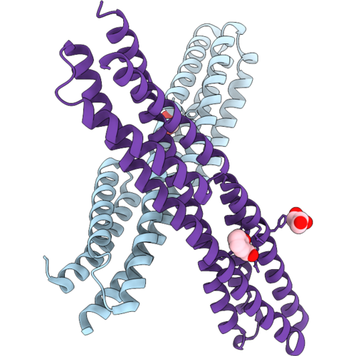
Structure Of The Ligand Binding Domain Of The Ancestral Reconstructed Pseudomonas Chemoreceptor Apcpi In Complex With Citrate
Organism: Pseudomonas sp. sid14000
Method: X-RAY DIFFRACTION Resolution:2.30 Å Release Date: 2026-09-02 Classification: LIPID BINDING PROTEIN Ligands: CIT |
Organism: Pseudomonas sp. sid14000
Method: X-RAY DIFFRACTION
Release Date: 2026-09-02
Ligands: CIT
 |
Structure Of The Ligand Binding Domain Of The Ancestral Reconstructed Pseudomonas Chemoreceptor Apcpi In Complex With Salicylate
Organism: Pseudomonas sp. sid14000
Method: X-RAY DIFFRACTION Resolution:2.80 Å Release Date: 2026-09-02 Classification: LIPID BINDING PROTEIN Ligands: GOL, SAL |
Organism: Pseudomonas sp. sid14000
Method: X-RAY DIFFRACTION
Release Date: 2026-09-02
Ligands: GOL, SAL
 |
Cytochrome P450 Cyp153A99 From Pseudomonas Sp. 19-Rlim, Complexed With Dehydroabietic Acid
Organism: Pseudomonas sp. 19-rlim
Method: X-RAY DIFFRACTION Resolution:2.10 Å Release Date: 2026-06-17 Classification: OXIDOREDUCTASE Ligands: A1ERW, HEM |
Organism: Pseudomonas sp. 19-rlim
Method: X-RAY DIFFRACTION
Release Date: 2026-06-17
Ligands: A1ERW, HEM
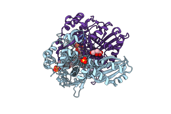 |
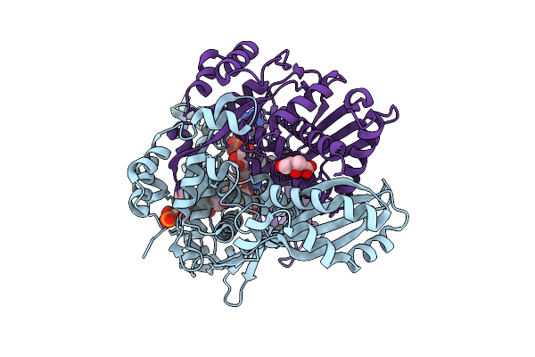
Structure Of Pmhmgr Bound To Mevalonate, Coa And Nad 2 Minutes 20 Seconds After Reaction Initiation At Ph 9
Organism: Pseudomonas sp. 'mevalonii'
Method: X-RAY DIFFRACTION Resolution:2.50 Å Release Date: 2026-04-08 Classification: OXIDOREDUCTASE/SUBSTRATE/INTERMEDIATE Ligands: COA, SO4, A1AFY, ZKE, NAI, MEV |
Organism: Pseudomonas sp. 'mevalonii'
Method: X-RAY DIFFRACTION
Release Date: 2026-04-08
Ligands: COA, SO4, A1AFY, ZKE, NAI, MEV
 |
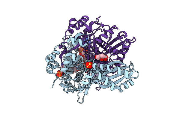
Structure Of Pmhmgr Bound To Mevalonate, Coa And Nad 1 Minute After Reaction Initiation At Ph 9
Organism: Pseudomonas sp. 'mevalonii'
Method: X-RAY DIFFRACTION Resolution:2.06 Å Release Date: 2026-03-18 Classification: OXIDOREDUCTASE/SUBSTRATE/INTERMEDIATE Ligands: COA, A1AFY, NAI, NAD, MEV |
Organism: Pseudomonas sp. 'mevalonii'
Method: X-RAY DIFFRACTION
Release Date: 2026-03-18
Ligands: COA, A1AFY, NAI, NAD, MEV
 |
Pmhmgr Bound To Mevalonate, Coa, And Nad, Buffer-Exchanged To Ammonium Acetate Environment At Ph 6.7
Organism: Pseudomonas sp. 'mevalonii'
Method: X-RAY DIFFRACTION Resolution:1.93 Å Release Date: 2026-03-18 Classification: OXIDOREDUCTASE Ligands: COA, MEV, SO4, NAD |
Organism: Pseudomonas sp. 'mevalonii'
Method: X-RAY DIFFRACTION
Release Date: 2026-03-18
Ligands: COA, MEV, SO4, NAD
 |
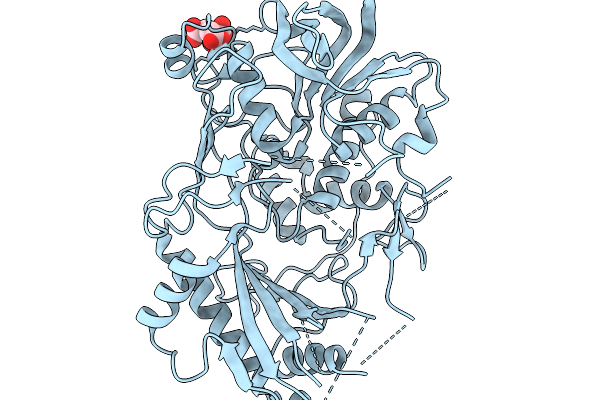
Structure Of A Rossmann Fold Domain From Pseudomonas
Organism: Pseudomonas sp. mwu12-2037
Method: X-RAY DIFFRACTION Resolution:2.92 Å Release Date: 2026-03-11 Classification: UNKNOWN FUNCTION Ligands: CA, EDO |
Organism: Pseudomonas sp. mwu12-2037
Method: X-RAY DIFFRACTION
Release Date: 2026-03-11
Ligands: CA, EDO
 |
Crystal Structure Of Choline Dehydrogenase Beta From Pseudomonas Denitrificans
Organism: Pseudomonas sp. atcc 13867
Method: X-RAY DIFFRACTION Resolution:1.80 Å Release Date: 2026-02-04 Classification: OXIDOREDUCTASE Ligands: CIT |
Organism: Pseudomonas sp. atcc 13867
Method: X-RAY DIFFRACTION
Release Date: 2026-02-04
Ligands: CIT
 |
Mg8, A Pet Hydrolase Enzyme From The Human Saliva Metagenome
Organism: Pseudomonas sp.
Method: X-RAY DIFFRACTION Resolution:1.20 Å Release Date: 2026-01-28 Classification: HYDROLASE Ligands: GOL, PEG, NA |
Organism: Pseudomonas sp.
Method: X-RAY DIFFRACTION
Release Date: 2026-01-28
Ligands: GOL, PEG, NA
 |
Crystal Structure Of C43S Terpredoxin, A [2Fe-2S] Ferredoxin From Pseudomonas Sp
Organism: Pseudomonas sp.
Method: X-RAY DIFFRACTION Resolution:2.10 Å Release Date: 2025-12-24 Classification: ELECTRON TRANSPORT Ligands: FES |
Organism: Pseudomonas sp.
Method: X-RAY DIFFRACTION
Release Date: 2025-12-24
Ligands: FES
 |
Crystal Structure Of Nahx From Pseudomonas Sp. Mc1
Organism: Pseudomonas sp. mc1
Method: X-RAY DIFFRACTION Resolution:1.70 Å Release Date: 2025-10-08 Classification: UNKNOWN FUNCTION |
Organism: Pseudomonas sp. mc1
Method: X-RAY DIFFRACTION
Release Date: 2025-10-08
 |
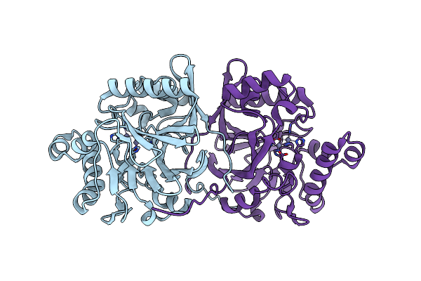
Structure Of The Functional Amyloid Fapc From Pseudomonas Sp.Uk4
Organism: Pseudomonas sp. uk4
Method: ELECTRON MICROSCOPY Release Date: 2025-08-06 Classification: PROTEIN FIBRIL |
Organism: Pseudomonas sp. uk4
Method: ELECTRON MICROSCOPY
Release Date: 2025-08-06
 |
Crystal Structure Of Methyl Parathion Hydrolase Mutant A127L/I145L/Q272D/S279A/S304P
Organism: Pseudomonas sp. wbc-3
Method: X-RAY DIFFRACTION Resolution:1.39 Å Release Date: 2025-06-11 Classification: HYDROLASE Ligands: ZN |
Organism: Pseudomonas sp. wbc-3
Method: X-RAY DIFFRACTION
Release Date: 2025-06-11
Ligands: ZN
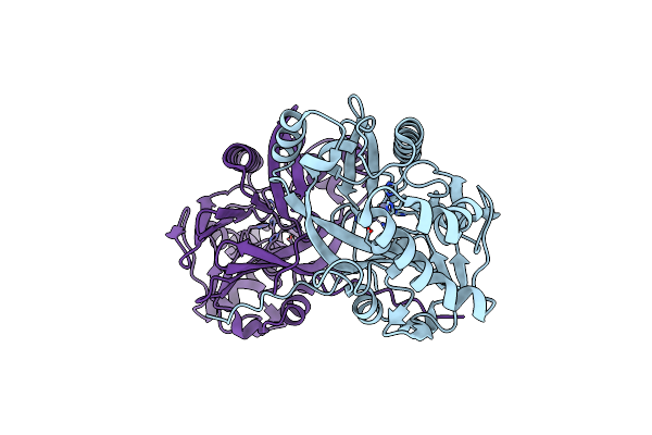 |
Crystal Structure Of Methyl Parathion Hydrolase Mutant A66D/I143V/I145L/Q272D/S279A/
Organism: Pseudomonas sp. wbc-3
Method: X-RAY DIFFRACTION Resolution:1.83 Å Release Date: 2025-06-11 Classification: HYDROLASE Ligands: ZN |
Organism: Pseudomonas sp. wbc-3
Method: X-RAY DIFFRACTION
Release Date: 2025-06-11
Ligands: ZN
 |
Crystal Structure Of The Enzyme-Product Complex Of L-Azetidine-2-Carboxylate Hydrolase
Organism: Pseudomonas sp. a2c
Method: X-RAY DIFFRACTION Resolution:0.93 Å Release Date: 2025-06-04 Classification: HYDROLASE Ligands: 42B, IMD, FMT, EDO, PEG, MG |
Organism: Pseudomonas sp. a2c
Method: X-RAY DIFFRACTION
Release Date: 2025-06-04
Ligands: 42B, IMD, FMT, EDO, PEG, MG
 |
Pinoresinol Hydroxylase From Pseudomonas Sp.
Organism: Pseudomonas sp.
Method: X-RAY DIFFRACTION Resolution:1.80 Å Release Date: 2024-12-11 Classification: FLAVOPROTEIN Ligands: ALA, FAD, GLY |
Organism: Pseudomonas sp.
Method: X-RAY DIFFRACTION
Release Date: 2024-12-11
Ligands: ALA, FAD, GLY
 |
Crystal Structure Of Acetylpolyamine Amidohydrolase (Apah) From Pseudomonas Sp. M30-35
Organism: Pseudomonas sp. m30-35
Method: X-RAY DIFFRACTION Resolution:2.72 Å Release Date: 2024-11-06 Classification: HYDROLASE Ligands: PGE, K, ZN, CL, ACT, PEG |
Organism: Pseudomonas sp. m30-35
Method: X-RAY DIFFRACTION
Release Date: 2024-11-06
Ligands: PGE, K, ZN, CL, ACT, PEG
 |
Nmr Structure Of Arthrofactin A In Micellar Dpc Solution
Organism: Pseudomonas sp. mis38
Method: SOLUTION NMR Release Date: 2024-08-14 Classification: SURFACTANT PROTEIN |
Organism: Pseudomonas sp. mis38
Method: SOLUTION NMR
Release Date: 2024-08-14