

Search Count: 3,350
 |
2-Heptyl-4(1H)-Quinolone Synthase Pqsbc In Complex With D3
Organism: Pseudomonas aeruginosa pao1
Method: X-RAY DIFFRACTION Resolution:2.24 Å Release Date: 2026-11-11 Classification: BIOSYNTHETIC PROTEIN Ligands: EDO, A1J5X Modified Residue(s): CSX |
Organism: Pseudomonas aeruginosa pao1
Method: X-RAY DIFFRACTION
Release Date: 2026-11-11
Ligands: EDO, A1J5X
 |
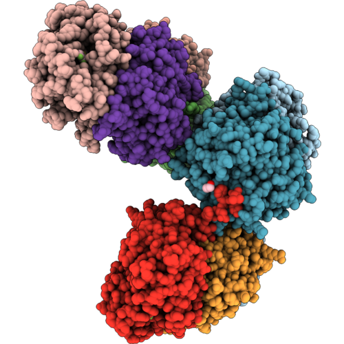
2-Heptyl-4(1H)-Quinolone Synthase Pqsbc In Complex With Compound 35
Organism: Pseudomonas aeruginosa pao1
Method: X-RAY DIFFRACTION Resolution:1.95 Å Release Date: 2026-11-11 Classification: BIOSYNTHETIC PROTEIN Ligands: A1J61, CL |
Organism: Pseudomonas aeruginosa pao1
Method: X-RAY DIFFRACTION
Release Date: 2026-11-11
Ligands: A1J61, CL
 |
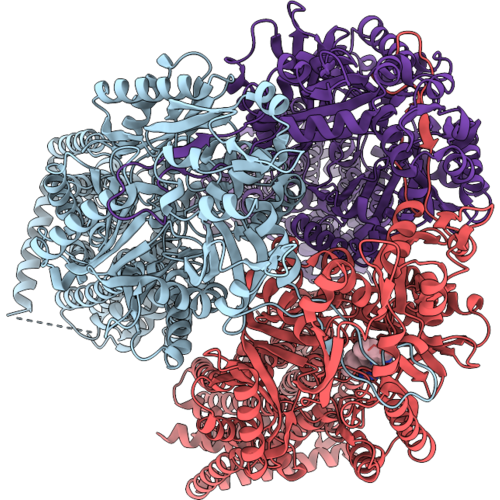
2-Heptyl-4(1H)-Quinolone Synthase Pqsbc In Complex With Compound 33
Organism: Pseudomonas aeruginosa pao1
Method: X-RAY DIFFRACTION Resolution:2.06 Å Release Date: 2026-11-11 Classification: BIOSYNTHETIC PROTEIN Ligands: EDO, A1J5V, CL |
Organism: Pseudomonas aeruginosa pao1
Method: X-RAY DIFFRACTION
Release Date: 2026-11-11
Ligands: EDO, A1J5V, CL
 |
Structure Of The Rnd Transporter Mexb In Complex With The Reference Inhibitor Phenylalanine-Arginine Beta-Naphthylamide.
Organism: Pseudomonas aeruginosa
Method: ELECTRON MICROSCOPY Release Date: 2026-11-11 Classification: ANTIMICROBIAL PROTEIN Ligands: MC2 |
Organism: Pseudomonas aeruginosa
Method: ELECTRON MICROSCOPY
Release Date: 2026-11-11
Ligands: MC2
 |
Structural And Functional Evaluation Of The Classical Rnd Efflux Pumps Inhibitor Pabn, A Systematic Study.
Organism: Pseudomonas aeruginosa
Method: X-RAY DIFFRACTION Resolution:3.56 Å Release Date: 2026-11-11 Classification: TRANSPORT PROTEIN Ligands: LMT, MC2, ACT |
Organism: Pseudomonas aeruginosa
Method: X-RAY DIFFRACTION
Release Date: 2026-11-11
Ligands: LMT, MC2, ACT
 |
Pseudomonas Aeruginosa Achc1 (Yfdx1) At Ph 8.5
Organism: Pseudomonas aeruginosa
Method: X-RAY DIFFRACTION Resolution:1.92 Å Release Date: 2026-11-04 Classification: CHAPERONE |
Organism: Pseudomonas aeruginosa
Method: X-RAY DIFFRACTION
Release Date: 2026-11-04
 |
Pseudomonas Aeruginosa Achc1 (Yfdx1) At Ph 3.0
Organism: Pseudomonas aeruginosa
Method: X-RAY DIFFRACTION Resolution:3.36 Å Release Date: 2026-11-04 Classification: CHAPERONE |
Organism: Pseudomonas aeruginosa
Method: X-RAY DIFFRACTION
Release Date: 2026-11-04
 |
Pseudomonas Aeruginosa Achc2 (Yfdx2) At Ph 7.0
Organism: Pseudomonas aeruginosa
Method: X-RAY DIFFRACTION Resolution:2.48 Å Release Date: 2026-11-04 Classification: CHAPERONE |
Organism: Pseudomonas aeruginosa
Method: X-RAY DIFFRACTION
Release Date: 2026-11-04
 |
Pseudomonas Aeruginosa Achc2 (Yfdx2) At Ph 4.0
Organism: Pseudomonas aeruginosa
Method: X-RAY DIFFRACTION Resolution:1.94 Å Release Date: 2026-11-04 Classification: CHAPERONE |
Organism: Pseudomonas aeruginosa
Method: X-RAY DIFFRACTION
Release Date: 2026-11-04
 |
Crystal Structure Of Paaldh Variant
Organism: Pseudomonas aeruginosa c1913c
Method: X-RAY DIFFRACTION Resolution:2.38 Å Release Date: 2026-11-04 Classification: OXIDOREDUCTASE Ligands: PEG |
Organism: Pseudomonas aeruginosa c1913c
Method: X-RAY DIFFRACTION
Release Date: 2026-11-04
Ligands: PEG
 |
Cupin Protein Glob From Pseudomonas Aeruginosa
Organism: Pseudomonas aeruginosa
Method: X-RAY DIFFRACTION Resolution:1.48 Å Release Date: 2026-11-04 Classification: UNKNOWN FUNCTION Ligands: MG, K |
Organism: Pseudomonas aeruginosa
Method: X-RAY DIFFRACTION
Release Date: 2026-11-04
Ligands: MG, K
 |
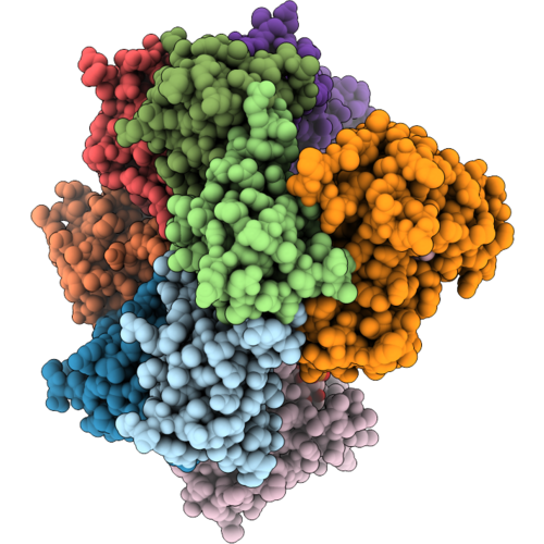
Pseudomonas Aeruginosa T6Ss Effector Tne3 In Complex With Its Cognate Immunity Protein Tni3
Organism: Pseudomonas aeruginosa
Method: X-RAY DIFFRACTION Resolution:2.35 Å Release Date: 2026-11-04 Classification: TOXIN |
Organism: Pseudomonas aeruginosa
Method: X-RAY DIFFRACTION
Release Date: 2026-11-04
 |
Crystal Structure Of The Pseudomonas Aeruginosa Mlac-Mlad Complex In Space Group P1
Organism: Pseudomonas aeruginosa pao1
Method: X-RAY DIFFRACTION Resolution:2.56 Å Release Date: 2026-10-28 Classification: LIPID TRANSPORT Ligands: LPP |
Organism: Pseudomonas aeruginosa pao1
Method: X-RAY DIFFRACTION
Release Date: 2026-10-28
Ligands: LPP
 |
Crystal Structure Of The Pseudomonas Aeruginosa Mlac-Mlad Complex In Space Group P41212
Organism: Pseudomonas aeruginosa pao1
Method: X-RAY DIFFRACTION Resolution:3.46 Å Release Date: 2026-10-28 Classification: LIPID TRANSPORT |
Organism: Pseudomonas aeruginosa pao1
Method: X-RAY DIFFRACTION
Release Date: 2026-10-28
 |
Structure Of Padxps In Complex With Small Molecule Inhibitor
Organism: Pseudomonas aeruginosa lesb58
Method: X-RAY DIFFRACTION Resolution:1.91 Å Release Date: 2026-10-28 Classification: ANTIBIOTIC Ligands: EPE, A1JB5, CA, PEG |
Organism: Pseudomonas aeruginosa lesb58
Method: X-RAY DIFFRACTION
Release Date: 2026-10-28
Ligands: EPE, A1JB5, CA, PEG
 |
Psl Polysaccharide Related Protein Structures
Organism: Pseudomonas aeruginosa
Method: ELECTRON MICROSCOPY Release Date: 2026-10-28 Classification: BIOSYNTHETIC PROTEIN |
Organism: Pseudomonas aeruginosa
Method: ELECTRON MICROSCOPY
Release Date: 2026-10-28
 |
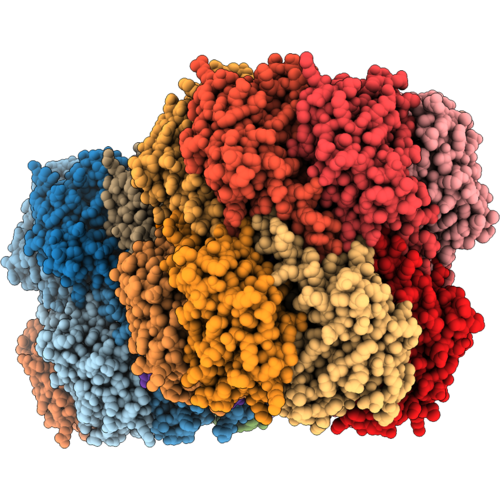
Crystal Structure Of Pseudomonas Aeruginosa Suhb Complexed With Phosphate, Myo-Inositol And Ca2+ At Active Site
Organism: Pseudomonas aeruginosa pao1
Method: X-RAY DIFFRACTION Resolution:2.30 Å Release Date: 2026-10-28 Classification: HYDROLASE Ligands: INS, ACT, CA, PO4, GOL |
Organism: Pseudomonas aeruginosa pao1
Method: X-RAY DIFFRACTION
Release Date: 2026-10-28
Ligands: INS, ACT, CA, PO4, GOL
 |
Structure Of Pseudomonas Aeruginosa Mutant Clpp2-R140A
Organism: Pseudomonas aeruginosa pao1
Method: X-RAY DIFFRACTION Resolution:3.00 Å Release Date: 2026-10-21 Classification: HYDROLASE |
Organism: Pseudomonas aeruginosa pao1
Method: X-RAY DIFFRACTION
Release Date: 2026-10-21
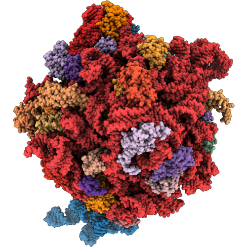 |
Structural Characterization Of Hibernating Ribosomes In The Gram-Negative Pathogen Pseudomonas Aeruginosa
Organism: Escherichia coli, Pseudomonas aeruginosa pa14
Method: ELECTRON MICROSCOPY Release Date: 2026-10-21 Classification: RIBOSOME Ligands: ZN, MG, K Modified Residue(s): PSU, G7M, 2MG, 5MC, 4OC, UR3, MA6, D2T, MEQ, 4D4, MS6 |
Organism: Escherichia coli, Pseudomonas aeruginosa pa14
Method: ELECTRON MICROSCOPY
Release Date: 2026-10-21
Ligands: ZN, MG, K
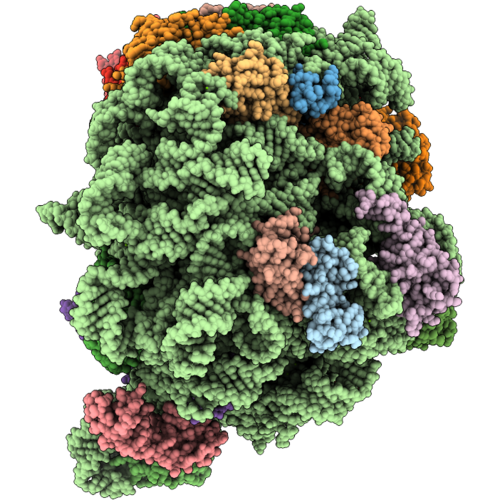 |
Pseudomonas Aeruginosa 50S Ribosome (L1 Stalk In 'Mid' Conformation)
|
Organism: Pseudomonas aeruginosa pao1
Method: ELECTRON MICROSCOPY
Release Date: 2026-10-21
Ligands: MG, ZN