

Search Count: 65
 |
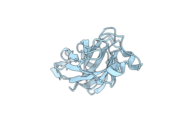
Crystal Structure Of Proteus Mirabilis Uree
Organism: Proteus mirabilis
Method: X-RAY DIFFRACTION Resolution:2.00 Å Release Date: 2026-03-25 Classification: METAL BINDING PROTEIN |
Organism: Proteus mirabilis
Method: X-RAY DIFFRACTION
Release Date: 2026-03-25
 |
Crystal Structure Of The Ucad Lectin-Binding Domain
Organism: Proteus mirabilis
Method: X-RAY DIFFRACTION Resolution:1.62 Å Release Date: 2022-07-06 Classification: SUGAR BINDING PROTEIN Ligands: CL |
Organism: Proteus mirabilis
Method: X-RAY DIFFRACTION
Release Date: 2022-07-06
Ligands: CL
 |
Crystal Structure Of The Ucad Lectin-Binding Domain In Complex With Fucose
Organism: Proteus mirabilis
Method: X-RAY DIFFRACTION Resolution:1.50 Å Release Date: 2022-07-06 Classification: SUGAR BINDING PROTEIN Ligands: FUL, CL |
Organism: Proteus mirabilis
Method: X-RAY DIFFRACTION
Release Date: 2022-07-06
Ligands: FUL, CL
 |
Crystal Structure Of The Ucad Lectin-Binding Domain In Complex With Glucose
Organism: Proteus mirabilis
Method: X-RAY DIFFRACTION Resolution:1.78 Å Release Date: 2022-07-06 Classification: SUGAR BINDING PROTEIN Ligands: BGC, CL |
Organism: Proteus mirabilis
Method: X-RAY DIFFRACTION
Release Date: 2022-07-06
Ligands: BGC, CL
 |
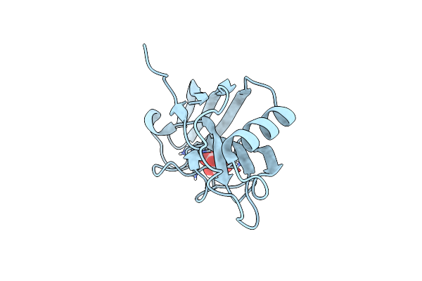
Crystal Structure Of The Ucad Lectin-Binding Domain In Complex With Galactose
Organism: Proteus mirabilis
Method: X-RAY DIFFRACTION Resolution:1.72 Å Release Date: 2022-07-06 Classification: SUGAR BINDING PROTEIN Ligands: GLA, CL |
Organism: Proteus mirabilis
Method: X-RAY DIFFRACTION
Release Date: 2022-07-06
Ligands: GLA, CL
 |
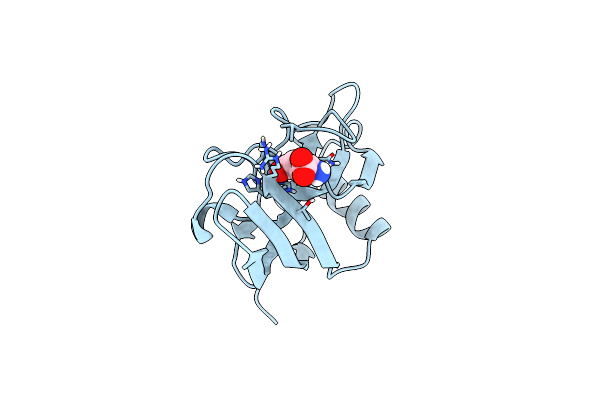
X-Ray Structure Of The Zn-Dependent Receptor-Binding Domain Of Proteus Mirabilis Mr/P Fimbrial Adhesin Mrph
Organism: Proteus mirabilis (strain hi4320)
Method: X-RAY DIFFRACTION Resolution:1.02 Å Release Date: 2020-08-19 Classification: CELL ADHESION Ligands: ZN, TLA |
Organism: Proteus mirabilis (strain hi4320)
Method: X-RAY DIFFRACTION
Release Date: 2020-08-19
Ligands: ZN, TLA
 |
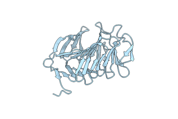
X-Ray Structure Of The Zn-Dependent Receptor-Binding Domain Of Proteus Mirabilis Mr/P Fimbrial Adhesin Mrph
Organism: Proteus mirabilis (strain hi4320)
Method: X-RAY DIFFRACTION Resolution:1.75 Å Release Date: 2020-08-19 Classification: CELL ADHESION Ligands: ZN, GLU |
Organism: Proteus mirabilis (strain hi4320)
Method: X-RAY DIFFRACTION
Release Date: 2020-08-19
Ligands: ZN, GLU
 |
P. Mirabilis Hemolysin A Mutant - F80L
Organism: Proteus mirabilis
Method: X-RAY DIFFRACTION Resolution:1.35 Å Release Date: 2020-08-05 Classification: TOXIN |
Organism: Proteus mirabilis
Method: X-RAY DIFFRACTION
Release Date: 2020-08-05
 |
P. Mirabilis Hemolysin A Mutant - Q125A
Organism: Proteus mirabilis
Method: X-RAY DIFFRACTION Resolution:1.17 Å Release Date: 2020-08-05 Classification: TOXIN Ligands: GOL |
Organism: Proteus mirabilis
Method: X-RAY DIFFRACTION
Release Date: 2020-08-05
Ligands: GOL
 |
P. Mirabilis Hemolysin A Mutant - Y134S
Organism: Proteus mirabilis
Method: X-RAY DIFFRACTION Resolution:1.54 Å Release Date: 2020-08-05 Classification: TOXIN |
Organism: Proteus mirabilis
Method: X-RAY DIFFRACTION
Release Date: 2020-08-05
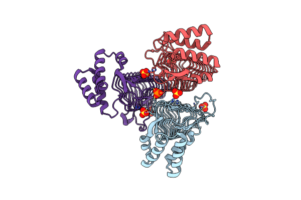 |
Crystal Structure Of The Acyl-Carrier-Protein Udp-N-Acetylglucosamine O-Acyltransferase Lpxa From Proteus Mirabilis
Organism: Proteus mirabilis (strain hi4320)
Method: X-RAY DIFFRACTION Resolution:2.19 Å Release Date: 2020-01-29 Classification: TRANSFERASE Ligands: SO4, PO3 |
Organism: Proteus mirabilis (strain hi4320)
Method: X-RAY DIFFRACTION
Release Date: 2020-01-29
Ligands: SO4, PO3
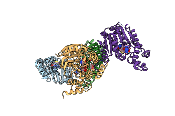 |
Crystal Structure Of Aminoglycoside-Resistance Methyltransferase Rmtc Bound To S-Adenosylhomocysteine (Sah)
Organism: Proteus mirabilis
Method: X-RAY DIFFRACTION Resolution:3.14 Å Release Date: 2019-10-16 Classification: TRANSFERASE Ligands: SAH |
Organism: Proteus mirabilis
Method: X-RAY DIFFRACTION
Release Date: 2019-10-16
Ligands: SAH
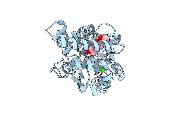 |
Proteus Mirabilis Lipase Mutant - I118V/E130G
Organism: Proteus mirabilis
Method: X-RAY DIFFRACTION Resolution:1.58 Å Release Date: 2019-07-24 Classification: HYDROLASE Ligands: CA, MPD |
Organism: Proteus mirabilis
Method: X-RAY DIFFRACTION
Release Date: 2019-07-24
Ligands: CA, MPD
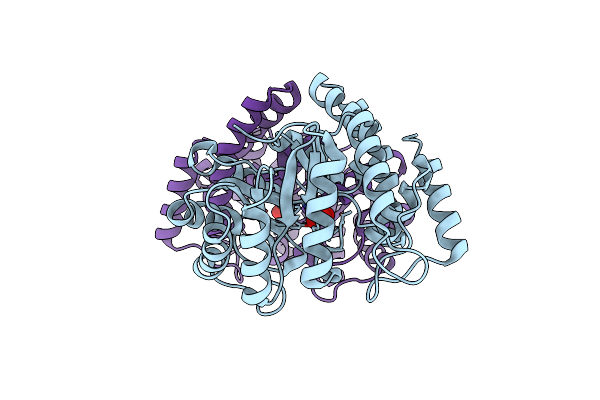 |
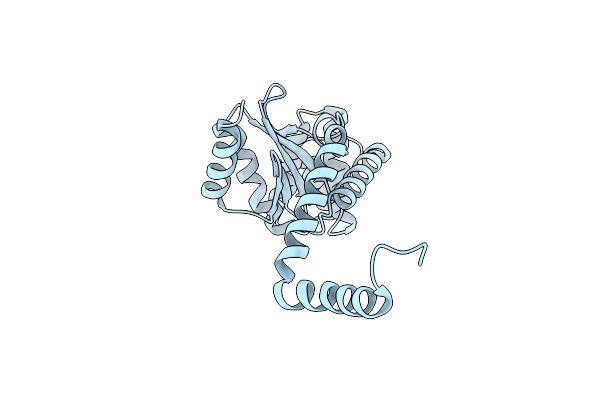
Proteus Mirabilis N-Acetylneuraminate Lyase
Organism: Proteus mirabilis (strain hi4320)
Method: X-RAY DIFFRACTION Resolution:1.56 Å Release Date: 2019-06-26 Classification: LYASE Ligands: SO4 |
Organism: Proteus mirabilis (strain hi4320)
Method: X-RAY DIFFRACTION
Release Date: 2019-06-26
Ligands: SO4
 |
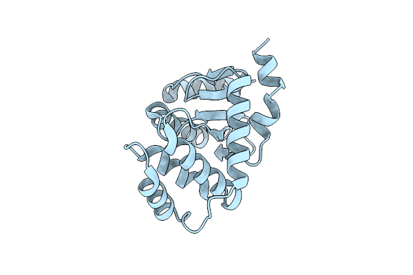
Proteus Mirabilis Scsc Linker (Residues 39-49) Deletion And N6K Mutant
Organism: Proteus mirabilis
Method: X-RAY DIFFRACTION Resolution:2.08 Å Release Date: 2019-03-06 Classification: ISOMERASE |
Organism: Proteus mirabilis
Method: X-RAY DIFFRACTION
Release Date: 2019-03-06
 |
Catalytic Domain Of Proteus Mirabilis Scsc
Organism: Proteus mirabilis
Method: X-RAY DIFFRACTION Resolution:2.15 Å Release Date: 2019-03-06 Classification: OXIDOREDUCTASE |
Organism: Proteus mirabilis
Method: X-RAY DIFFRACTION
Release Date: 2019-03-06
 |
Proteus Mirabilis Ambient Temperature Fimbriae Adhesin Atfe
Organism: Proteus mirabilis hi4320
Method: X-RAY DIFFRACTION Resolution:1.58 Å Release Date: 2018-11-07 Classification: CELL ADHESION Ligands: GOL, PO4 |
Organism: Proteus mirabilis hi4320
Method: X-RAY DIFFRACTION
Release Date: 2018-11-07
Ligands: GOL, PO4
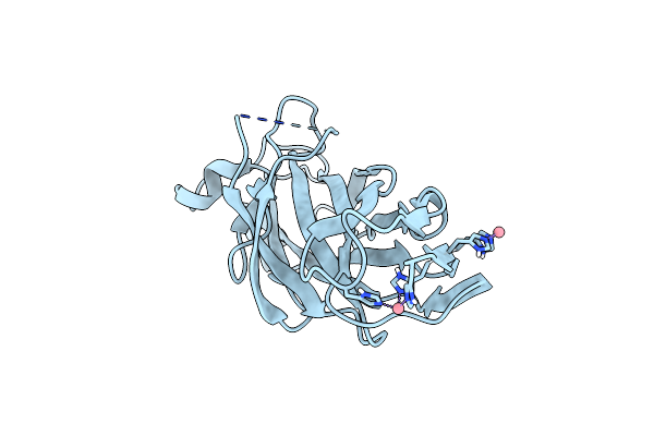 |
Receptor-Binding Domain Of Proteus Mirabilis Uroepithelial Cell Adhesin Ucad21-211
Organism: Proteus mirabilis
Method: X-RAY DIFFRACTION Resolution:1.70 Å Release Date: 2018-11-07 Classification: CELL ADHESION Ligands: CO |
Organism: Proteus mirabilis
Method: X-RAY DIFFRACTION
Release Date: 2018-11-07
Ligands: CO
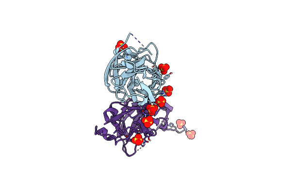 |
Receptor-Binding Domain Of Proteus Mirabilis Uroepithelial Cell Adhesin Ucad21-217
Organism: Proteus mirabilis
Method: X-RAY DIFFRACTION Resolution:1.50 Å Release Date: 2018-11-07 Classification: CELL ADHESION Ligands: SO4 |
Organism: Proteus mirabilis
Method: X-RAY DIFFRACTION
Release Date: 2018-11-07
Ligands: SO4
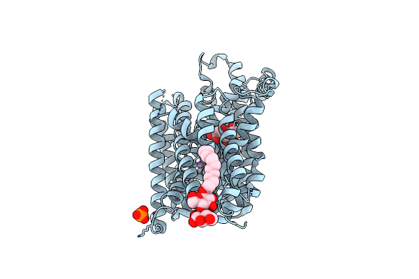 |
Substrate-Bound Outward-Open State Of A Na+-Coupled Sialic Acid Symporter Reveals A Novel Na+-Site
Organism: Proteus mirabilis (strain hi4320)
Method: X-RAY DIFFRACTION Resolution:1.95 Å Release Date: 2018-04-04 Classification: MEMBRANE PROTEIN |
Organism: Proteus mirabilis (strain hi4320)
Method: X-RAY DIFFRACTION
Release Date: 2018-04-04