
Search Count: 6,400
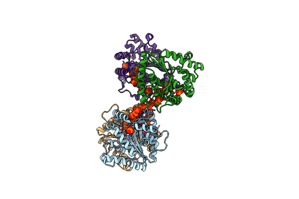 |
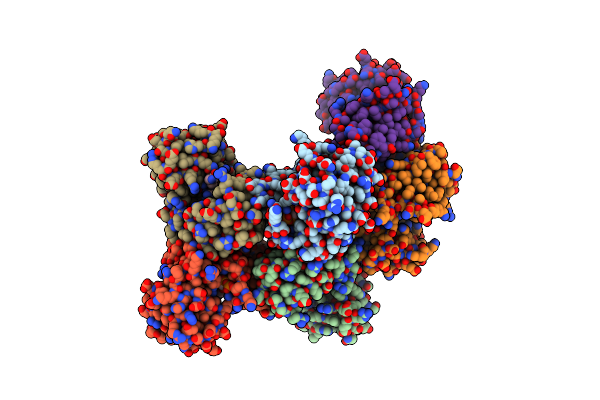
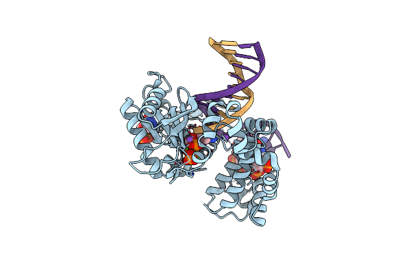
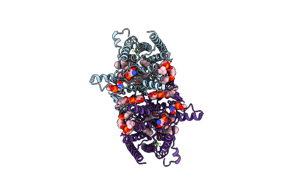
Structure Of Polyphosphate Kinase 2 Mutant D117N From Francisella Tularensis With Polyphosphate
Organism: Francisella tularensis subsp. tularensis (strain schu s4 / schu 4)
Method: X-RAY DIFFRACTION Resolution:2.31 Å Release Date: 2017-10-11 Classification: TRANSFERASE Ligands: PO4, 6YX, 6YY, CL, 6YW |
 |
Crystal Structure Of Ppk2 Class Iii From Erysipelotrichaceae Bacterium In Complex With Appch2P And Polyphosphate
Organism: Erysipelotrichaceae bacterium
Method: X-RAY DIFFRACTION Resolution:1.70 Å Release Date: 2025-07-23 Classification: TRANSFERASE Ligands: ACP, 6YY, GOL, BEZ, MG, MPD, A1I4D |
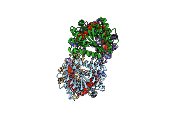 |
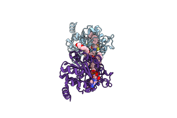
Structure Of Polyphosphate Kinase 2 From Francisella Tularensis With Amppch2Ppp And Polyphosphate
Organism: Francisella tularensis subsp. tularensis (strain schu s4 / schu 4)
Method: X-RAY DIFFRACTION Resolution:1.92 Å Release Date: 2017-10-11 Classification: TRANSFERASE Ligands: CL, MG, 6YZ, 6YW |
 |
Kod-H4 Dna Polymerase Mutant In A Ternary Complex Containing Six Hna Nucleotides And A Non-Hydrolyzable Triphosphate
Organism: Thermococcus kodakarensis kod1, Synthetic construct
Method: X-RAY DIFFRACTION Resolution:2.27 Å Release Date: 2024-11-13 Classification: TRANSFERASE Ligands: EDO, MG, XG4 |
 |
Organism: Rhodobacteraceae bacterium qy30
Method: X-RAY DIFFRACTION Resolution:2.41 Å Release Date: 2025-05-07 Classification: ANTIVIRAL PROTEIN Ligands: A1A3G, F2A, MN, A1A3H, D5M |
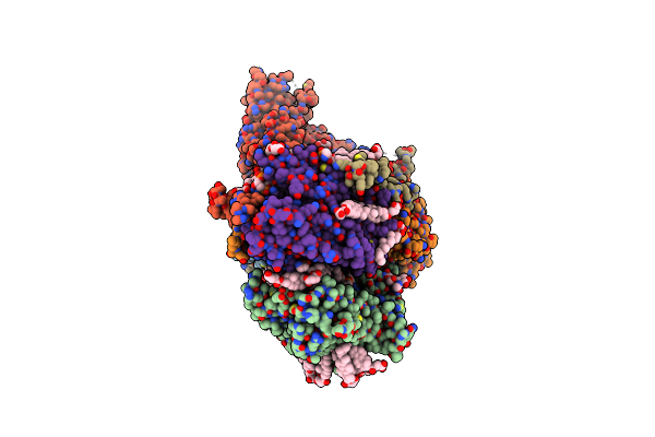 |
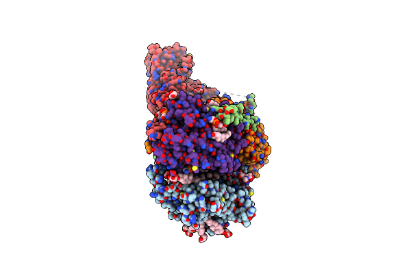
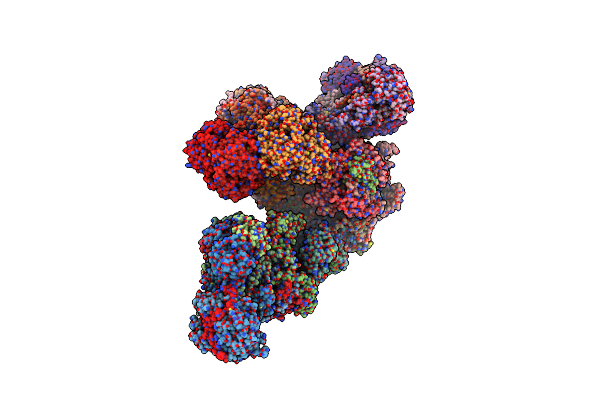
Cryo-Em Structure Of Gpi-T (Inactive Mutant) With Gpi And Proulbp2, A Proprotein Substrate
Organism: Homo sapiens, Clavularia sp.
Method: ELECTRON MICROSCOPY Release Date: 2023-08-16 Classification: MEMBRANE PROTEIN Ligands: AJP, 6OU, Y01, MG, 05E, 80Y, CA, LBN, NAG, 81Q, PA1, 80T |
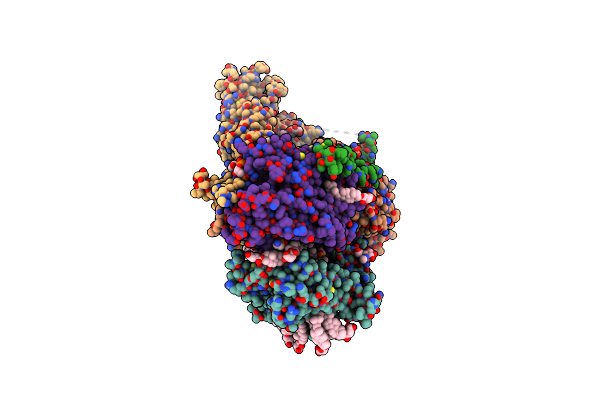 |
Organism: Homo sapiens, Clavularia sp., Synthetic construct
Method: ELECTRON MICROSCOPY Release Date: 2023-08-16 Classification: MEMBRANE PROTEIN Ligands: Y01, MG, 6OU, AJP, CA, NAG, LBN, 80T, 80Y, PA1, 05E, 81Q |
 |
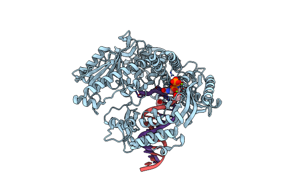
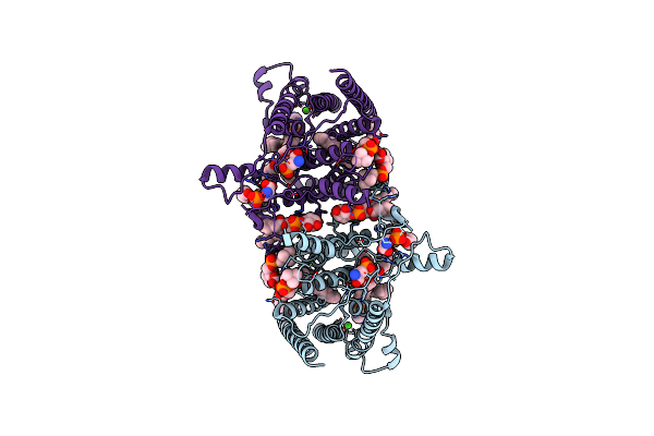
Ap4G Bound In De Novo Transcription Initiation T. Thermophilus Rna Polymerase Complex With Tc Dna Template
Organism: Thermus thermophilus hb8, Synthetic construct
Method: ELECTRON MICROSCOPY Resolution:3.00 Å Release Date: 2025-12-17 Classification: TRANSCRIPTION Ligands: MG, ZN, 2TM, A1IF9 |
 |
Ternary Complex Crystal Structure Of Dna Polymerase Beta With A Dideoxy Terminated Primer With Chcl (R &Amp; S Isomers, Beta, Gamma Dctp Analogue
Organism: Homo sapiens
Method: X-RAY DIFFRACTION Resolution:2.00 Å Release Date: 2018-06-20 Classification: transcription/dna Ligands: MG, NA, FF7, DJJ, DC, CL |
 |
Ternary Complex Crystal Structure Of Dna Polymerase Beta With A Dideoxy Terminated Primer With Chf (R & S Isomers), Beta, Gamma Dctp Analogue
Organism: Homo sapiens
Method: X-RAY DIFFRACTION Resolution:1.85 Å Release Date: 2018-06-20 Classification: transcription/dna Ligands: MG, NA, CL, F3C, FF4, DC |
 |
Cryo-Em Structure Of The Freshwater Actinorhodopsin, Rhodoluna Lacicola (Rlactr)
Organism: Rhodoluna lacicola
Method: ELECTRON MICROSCOPY Release Date: 2026-03-18 Classification: PROTON TRANSPORT Ligands: A1INB, A1IVO, A1INC, RET |
 |
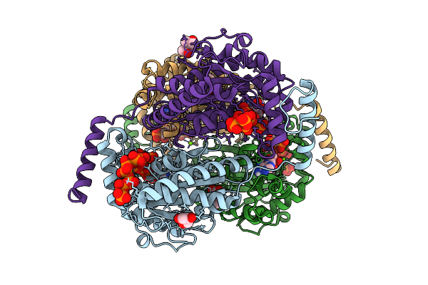
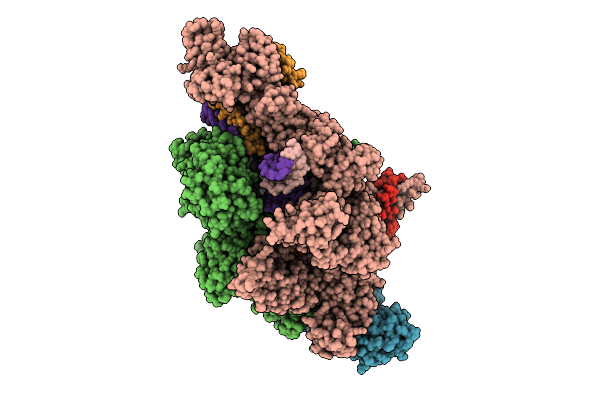
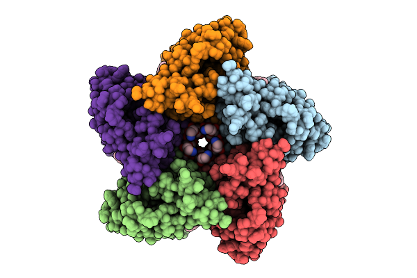
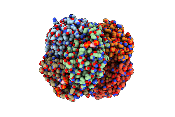
Cryo-Em Structure Of The Human Glycosylphosphatidylinositol Transamidase Complex At 2.53 Angstrom Resolution
Organism: Homo sapiens
Method: ELECTRON MICROSCOPY Resolution:2.53 Å Release Date: 2022-04-27 Classification: TRANSFERASE Ligands: Y01, AJP, BJR, PEE, CA, P5S, NAG, CLR, 05E, PA1, CDL, DKB, 06O |
 |
Organism: Homo sapiens
Method: ELECTRON MICROSCOPY Release Date: 2024-09-04 Classification: MEMBRANE PROTEIN Ligands: P5S, LBN, A1AIS, 6OU, CA |
 |
Organism: Homo sapiens
Method: ELECTRON MICROSCOPY Release Date: 2024-09-04 Classification: MEMBRANE PROTEIN Ligands: P5S, LBN, A1AIS, 6OU, CA, A1AIT |
 |
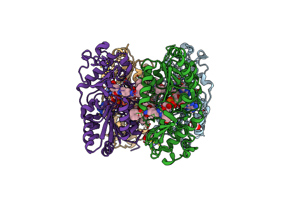
In-Situ Structure Of Typea Supercomplex With Lipids In Respiratory Chain (Composite)
|
 |
Methylmalonyl-Coa Decarboxylase In Complex With 2-Sulfonate-Propionyl-Oxa(Dethia)-Coa
Organism: Escherichia coli (strain k12)
Method: X-RAY DIFFRACTION Resolution:1.70 Å Release Date: 2019-04-10 Classification: LYASE Ligands: LCV, SO5, IMD |
 |
Organism: Streptomyces atroolivaceus
Method: X-RAY DIFFRACTION Resolution:1.55 Å Release Date: 2020-06-10 Classification: transferase/transferase inhibitor Ligands: SO5, LCV, SO4 |
 |
Crystal Structure Of Yfih With C107A Mutation In Complex With Udp-Murnac-L-Serine
Organism: Escherichia coli
Method: X-RAY DIFFRACTION Resolution:1.86 Å Release Date: 2021-12-29 Classification: HYDROLASE Ligands: EPZ, SO4, ACT, TRS, 87E |
 |
Crystal Structure Of Human Sirtuin 3 Fragment (Residues 118-399) Bound To Intermediates From Reaction With Nad And Inhibitor Nh6-10
Organism: Homo sapiens
Method: X-RAY DIFFRACTION Resolution:1.95 Å Release Date: 2024-08-07 Classification: HYDROLASE Ligands: ZN, 5IA, PG4, 5I7 |
 |
Zebrafish Sirt5 In Complex With Stalled Peptidylimidate And Bicyclic Intermediate Of Inhibitory Compound 29
Organism: Danio rerio
Method: X-RAY DIFFRACTION Resolution:2.40 Å Release Date: 2017-11-01 Classification: SIGNALING PROTEIN Ligands: ZN, EDO, BV8, BVT |