

Search Count: 71
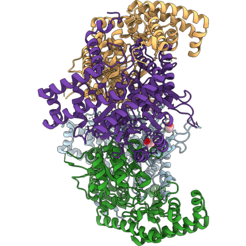 |
Structure Of The Pathogenic Variant G423S Of Human Shmt2
Organism: Homo sapiens
Method: X-RAY DIFFRACTION Resolution:1.80 Å Release Date: 2026-09-02 Classification: TRANSFERASE Ligands: BU1, PDO, 1BO, EDO |
Organism: Homo sapiens
Method: X-RAY DIFFRACTION
Release Date: 2026-09-02
Ligands: BU1, PDO, 1BO, EDO
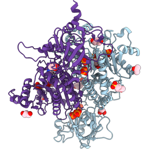 |
Plasmodium Vivax Aspartyl-Trna Synthetase In Complex With Amp, Po4, Mops And Alcohols
Organism: Plasmodium vivax
Method: X-RAY DIFFRACTION Resolution:2.30 Å Release Date: 2026-09-02 Classification: LIGASE Ligands: AMP, PO4, MPO, PGO, PDO, HEZ, CL |
Organism: Plasmodium vivax
Method: X-RAY DIFFRACTION
Release Date: 2026-09-02
Ligands: AMP, PO4, MPO, PGO, PDO, HEZ, CL
 |
Crystal Structure Of T. Brucei Eif4E5 In Complex With Eif4G1 Peptide
Organism: Trypanosoma brucei brucei treu927
Method: X-RAY DIFFRACTION Resolution:1.66 Å Release Date: 2026-01-21 Classification: TRANSLATION Ligands: PDO, 1BO, NA |
Organism: Trypanosoma brucei brucei treu927
Method: X-RAY DIFFRACTION
Release Date: 2026-01-21
Ligands: PDO, 1BO, NA
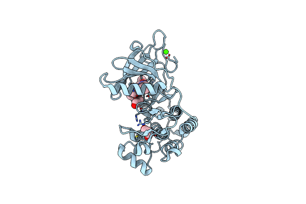 |
Co-Crystal Structure Of The Kinase Domain Of Egfr With Non-Covalent Osimertinib
Organism: Homo sapiens
Method: X-RAY DIFFRACTION Resolution:2.31 Å Release Date: 2025-05-28 Classification: TRANSFERASE Ligands: PDO, Q6K, CA |
Organism: Homo sapiens
Method: X-RAY DIFFRACTION
Release Date: 2025-05-28
Ligands: PDO, Q6K, CA
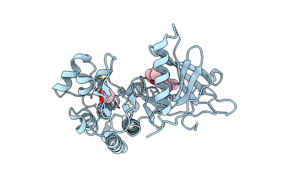 |
Crystal Structure Of The Kinase Domain Of Egfr Soaked With Non-Covalent Osimertinib
Organism: Homo sapiens
Method: X-RAY DIFFRACTION Resolution:2.55 Å Release Date: 2025-05-28 Classification: TRANSFERASE Ligands: PDO, Q6K |
Organism: Homo sapiens
Method: X-RAY DIFFRACTION
Release Date: 2025-05-28
Ligands: PDO, Q6K
 |
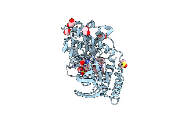
Crystal Structure Of Rice Dwarf14 In Complex With Cyclo(L-Leu-L-Pro)
Organism: Oryza sativa japonica group
Method: X-RAY DIFFRACTION Resolution:1.50 Å Release Date: 2025-05-21 Classification: HYDROLASE Ligands: A1EBP, GOL, BEZ, PDO, BU1, PGO |
Organism: Oryza sativa japonica group
Method: X-RAY DIFFRACTION
Release Date: 2025-05-21
Ligands: A1EBP, GOL, BEZ, PDO, BU1, PGO
 |
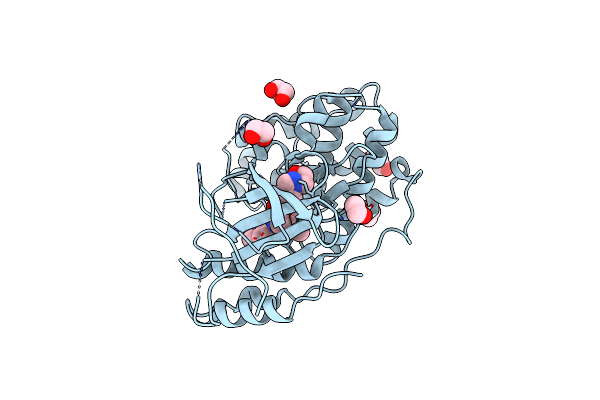
Dual Phosphorylated Human P38 Alpha Bound To Pexmetinib
Organism: Homo sapiens
Method: X-RAY DIFFRACTION Resolution:1.95 Å Release Date: 2024-11-13 Classification: TRANSFERASE Ligands: SO4, EDO, GOL, DMS, PDO, A1AWV |
Organism: Homo sapiens
Method: X-RAY DIFFRACTION
Release Date: 2024-11-13
Ligands: SO4, EDO, GOL, DMS, PDO, A1AWV
 |
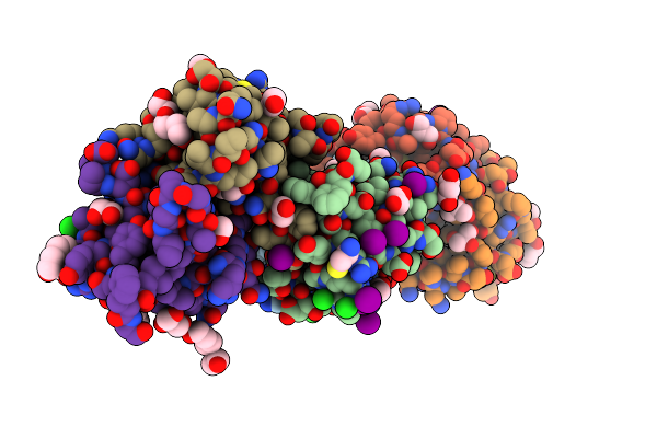
Unphosphorylated Human P38 Alpha Bound To Pexmetinib
Organism: Homo sapiens
Method: X-RAY DIFFRACTION Resolution:3.30 Å Release Date: 2024-11-13 Classification: TRANSFERASE Ligands: EDO, A1AWV, PDO |
Organism: Homo sapiens
Method: X-RAY DIFFRACTION
Release Date: 2024-11-13
Ligands: EDO, A1AWV, PDO
 |
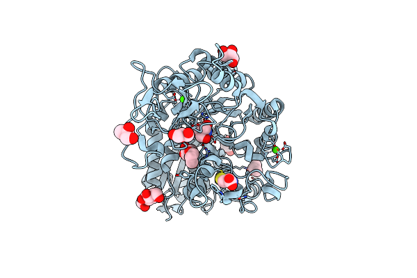
The Crystal Structure Of Protein A21, A Component Of The Conserved Poxvirus Entry-Fusion Complex
Organism: Vaccinia virus western reserve
Method: X-RAY DIFFRACTION Resolution:2.30 Å Release Date: 2024-09-04 Classification: VIRAL PROTEIN Ligands: EOH, EDO, MOH, IOD, CL, PDO, PGR, NA, PG0, DHL, PGE, PEG, POL, GOL, IPA |
 |
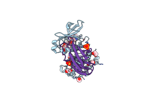
Structure Of A Salivary Alpha-Glucosidase From The Mosquito Vector Aedes Aegypti.
Organism: Aedes aegypti
Method: X-RAY DIFFRACTION Resolution:2.32 Å Release Date: 2024-03-20 Classification: HYDROLASE Ligands: NAG, CYS, GOL, TRS, IPA, PDO, CA, CL |
Organism: Aedes aegypti
Method: X-RAY DIFFRACTION
Release Date: 2024-03-20
Ligands: NAG, CYS, GOL, TRS, IPA, PDO, CA, CL
 |
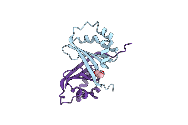
Aura Bound To Danusertib And Activating Monobody Mb1
Organism: Homo sapiens, Synthetic construct
Method: X-RAY DIFFRACTION Resolution:2.60 Å Release Date: 2023-09-06 Classification: TRANSFERASE Ligands: EDO, SO4, MES, PO4, PDO, 627 |
Organism: Homo sapiens, Synthetic construct
Method: X-RAY DIFFRACTION
Release Date: 2023-09-06
Ligands: EDO, SO4, MES, PO4, PDO, 627
 |
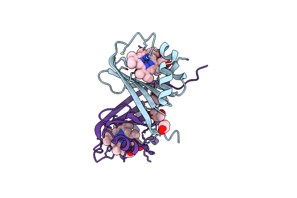
Crystal Structure Of Isdg From Bacillus Cereus
Organism: Bacillus cereus atcc 14579
Method: X-RAY DIFFRACTION Resolution:1.90 Å Release Date: 2023-07-12 Classification: OXIDOREDUCTASE Ligands: PDO |
Organism: Bacillus cereus atcc 14579
Method: X-RAY DIFFRACTION
Release Date: 2023-07-12
Ligands: PDO
 |
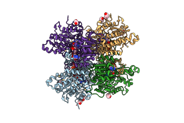
Crystal Structure Of Isdg From Bacillus Cereus In Complex With Heme
Organism: Bacillus cereus atcc 14579
Method: X-RAY DIFFRACTION Resolution:2.00 Å Release Date: 2023-07-12 Classification: OXIDOREDUCTASE Ligands: HEM, PGO, BU1, PDO |
Organism: Bacillus cereus atcc 14579
Method: X-RAY DIFFRACTION
Release Date: 2023-07-12
Ligands: HEM, PGO, BU1, PDO
 |
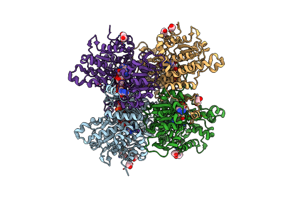
Crystal Structure Of Pseudomonas Aeruginosa S-Adenosyl-L-Homocysteine Hydrolase Inhibited By Cd2+ Ions
Organism: Pseudomonas aeruginosa pao1
Method: X-RAY DIFFRACTION Resolution:1.87 Å Release Date: 2023-04-19 Classification: HYDROLASE Ligands: NAD, ADN, BU1, PDO, K, CD, HEZ, CL |
Organism: Pseudomonas aeruginosa pao1
Method: X-RAY DIFFRACTION
Release Date: 2023-04-19
Ligands: NAD, ADN, BU1, PDO, K, CD, HEZ, CL
 |
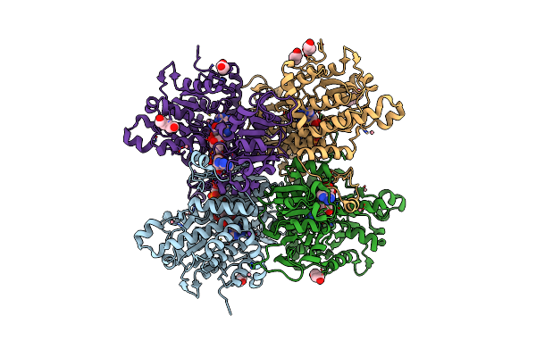
Crystal Structure Of Pseudomonas Aeruginosa S-Adenosyl-L-Homocysteine Hydrolase Inhibited By Hg2+ Ions
Organism: Pseudomonas aeruginosa pao1
Method: X-RAY DIFFRACTION Resolution:1.56 Å Release Date: 2023-04-19 Classification: HYDROLASE Ligands: NAD, ADN, BU1, PDO, K, HG, CL |
Organism: Pseudomonas aeruginosa pao1
Method: X-RAY DIFFRACTION
Release Date: 2023-04-19
Ligands: NAD, ADN, BU1, PDO, K, HG, CL
 |
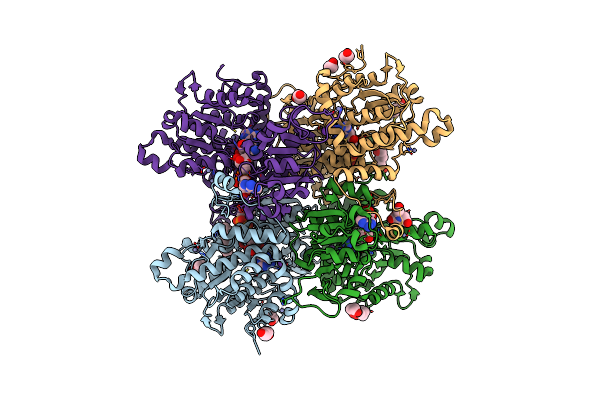
Crystal Structure Of Pseudomonas Aeruginosa S-Adenosyl-L-Homocysteine Hydrolase Inhibited By Co2+ Ions.
Organism: Pseudomonas aeruginosa pao1
Method: X-RAY DIFFRACTION Resolution:2.16 Å Release Date: 2023-04-19 Classification: HYDROLASE Ligands: NAD, ADN, PDO, K, CO, BU1, HEZ, CL |
Organism: Pseudomonas aeruginosa pao1
Method: X-RAY DIFFRACTION
Release Date: 2023-04-19
Ligands: NAD, ADN, PDO, K, CO, BU1, HEZ, CL
 |
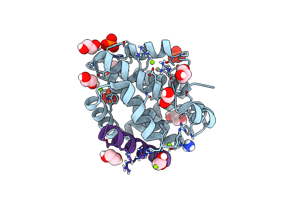
Crystal Structure Of Pseudomonas Aeruginosa S-Adenosyl-L-Homocysteine Hydrolase Inhibited By Zn2+ Ions
Organism: Pseudomonas aeruginosa pao1
Method: X-RAY DIFFRACTION Resolution:1.90 Å Release Date: 2023-04-19 Classification: HYDROLASE Ligands: NAD, ADN, BU1, PG4, K, ZN, BCN, HEZ, PDO, CL |
Organism: Pseudomonas aeruginosa pao1
Method: X-RAY DIFFRACTION
Release Date: 2023-04-19
Ligands: NAD, ADN, BU1, PG4, K, ZN, BCN, HEZ, PDO, CL
 |
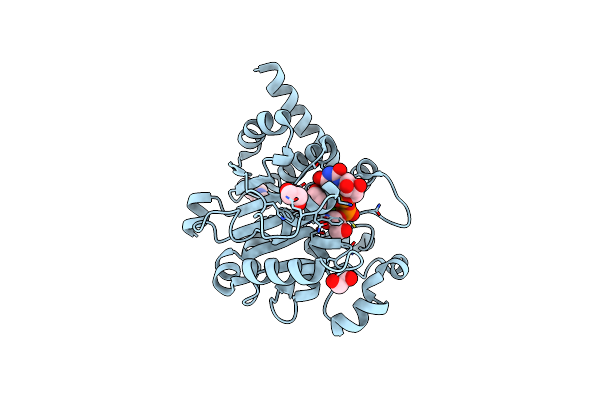
Epstein-Barr Virus Encoded Apoptosis Regulator Bhrf1 In Complex With Puma Bh3
Organism: Epstein-barr virus (strain b95-8), Homo sapiens
Method: X-RAY DIFFRACTION Resolution:2.00 Å Release Date: 2022-08-10 Classification: APOPTOSIS Ligands: PO4, EDO, NH4, PDO, MG, CL |
Organism: Epstein-barr virus (strain b95-8), Homo sapiens
Method: X-RAY DIFFRACTION
Release Date: 2022-08-10
Ligands: PO4, EDO, NH4, PDO, MG, CL
 |
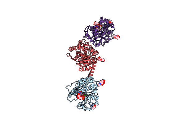
Catalytic Domain Of Udp-Glucose Glycoprotein Glucosyltransferase From Chaetomium Thermophilum In Complex With Udp
Organism: Chaetomium thermophilum
Method: X-RAY DIFFRACTION Resolution:1.82 Å Release Date: 2022-06-22 Classification: TRANSFERASE Ligands: UDP, NAG, CA, PDO, EDO |
Organism: Chaetomium thermophilum
Method: X-RAY DIFFRACTION
Release Date: 2022-06-22
Ligands: UDP, NAG, CA, PDO, EDO
 |
Catalytic Domain Of Udp-Glucose Glycoprotein Glucosyltransferase From Chaetomium Thermophilum In Complex With Udp-2-Deoxy-2-Fluoro-D-Glucose
Organism: Chaetomium thermophilum
Method: X-RAY DIFFRACTION Resolution:2.05 Å Release Date: 2022-06-22 Classification: TRANSFERASE Ligands: NAG, U2F, CA, PDO |
Organism: Chaetomium thermophilum
Method: X-RAY DIFFRACTION
Release Date: 2022-06-22
Ligands: NAG, U2F, CA, PDO