

Search Count: 65
 |
Crystal Structure Of An 8-Repeat Consensus Tpr Superhelix With Lead
Organism: Synthetic construct
Method: X-RAY DIFFRACTION Resolution:1.68 Å Release Date: 2024-03-13 Classification: DE NOVO PROTEIN Ligands: ACT, MPD, CL, PB |
Organism: Synthetic construct
Method: X-RAY DIFFRACTION
Release Date: 2024-03-13
Ligands: ACT, MPD, CL, PB
 |
Porous Framework Formed By Assembly Of A Bipyridyl-Conjugated Helical Peptide
Organism: Synthetic construct
Method: X-RAY DIFFRACTION Resolution:1.00 Å Release Date: 2023-11-15 Classification: DE NOVO PROTEIN Ligands: PB |
Organism: Synthetic construct
Method: X-RAY DIFFRACTION
Release Date: 2023-11-15
Ligands: PB
 |
Porous Framework Formed By Assembly Of A Bipyridyl-Conjugated Helical Peptide
Organism: Synthetic construct
Method: X-RAY DIFFRACTION Resolution:1.04 Å Release Date: 2023-11-15 Classification: DE NOVO PROTEIN Ligands: PB, ACT |
Organism: Synthetic construct
Method: X-RAY DIFFRACTION
Release Date: 2023-11-15
Ligands: PB, ACT
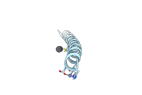 |
Crystal Structure Of A De Novo Three-Stranded Coiled Coil Peptide Containing Trigonal Pyrmidal Pb(Ii) Complexes In The Dual Tris-Thiolate Site
Organism: Synthetic construct
Method: X-RAY DIFFRACTION Resolution:1.29 Å Release Date: 2022-06-01 Classification: BIOSYNTHETIC PROTEIN Ligands: ZN, PB, CL |
Organism: Synthetic construct
Method: X-RAY DIFFRACTION
Release Date: 2022-06-01
Ligands: ZN, PB, CL
 |
The Tba-Pb2+ Complex In P41212 Space Group
Organism: Synthetic construct
Method: X-RAY DIFFRACTION Resolution:1.40 Å Release Date: 2021-09-22 Classification: DNA Ligands: PB |
Organism: Synthetic construct
Method: X-RAY DIFFRACTION
Release Date: 2021-09-22
Ligands: PB
 |
The Tba-Pb2+ Complex In P41212 Space Group
Organism: Synthetic construct
Method: X-RAY DIFFRACTION Resolution:1.71 Å Release Date: 2021-09-22 Classification: DNA Ligands: PB |
Organism: Synthetic construct
Method: X-RAY DIFFRACTION
Release Date: 2021-09-22
Ligands: PB
 |
The Pb2+ Complexed Structure Of Tba G8C Mutant
Organism: Synthetic construct
Method: X-RAY DIFFRACTION Resolution:2.12 Å Release Date: 2021-09-22 Classification: DNA Ligands: PB |
Organism: Synthetic construct
Method: X-RAY DIFFRACTION
Release Date: 2021-09-22
Ligands: PB
 |
Mouse Pkc C1B And C2 Domains
Organism: Mus musculus
Method: X-RAY DIFFRACTION Resolution:1.20 Å Release Date: 2020-12-09 Classification: SIGNALING PROTEIN Ligands: PB, SO4, CIT, ZN |
Organism: Mus musculus
Method: X-RAY DIFFRACTION
Release Date: 2020-12-09
Ligands: PB, SO4, CIT, ZN
 |
Crystal Structure Of Tris-Thiolate Pb(Ii) Complex With Adjacent Water In A De Novo Three Stranded Coiled Coil Peptide
Organism: Synthetic construct
Method: X-RAY DIFFRACTION Resolution:1.50 Å Release Date: 2020-03-04 Classification: DE NOVO PROTEIN Ligands: PB, ZN |
Organism: Synthetic construct
Method: X-RAY DIFFRACTION
Release Date: 2020-03-04
Ligands: PB, ZN
 |
Crystal Structure Of A Trigonal Pyramidal Pb(Ii)S3 Complex In A Three-Stranded Coiled Coil Peptide
Organism: Synthetic construct
Method: X-RAY DIFFRACTION Resolution:1.77 Å Release Date: 2020-02-26 Classification: DE NOVO PROTEIN Ligands: ZN, PB |
Organism: Synthetic construct
Method: X-RAY DIFFRACTION
Release Date: 2020-02-26
Ligands: ZN, PB
 |
Crystal Structure Of A Novel Dna Quadruplex
Organism: Synthetic construct
Method: X-RAY DIFFRACTION Resolution:1.45 Å Release Date: 2019-03-06 Classification: DNA Ligands: LI, NA, PB, NH4 |
Organism: Synthetic construct
Method: X-RAY DIFFRACTION
Release Date: 2019-03-06
Ligands: LI, NA, PB, NH4
 |
Synaptotagmin 1 C2A Domain, Lead-Bound
Organism: Mus musculus
Method: X-RAY DIFFRACTION Resolution:1.38 Å Release Date: 2018-04-11 Classification: METAL BINDING PROTEIN Ligands: PB, SO4 |
Organism: Mus musculus
Method: X-RAY DIFFRACTION
Release Date: 2018-04-11
Ligands: PB, SO4
 |
Synaptotagmin 1 C2B Domain, Lead-Bound (Low Occupancy)
Organism: Mus musculus
Method: X-RAY DIFFRACTION Resolution:1.41 Å Release Date: 2018-04-11 Classification: METAL BINDING PROTEIN Ligands: PB |
Organism: Mus musculus
Method: X-RAY DIFFRACTION
Release Date: 2018-04-11
Ligands: PB
 |
Synaptotagmin 1 C2B Domain, Lead-Bound (High Occupancy)
Organism: Mus musculus
Method: X-RAY DIFFRACTION Resolution:1.82 Å Release Date: 2018-04-11 Classification: METAL BINDING PROTEIN Ligands: PB |
Organism: Mus musculus
Method: X-RAY DIFFRACTION
Release Date: 2018-04-11
Ligands: PB
 |
Crystal Structure Of Asfvpolx In Complex With Dna Enzyme And Pb.
Organism: African swine fever virus (strain badajoz 1971 vero-adapted), Synthetic construct
Method: X-RAY DIFFRACTION Resolution:2.55 Å Release Date: 2018-01-24 Classification: TRANSFERASE/DNA Ligands: PO4, PB |
Organism: African swine fever virus (strain badajoz 1971 vero-adapted), Synthetic construct
Method: X-RAY DIFFRACTION
Release Date: 2018-01-24
Ligands: PO4, PB
 |
Crystal Structure Of The Transcription Regulator Pbrr691 From Ralstonia Metallidurans Ch34 In Complex With Lead(Ii)
Organism: Cupriavidus metallidurans ch34
Method: X-RAY DIFFRACTION Resolution:2.01 Å Release Date: 2016-12-28 Classification: TRANSCRIPTION Ligands: PB |
Organism: Cupriavidus metallidurans ch34
Method: X-RAY DIFFRACTION
Release Date: 2016-12-28
Ligands: PB
 |
Haloferax Volcanii Malate Synthase Lead(Ii) Complex
Organism: Haloferax volcanii (strain atcc 29605 / dsm 3757 / jcm 8879 / nbrc 14742 / ncimb 2012 / vkm b-1768 / ds2)
Method: X-RAY DIFFRACTION Resolution:2.10 Å Release Date: 2016-11-09 Classification: TRANSFERASE Ligands: ACT, GLV, PB, K, CL, NA |
Organism: Haloferax volcanii (strain atcc 29605 / dsm 3757 / jcm 8879 / nbrc 14742 / ncimb 2012 / vkm b-1768 / ds2)
Method: X-RAY DIFFRACTION
Release Date: 2016-11-09
Ligands: ACT, GLV, PB, K, CL, NA
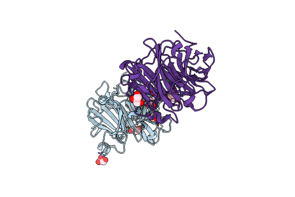 |
Catalytic Domain Of Coprinopsis Cinerea Gh62 Alpha-L-Arabinofuranosidase Complexed With Pb
Organism: Coprinopsis cinerea (strain okayama-7 / 130 / atcc mya-4618 / fgsc 9003)
Method: X-RAY DIFFRACTION Resolution:1.48 Å Release Date: 2016-09-07 Classification: HYDROLASE Ligands: PB, CA, GOL |
Organism: Coprinopsis cinerea (strain okayama-7 / 130 / atcc mya-4618 / fgsc 9003)
Method: X-RAY DIFFRACTION
Release Date: 2016-09-07
Ligands: PB, CA, GOL
 |
Crystal Structure Of A Tris-Thiolate Pb(Ii) Complex In A De Novo Three-Stranded Coiled Coil Peptide
Organism: Synthetic construct
Method: X-RAY DIFFRACTION Resolution:2.13 Å Release Date: 2016-08-31 Classification: DE NOVO PROTEIN Ligands: ZN, PB, CL |
Organism: Synthetic construct
Method: X-RAY DIFFRACTION
Release Date: 2016-08-31
Ligands: ZN, PB, CL
 |
Elongation Factor Tu:Ts Complex With A Bound Phosphate
Organism: Escherichia coli
Method: X-RAY DIFFRACTION Resolution:1.95 Å Release Date: 2015-05-06 Classification: TRANSLATION Ligands: PO4, GOL, PB, TRS |
Organism: Escherichia coli
Method: X-RAY DIFFRACTION
Release Date: 2015-05-06
Ligands: PO4, GOL, PB, TRS