
Search Count: 1,471
 |
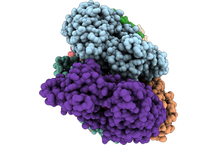
Crystal Structure Of A Human Leukocyte Antigen-A (Hla-G) Blocking Antibody
Organism: Homo sapiens, Oryctolagus cuniculus
Method: X-RAY DIFFRACTION Resolution:3.45 Å Release Date: 2026-07-22 Classification: IMMUNE SYSTEM |
Organism: Homo sapiens, Oryctolagus cuniculus
Method: X-RAY DIFFRACTION
Release Date: 2026-07-22
 |
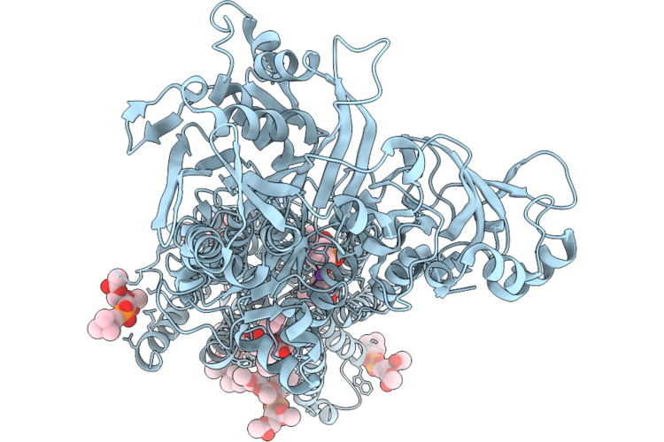
Crystal Structure Of Sr Ca2+-Atpase In E2(Tg) With Bound Ahq
Organism: Oryctolagus cuniculus
Method: X-RAY DIFFRACTION Resolution:2.12 Å Release Date: 2026-07-15 Classification: MEMBRANE PROTEIN |
Organism: Oryctolagus cuniculus
Method: X-RAY DIFFRACTION
Release Date: 2026-07-15
 |
Crystal Structure Of Sr Ca2+-Atpase In E2(Tg) With Bound B1Hq
Organism: Oryctolagus cuniculus
Method: X-RAY DIFFRACTION Resolution:2.10 Å Release Date: 2026-07-15 Classification: MEMBRANE PROTEIN |
Organism: Oryctolagus cuniculus
Method: X-RAY DIFFRACTION
Release Date: 2026-07-15
 |
Crystal Structure Of Sr Ca2+-Atpase In E2(Tg) With Bound Ba
Organism: Oryctolagus cuniculus
Method: X-RAY DIFFRACTION Resolution:2.50 Å Release Date: 2026-07-15 Classification: MEMBRANE PROTEIN |
Organism: Oryctolagus cuniculus
Method: X-RAY DIFFRACTION
Release Date: 2026-07-15
 |
Crystal Structure Of Sr Ca2+-Atpase In E2(Tg) With Bound Bc
Organism: Oryctolagus cuniculus
Method: X-RAY DIFFRACTION Resolution:2.60 Å Release Date: 2026-07-15 Classification: MEMBRANE PROTEIN |
Organism: Oryctolagus cuniculus
Method: X-RAY DIFFRACTION
Release Date: 2026-07-15
 |
Crystal Structure Of Sr Ca2+-Atpase In E2(Tg) With Bound Bmp
Organism: Oryctolagus cuniculus
Method: X-RAY DIFFRACTION Resolution:2.10 Å Release Date: 2026-07-15 Classification: MEMBRANE PROTEIN |
Organism: Oryctolagus cuniculus
Method: X-RAY DIFFRACTION
Release Date: 2026-07-15
 |
Crystal Structure Of Sr Ca2+-Atpase In E2(Tg) With Bound Ipp
Organism: Oryctolagus cuniculus
Method: X-RAY DIFFRACTION Resolution:2.00 Å Release Date: 2026-07-15 Classification: MEMBRANE PROTEIN |
Organism: Oryctolagus cuniculus
Method: X-RAY DIFFRACTION
Release Date: 2026-07-15
 |
Crystal Structure Of Sr Ca2+-Atpase In E2(Tg) With Bound Mpq
Organism: Oryctolagus cuniculus
Method: X-RAY DIFFRACTION Resolution:2.20 Å Release Date: 2026-07-15 Classification: MEMBRANE PROTEIN |
Organism: Oryctolagus cuniculus
Method: X-RAY DIFFRACTION
Release Date: 2026-07-15
 |
Crystal Structure Of Sr Ca2+-Atpase In E2(Tg) With Bound Phq
Organism: Oryctolagus cuniculus
Method: X-RAY DIFFRACTION Resolution:2.20 Å Release Date: 2026-07-15 Classification: MEMBRANE PROTEIN |
Organism: Oryctolagus cuniculus
Method: X-RAY DIFFRACTION
Release Date: 2026-07-15
 |
Gelsolin Domain G2 Transitionally Bound To F-Actin
Organism: Homo sapiens, Oryctolagus cuniculus
Method: ELECTRON MICROSCOPY Resolution:2.86 Å Release Date: 2026-07-08 Classification: STRUCTURAL PROTEIN Ligands: ADP, MG, CA |
Organism: Homo sapiens, Oryctolagus cuniculus
Method: ELECTRON MICROSCOPY
Release Date: 2026-07-08
Ligands: ADP, MG, CA
 |
Gelsolin Domain G2G3 Transitionally Bound To F-Actin
Organism: Homo sapiens, Oryctolagus cuniculus
Method: ELECTRON MICROSCOPY Resolution:3.03 Å Release Date: 2026-07-08 Classification: STRUCTURAL PROTEIN Ligands: ADP, MG, CA |
Organism: Homo sapiens, Oryctolagus cuniculus
Method: ELECTRON MICROSCOPY
Release Date: 2026-07-08
Ligands: ADP, MG, CA
 |
Gelsolin Domain G2G3 Fully Bound To F-Actin
Organism: Homo sapiens, Oryctolagus cuniculus
Method: ELECTRON MICROSCOPY Resolution:2.76 Å Release Date: 2026-07-08 Classification: STRUCTURAL PROTEIN Ligands: ADP, MG, CA |
Organism: Homo sapiens, Oryctolagus cuniculus
Method: ELECTRON MICROSCOPY
Release Date: 2026-07-08
Ligands: ADP, MG, CA
 |
Two Gelsolin Domains G2G3 Bound To F-Actin
Organism: Homo sapiens, Oryctolagus cuniculus
Method: ELECTRON MICROSCOPY Resolution:3.44 Å Release Date: 2026-07-08 Classification: STRUCTURAL PROTEIN Ligands: ADP, MG, CA |
Organism: Homo sapiens, Oryctolagus cuniculus
Method: ELECTRON MICROSCOPY
Release Date: 2026-07-08
Ligands: ADP, MG, CA
 |
Cryo-Em Structure Of F-Actin In The Adp State
Organism: Oryctolagus cuniculus
Method: ELECTRON MICROSCOPY Resolution:2.06 Å Release Date: 2026-06-24 Classification: STRUCTURAL PROTEIN Ligands: ADP, MG |
Organism: Oryctolagus cuniculus
Method: ELECTRON MICROSCOPY
Release Date: 2026-06-24
Ligands: ADP, MG
 |
One Lmod2 At The Pointed End Of F-Actin
Organism: Homo sapiens, Oryctolagus cuniculus
Method: ELECTRON MICROSCOPY Release Date: 2026-06-24 Classification: STRUCTURAL PROTEIN Ligands: ADP, MG |
Organism: Homo sapiens, Oryctolagus cuniculus
Method: ELECTRON MICROSCOPY
Release Date: 2026-06-24
Ligands: ADP, MG
 |
One Lmod2 And Incoming Actin At The Pointed End Of F-Actin
Organism: Homo sapiens, Oryctolagus cuniculus
Method: ELECTRON MICROSCOPY Resolution:4.06 Å Release Date: 2026-06-24 Classification: STRUCTURAL PROTEIN Ligands: ADP, MG, ATP |
Organism: Homo sapiens, Oryctolagus cuniculus
Method: ELECTRON MICROSCOPY
Release Date: 2026-06-24
Ligands: ADP, MG, ATP
 |
Two Lmod2S And Incoming Actin At The Pointed End Of F-Actin
Organism: Homo sapiens, Oryctolagus cuniculus
Method: ELECTRON MICROSCOPY Release Date: 2026-06-24 Classification: STRUCTURAL PROTEIN Ligands: ADP, MG, ATP |
Organism: Homo sapiens, Oryctolagus cuniculus
Method: ELECTRON MICROSCOPY
Release Date: 2026-06-24
Ligands: ADP, MG, ATP
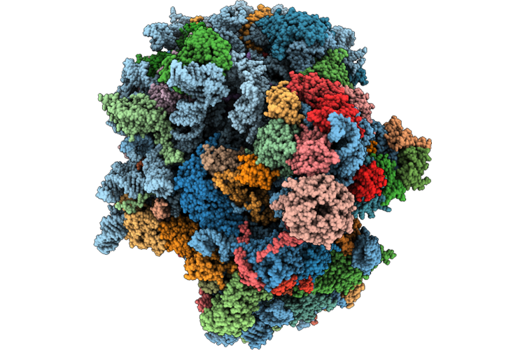 |
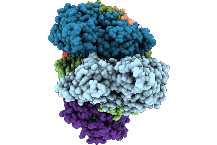
Nediv Ires (A Site) In Complex With Rabbit 80S Ribosome
Organism: Oryctolagus cuniculus
Method: ELECTRON MICROSCOPY Release Date: 2026-06-17 Classification: RIBOSOME Ligands: MG, ZN |
Organism: Oryctolagus cuniculus
Method: ELECTRON MICROSCOPY
Release Date: 2026-06-17
Ligands: MG, ZN
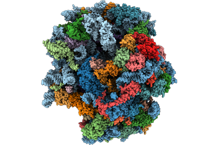 |
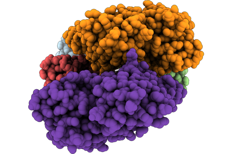
Nediv Ires (P Site) In Complex With Rabbit 80S Ribosome
Organism: Nedicistrovirus tfn-2012, Oryctolagus cuniculus
Method: ELECTRON MICROSCOPY Resolution:3.00 Å Release Date: 2026-06-17 Classification: RIBOSOME Ligands: MG, ZN |
Organism: Nedicistrovirus tfn-2012, Oryctolagus cuniculus
Method: ELECTRON MICROSCOPY
Release Date: 2026-06-17
Ligands: MG, ZN
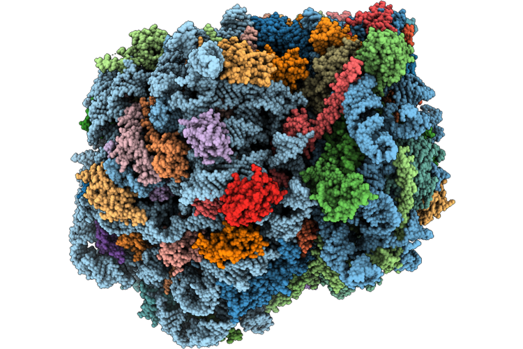 |
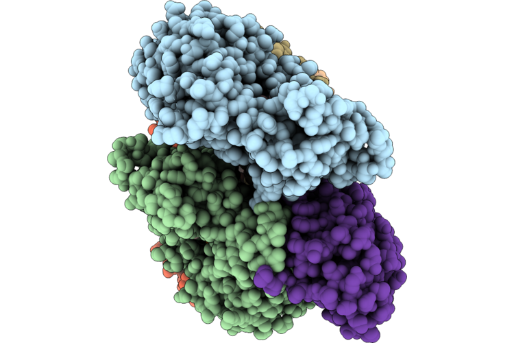
Rabbit Ribosomal 80S Elongation Complex With Eef2, Partial P Site Ala-Trna, E Site Ala-Trna On Nediv Orf
Organism: Oryctolagus cuniculus, Nedicistrovirus tfn-2012
Method: ELECTRON MICROSCOPY Release Date: 2026-06-17 Classification: RIBOSOME Ligands: MG, ZN |
Organism: Oryctolagus cuniculus, Nedicistrovirus tfn-2012
Method: ELECTRON MICROSCOPY
Release Date: 2026-06-17
Ligands: MG, ZN