
Search Count: 43
All
Selected
 |
Organism: Homo sapiens
Method: X-RAY DIFFRACTION Resolution:1.26 Å Release Date: 2023-11-22 Classification: PEPTIDE BINDING PROTEIN Ligands: ZJ9, EDO, DMS, PO4 |
 |
Organism: Homo sapiens
Method: X-RAY DIFFRACTION Resolution:1.70 Å Release Date: 2023-11-22 Classification: PEPTIDE BINDING PROTEIN Ligands: EDO, DMS, ZJF, SO4 |
 |
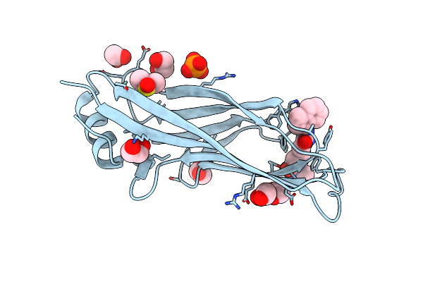
Crystal Structure Of Human Yeats4 In Complex With Pfizer Small Molecule Compound 3B
Organism: Homo sapiens
Method: X-RAY DIFFRACTION Resolution:2.58 Å Release Date: 2023-01-11 Classification: PROTEIN BINDING Ligands: SJI |
 |
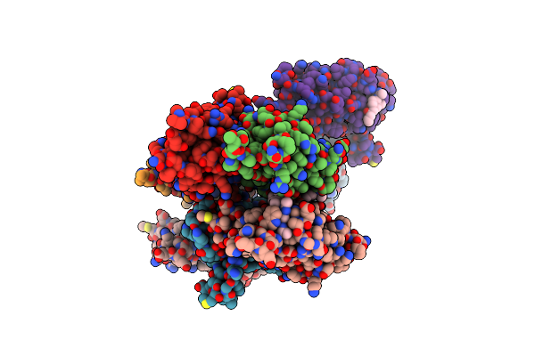
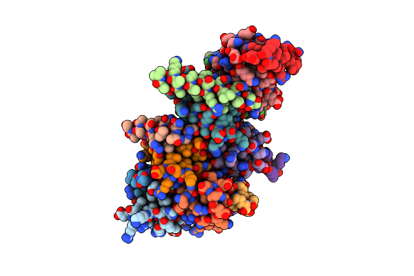
Crystal Structure Of A Trapped Pab-Agog/Double-Standed Dna Covalent Intermediate (Dna Containing Adenine Opposite To Lesion)
Organism: Pyrococcus abyssi, Synthetic construct
Method: X-RAY DIFFRACTION Resolution:1.33 Å Release Date: 2022-08-10 Classification: HYDROLASE Ligands: MPD |
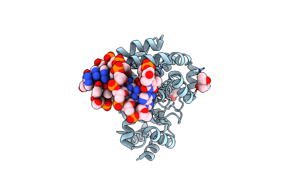 |
Crystal Structure Of Pyrococcus Abyssi 8-Oxoguanine Dna Glycosylase (Pabagog) In Complex With Dsdna Containing Cytosine Opposite To 8-Oxog
Organism: Pyrococcus abyssi (strain ge5 / orsay), Synthetic construct
Method: X-RAY DIFFRACTION Resolution:1.25 Å Release Date: 2022-08-03 Classification: HYDROLASE Ligands: MPD |
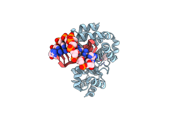 |
Crystal Structure Of A Trapped Pab-Agog/Double-Standed Dna Covalent Intermediate (Dna Containing Cytosine Opposite To Lesion)
Organism: Pyrococcus abyssi (strain ge5 / orsay), Synthetic construct
Method: X-RAY DIFFRACTION Resolution:1.70 Å Release Date: 2022-07-13 Classification: HYDROLASE Ligands: MPD |
 |
Crystal Structure Of A Trapped Pab-Agog/Double-Standed Dna Covalent Intermediate (Dna Containing Thymine Opposite To Lesion)
Organism: Pyrococcus abyssi (strain ge5 / orsay), Synthetic construct
Method: X-RAY DIFFRACTION Resolution:1.12 Å Release Date: 2022-07-13 Classification: HYDROLASE Ligands: MPD, NA |
 |
Crystal Structure Of Tga-Agog, An 8-Oxoguanine Dna Glycosylase From Thermococcus Gammatolerans
Organism: Thermococcus gammatolerans (strain dsm 15229 / jcm 11827 / ej3)
Method: X-RAY DIFFRACTION Resolution:1.49 Å Release Date: 2022-06-22 Classification: HYDROLASE Ligands: CL, GOL |
 |
Crystal Structure Of A Trapped Pab-Agog/Single-Standed Dna Covalent Intermediate
Organism: Pyrococcus abyssi (strain ge5 / orsay), Synthetic construct
Method: X-RAY DIFFRACTION Resolution:2.04 Å Release Date: 2022-06-22 Classification: HYDROLASE Ligands: PO4, K |
 |
Crystal Structure Of Pab-Agog, An 8-Oxoguanine Dna Glycosylase From Pyrococcus Abyssi
Organism: Pyrococcus abyssi (strain ge5 / orsay)
Method: X-RAY DIFFRACTION Resolution:1.10 Å Release Date: 2022-06-01 Classification: HYDROLASE Ligands: EDO, CIT, CL, NA |
 |
Organism: Pyrococcus abyssi (strain ge5 / orsay)
Method: X-RAY DIFFRACTION Resolution:1.65 Å Release Date: 2022-06-01 Classification: HYDROLASE Ligands: 8HG, EDO |
 |
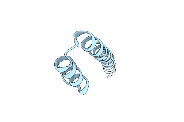
C Terminal Domain Of Nipah Virus Phosphoprotein Fused To The Ntail Alpha More Of The Nucleoprotein.
Organism: Nipah virus, Nipah henipavirus
Method: X-RAY DIFFRACTION Resolution:2.79 Å Release Date: 2022-04-20 Classification: VIRAL PROTEIN |
 |
Organism: Nipah virus
Method: X-RAY DIFFRACTION Resolution:2.10 Å Release Date: 2022-04-20 Classification: VIRAL PROTEIN |
 |
Organism: Toxoplasma gondii (strain atcc 50611 / me49)
Method: X-RAY DIFFRACTION Resolution:1.23 Å Release Date: 2021-07-21 Classification: RNA BINDING PROTEIN Ligands: IPA |
 |
Organism: Toxoplasma gondii (strain atcc 50611 / me49)
Method: X-RAY DIFFRACTION Resolution:1.35 Å Release Date: 2021-07-21 Classification: RNA BINDING PROTEIN Ligands: PG4, OYK |
 |
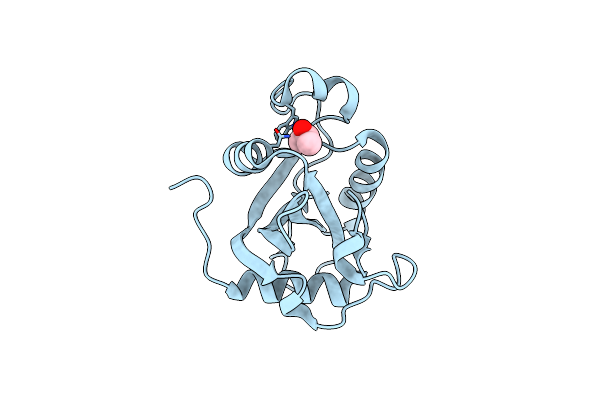
Crystal Structure Of The Toxoplasma Cpsf4 Yth-Domain In Complex With A 7 Mer M6A-Modified Rna
Organism: Toxoplasma gondii (strain atcc 50611 / me49), Synthetic construct
Method: X-RAY DIFFRACTION Resolution:1.38 Å Release Date: 2021-07-21 Classification: RNA BINDING PROTEIN Ligands: TOE, 6MD |
 |
Organism: Homo sapiens
Method: X-RAY DIFFRACTION Resolution:1.60 Å Release Date: 2019-07-17 Classification: CELL CYCLE Ligands: MPD, DMS, JC5, GLY, PO4, GOL |
 |
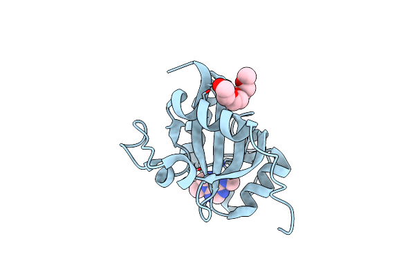
Crystal Structure Of Spindlin1 In Complex With The Methyltransferase Inhibitor A366
Organism: Homo sapiens
Method: X-RAY DIFFRACTION Resolution:1.52 Å Release Date: 2018-12-26 Classification: GENE REGULATION Ligands: 2OD, EDO, DMS, MPD, GOL, NA |
 |
Organism: Homo sapiens
Method: X-RAY DIFFRACTION Resolution:1.76 Å Release Date: 2018-12-05 Classification: CELL CYCLE Ligands: H7T, DMS, GLY, PO4 |
 |
Organism: Homo sapiens
Method: X-RAY DIFFRACTION Resolution:1.58 Å Release Date: 2018-12-05 Classification: CELL CYCLE Ligands: MPD, MRD, DMS, H7Q, CL |