

Search Count: 44
 |
Crystal Structure Of Mycobacterium Tuberculosis Rv2514C-Rv2515C Toxin Antitoxin System.
Organism: Mycobacterium tuberculosis h37rv
Method: X-RAY DIFFRACTION Resolution:3.20 Å Release Date: 2026-07-01 Classification: TOXIN Ligands: ZN, MG |
Organism: Mycobacterium tuberculosis h37rv
Method: X-RAY DIFFRACTION
Release Date: 2026-07-01
Ligands: ZN, MG
 |
Crystal Structure Of Rv2514C From Mycobacterium Tuberculosis
Organism: Mycobacterium tuberculosis
Method: X-RAY DIFFRACTION Resolution:2.60 Å Release Date: 2026-07-01 Classification: TOXIN Ligands: SO4, GOL |
Organism: Mycobacterium tuberculosis
Method: X-RAY DIFFRACTION
Release Date: 2026-07-01
Ligands: SO4, GOL
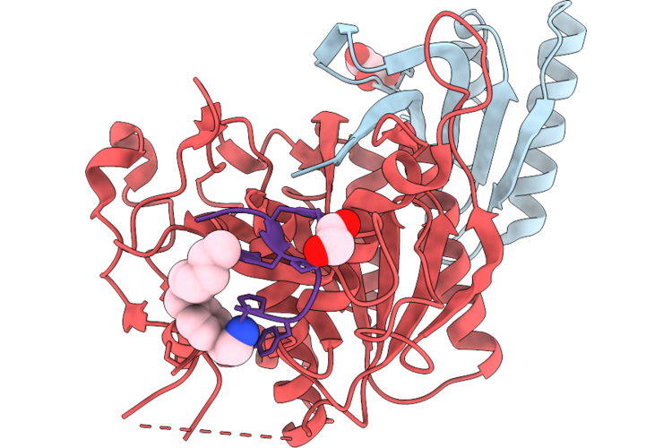 |
Pcsk9 In Complex With Cyclic Peptide 21
Organism: Homo sapiens, Synthetic construct
Method: X-RAY DIFFRACTION Resolution:1.83 Å Release Date: 2026-06-03 Classification: HYDROLASE Ligands: GOL, A1C8P, GOA |
Organism: Homo sapiens, Synthetic construct
Method: X-RAY DIFFRACTION
Release Date: 2026-06-03
Ligands: GOL, A1C8P, GOA
 |
Crystal Structure Of Vaborbactam In Complex With Sme-1 Class A Carbapenemase
Organism: Serratia marcescens
Method: X-RAY DIFFRACTION Resolution:2.00 Å Release Date: 2025-11-19 Classification: HYDROLASE Ligands: 4D6, PG4, EDO |
Organism: Serratia marcescens
Method: X-RAY DIFFRACTION
Release Date: 2025-11-19
Ligands: 4D6, PG4, EDO
 |
Crystal Structure Of Taniborbactam In Complex With Sme-1 Class A Carbapenemase
Organism: Serratia marcescens
Method: X-RAY DIFFRACTION Resolution:1.93 Å Release Date: 2025-11-19 Classification: HYDROLASE Ligands: KJK, PG4 |
Organism: Serratia marcescens
Method: X-RAY DIFFRACTION
Release Date: 2025-11-19
Ligands: KJK, PG4
 |
Crystal Structure Of Ledaborbactam In Complex With Sme-1 Class A Carbapenemase
Organism: Serratia marcescens
Method: X-RAY DIFFRACTION Resolution:2.20 Å Release Date: 2025-11-19 Classification: HYDROLASE Ligands: A1MBD, CL |
Organism: Serratia marcescens
Method: X-RAY DIFFRACTION
Release Date: 2025-11-19
Ligands: A1MBD, CL
 |
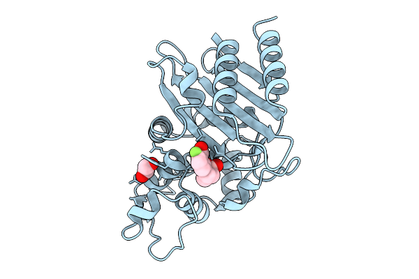
Crystal Structure Of 2R, 3S-Xeruborbactam With Sme-1
Organism: Serratia marcescens
Method: X-RAY DIFFRACTION Resolution:2.20 Å Release Date: 2025-10-08 Classification: HYDROLASE Ligands: A1MAF, PEG, PG4, CL, NA |
Organism: Serratia marcescens
Method: X-RAY DIFFRACTION
Release Date: 2025-10-08
Ligands: A1MAF, PEG, PG4, CL, NA
 |
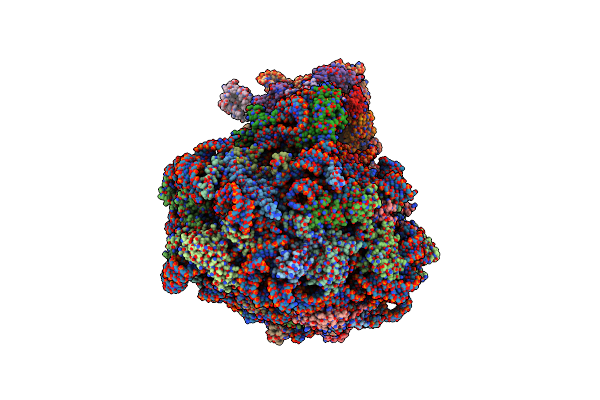
Crystal Structure Of Xeruborbactam (Qpx7728) In Complex With Sme-1 Class A Carbapenemase
Organism: Serratia marcescens
Method: X-RAY DIFFRACTION Resolution:2.40 Å Release Date: 2025-10-08 Classification: HYDROLASE Ligands: EDO, RM9, CL |
Organism: Serratia marcescens
Method: X-RAY DIFFRACTION
Release Date: 2025-10-08
Ligands: EDO, RM9, CL
 |
Single Particle Cryoem Structure Of The Pf80S Ribosome In Non-Rotated Pre State (Nrt A-P-E)
Organism: Plasmodium falciparum 3d7
Method: ELECTRON MICROSCOPY Release Date: 2025-05-28 Classification: RIBOSOME |
Organism: Plasmodium falciparum 3d7
Method: ELECTRON MICROSCOPY
Release Date: 2025-05-28
 |
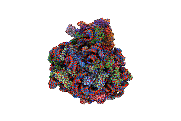
Single Particle Cryoem Structure Of The Pf80S Ribosome In The Post State (Nrt With P- And E-Site Trna)
Organism: Plasmodium falciparum 3d7
Method: ELECTRON MICROSCOPY Release Date: 2025-05-28 Classification: RIBOSOME |
Organism: Plasmodium falciparum 3d7
Method: ELECTRON MICROSCOPY
Release Date: 2025-05-28
 |
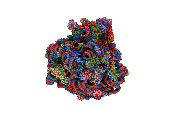
Single Particle Cryoem Structure Of The Pf80S Ribosome In The Unloaded State (Nrt With E-Site Trna)
Organism: Plasmodium falciparum 3d7
Method: ELECTRON MICROSCOPY Release Date: 2025-05-28 Classification: RIBOSOME |
Organism: Plasmodium falciparum 3d7
Method: ELECTRON MICROSCOPY
Release Date: 2025-05-28
 |
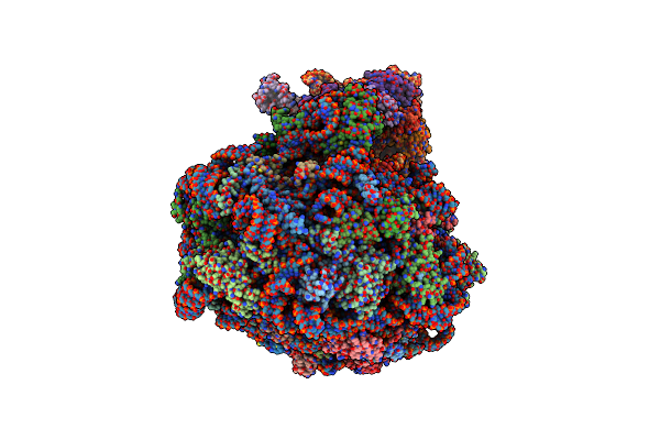
Single Particle Cryoem Structure Of The Pf80S Ribosome In The Rotated-2 Pre State (Rt State With P And E-Site Trna)
Organism: Plasmodium falciparum 3d7
Method: ELECTRON MICROSCOPY Release Date: 2025-05-28 Classification: RIBOSOME |
Organism: Plasmodium falciparum 3d7
Method: ELECTRON MICROSCOPY
Release Date: 2025-05-28
 |
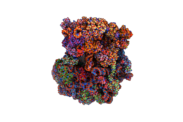
Single Particle Cryoem Structure Of The Pf80S Ribosome In Rotated State With E-Site Trna
Organism: Plasmodium falciparum 3d7
Method: ELECTRON MICROSCOPY Release Date: 2025-05-28 Classification: RIBOSOME |
Organism: Plasmodium falciparum 3d7
Method: ELECTRON MICROSCOPY
Release Date: 2025-05-28
 |
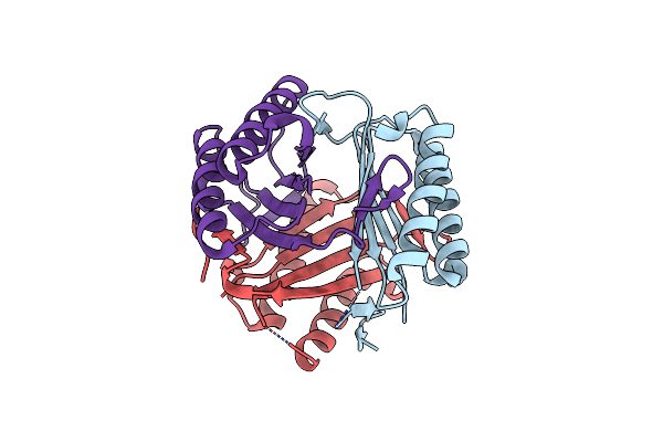
Subtomogram Averaged Consensus Structure Of The Malarial 80S Ribosome In Plasmodium Falciparum-Infected Human Erythrocytes
Organism: Plasmodium falciparum 3d7
Method: ELECTRON MICROSCOPY Release Date: 2024-08-14 Classification: RIBOSOME |
Organism: Plasmodium falciparum 3d7
Method: ELECTRON MICROSCOPY
Release Date: 2024-08-14
 |
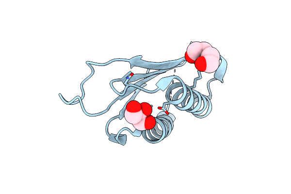
Crystal Structure Of Macrophage Migration Inhibitory Factor (Mif) From Trichomonas Vaginalis (I4122 Form)
Organism: Trichomonas vaginalis g3
Method: X-RAY DIFFRACTION Resolution:2.55 Å Release Date: 2024-03-20 Classification: CYTOKINE Ligands: CL |
Organism: Trichomonas vaginalis g3
Method: X-RAY DIFFRACTION
Release Date: 2024-03-20
Ligands: CL
 |
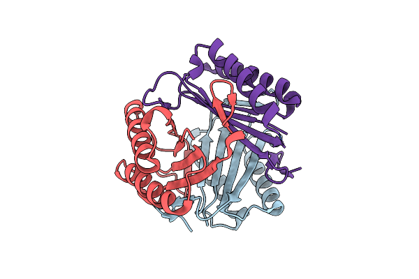
Crystal Structure Of Macrophage Migration Inhibitory Factor From Plasmodium Vivax
Organism: Plasmodium vivax sal-1
Method: X-RAY DIFFRACTION Resolution:1.80 Å Release Date: 2024-03-20 Classification: CYTOKINE Ligands: GOL, PEG |
Organism: Plasmodium vivax sal-1
Method: X-RAY DIFFRACTION
Release Date: 2024-03-20
Ligands: GOL, PEG
 |
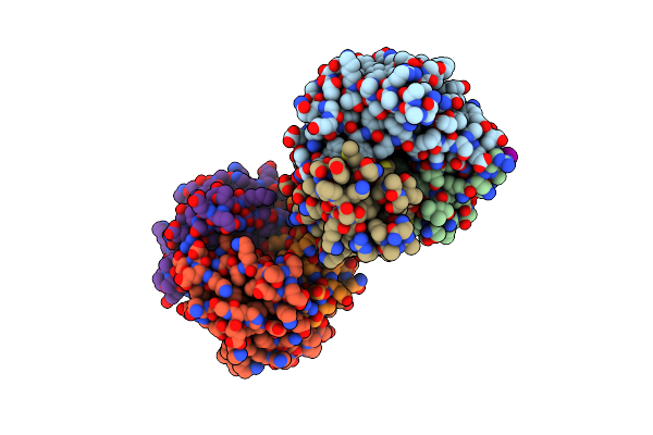
Crystal Structure Of Macrophage Migration Inhibitory Factor (Mif) From Trichomonas Vaginalis (Apo, P41212 Form)
Organism: Trichomonas vaginalis
Method: X-RAY DIFFRACTION Resolution:2.40 Å Release Date: 2023-12-06 Classification: CYTOKINE |
Organism: Trichomonas vaginalis
Method: X-RAY DIFFRACTION
Release Date: 2023-12-06
 |
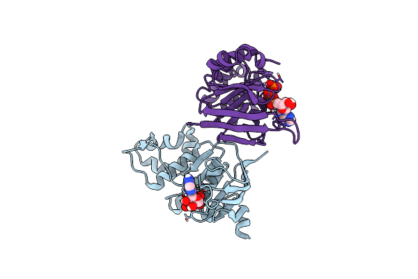
Crystal Structure Of Macrophage Migration Inhibitory Factor (Mif) From Trichomonas Vaginalis (I41 Form)
Organism: Trichomonas vaginalis g3
Method: X-RAY DIFFRACTION Resolution:1.90 Å Release Date: 2023-11-08 Classification: CYTOKINE Ligands: IOD, PYR |
Organism: Trichomonas vaginalis g3
Method: X-RAY DIFFRACTION
Release Date: 2023-11-08
Ligands: IOD, PYR
 |
Human Cftr First Nucleotide Binding Domain With Df508/V510D
Organism: Homo sapiens
Method: X-RAY DIFFRACTION Resolution:1.86 Å Release Date: 2021-04-07 Classification: HYDROLASE Ligands: ATP, MG |
Organism: Homo sapiens
Method: X-RAY DIFFRACTION
Release Date: 2021-04-07
Ligands: ATP, MG
 |
Pksg, A Hmg-Coa Synthase From Bacillus Subtilis
Organism: Bacillus subtilis (strain 168)
Method: X-RAY DIFFRACTION Resolution:2.47 Å Release Date: 2016-06-29 Classification: TRANSFERASE Ligands: PEG |
Organism: Bacillus subtilis (strain 168)
Method: X-RAY DIFFRACTION
Release Date: 2016-06-29
Ligands: PEG