

Search Count: 334
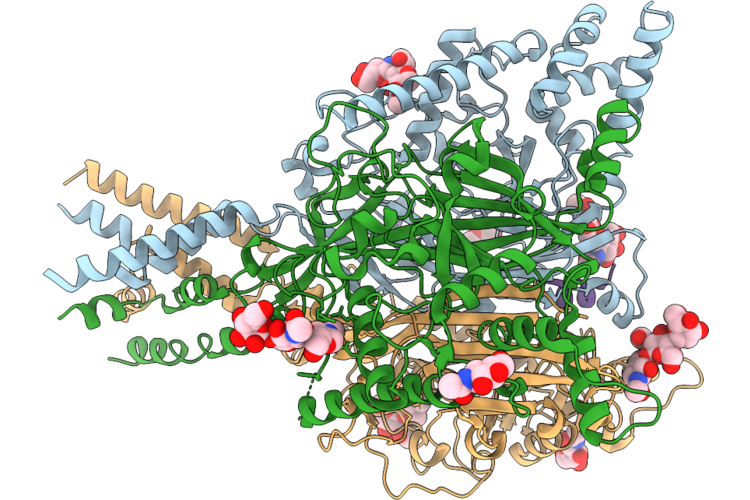 |
Heterotrimeric Enac Channel Complex Ppk1/Ppk26/Ppk26 With Vulnusin
Organism: Drosophila melanogaster
Method: ELECTRON MICROSCOPY Resolution:2.86 Å Release Date: 2026-08-19 Classification: NEUROPEPTIDE Ligands: NAG, NH4 |
Organism: Drosophila melanogaster
Method: ELECTRON MICROSCOPY
Release Date: 2026-08-19
Ligands: NAG, NH4
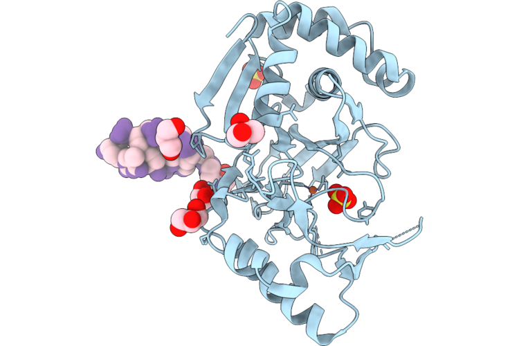 |
Glutarate L-2-Hydroxylase Q184C, K270A Mutant-5'-Mal-C2-Agct Dna Conjugate
Organism: Escherichia coli, Synthetic construct
Method: X-RAY DIFFRACTION Resolution:1.77 Å Release Date: 2026-07-29 Classification: METAL BINDING PROTEIN Ligands: NH4, GOL, FE2, SO4, A1BWE |
Organism: Escherichia coli, Synthetic construct
Method: X-RAY DIFFRACTION
Release Date: 2026-07-29
Ligands: NH4, GOL, FE2, SO4, A1BWE
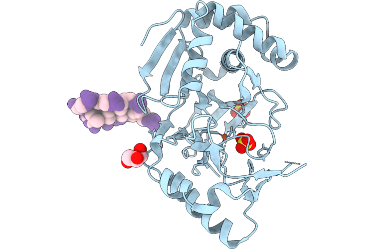 |
Glutarate L-2-Hydroxylase N182A, Q184C, K270A Mutant-5'-Mal-C2-Agct Dna Conjugate
Organism: Escherichia coli, Synthetic construct
Method: X-RAY DIFFRACTION Resolution:1.42 Å Release Date: 2026-07-29 Classification: METAL BINDING PROTEIN Ligands: NH4, GOL, FE2, SO4 |
Organism: Escherichia coli, Synthetic construct
Method: X-RAY DIFFRACTION
Release Date: 2026-07-29
Ligands: NH4, GOL, FE2, SO4
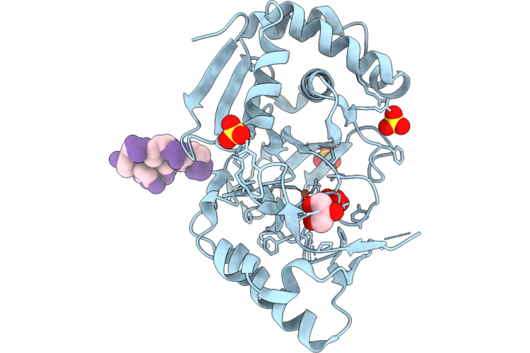 |
Glutarate L-2-Hydroxylase Q184C Mutant-5'-Mal-C2-Agct Dna Conjugate At 2.01 Angstrom Resolution
Organism: Escherichia coli, Synthetic construct
Method: X-RAY DIFFRACTION Resolution:2.01 Å Release Date: 2026-07-29 Classification: METAL BINDING PROTEIN Ligands: NH4, GOL, FE2, SO4 |
Organism: Escherichia coli, Synthetic construct
Method: X-RAY DIFFRACTION
Release Date: 2026-07-29
Ligands: NH4, GOL, FE2, SO4
 |
Crystal Structure Of N-Methylhydantoinase In Complex With 1-Methylimidazolidine-2,4-Dione
Organism: Glutamicibacter protophormiae
Method: X-RAY DIFFRACTION Resolution:2.07 Å Release Date: 2026-04-15 Classification: HYDROLASE Ligands: CA, A1BC1, NH4, BTB |
Organism: Glutamicibacter protophormiae
Method: X-RAY DIFFRACTION
Release Date: 2026-04-15
Ligands: CA, A1BC1, NH4, BTB
 |
Crystal Structure Of N-Methylhydantoinase In Complex With 1-Methylimidazolidine-2,4-Dione, Iodide Soak
Organism: Glutamicibacter protophormiae
Method: X-RAY DIFFRACTION Resolution:2.62 Å Release Date: 2026-04-15 Classification: HYDROLASE Ligands: CA, A1BC1, NH4, IOD, BTB |
Organism: Glutamicibacter protophormiae
Method: X-RAY DIFFRACTION
Release Date: 2026-04-15
Ligands: CA, A1BC1, NH4, IOD, BTB
 |
Crystal Structure Of N-Methylhydantoinase In Complex With Mg-Adpnp
Organism: Glutamicibacter protophormiae
Method: X-RAY DIFFRACTION Resolution:3.13 Å Release Date: 2026-04-15 Classification: HYDROLASE Ligands: ANP, MG, NH4 |
Organism: Glutamicibacter protophormiae
Method: X-RAY DIFFRACTION
Release Date: 2026-04-15
Ligands: ANP, MG, NH4
 |
Crystal Structure Of N-Methylhydantoinase In Complex With 1-Methylimidazolidine-2,4-Dione, C2221 Form
Organism: Glutamicibacter protophormiae
Method: X-RAY DIFFRACTION Resolution:2.80 Å Release Date: 2026-04-15 Classification: HYDROLASE Ligands: CA, A1BC1, NH4, MES, SO4 |
Organism: Glutamicibacter protophormiae
Method: X-RAY DIFFRACTION
Release Date: 2026-04-15
Ligands: CA, A1BC1, NH4, MES, SO4
 |
Crystal Structure Of N-Methylhydantoinase In Complex With Zn2+ And Adpnp
Organism: Glutamicibacter protophormiae
Method: X-RAY DIFFRACTION Resolution:2.58 Å Release Date: 2026-04-15 Classification: HYDROLASE Ligands: ANP, ZN, NH4, ACT |
Organism: Glutamicibacter protophormiae
Method: X-RAY DIFFRACTION
Release Date: 2026-04-15
Ligands: ANP, ZN, NH4, ACT
 |
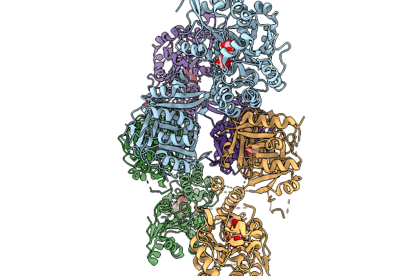
Crystal Structure Of N-Methylhydantoinase In Complex With 1-Methylimidazolidine-2,4-Dione, C-Terminal Residues Visible
Organism: Glutamicibacter protophormiae
Method: X-RAY DIFFRACTION Resolution:2.07 Å Release Date: 2026-04-15 Classification: HYDROLASE Ligands: CA, A1BC1, BTB, NH4, SO4 |
Organism: Glutamicibacter protophormiae
Method: X-RAY DIFFRACTION
Release Date: 2026-04-15
Ligands: CA, A1BC1, BTB, NH4, SO4
 |
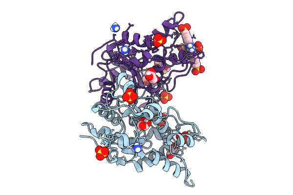
Crystal Structure Of Human Castor1 In Apo Form
Organism: Escherichia coli (strain k12), Homo sapiens
Method: X-RAY DIFFRACTION Resolution:3.08 Å Release Date: 2026-03-25 Classification: SIGNALING PROTEIN Ligands: NH4, ACY |
Organism: Escherichia coli (strain k12), Homo sapiens
Method: X-RAY DIFFRACTION
Release Date: 2026-03-25
Ligands: NH4, ACY
 |
The Mtrex1-Nsc 37204 Complex Structure By Soaking In Soaking Condition 3 (Nsc 37204 Complex 2)
Organism: Mus musculus
Method: X-RAY DIFFRACTION Resolution:1.70 Å Release Date: 2026-01-28 Classification: HYDROLASE Ligands: A1L5Z, GOL, SO4, NH4 |
Organism: Mus musculus
Method: X-RAY DIFFRACTION
Release Date: 2026-01-28
Ligands: A1L5Z, GOL, SO4, NH4
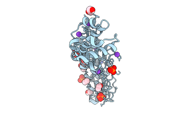 |
A Darpin Fused To The 1Tel Crystallization Chaperone Via A Direct Helical Fusion
Organism: Homo sapiens, Synthetic construct
Method: X-RAY DIFFRACTION Resolution:1.24 Å Release Date: 2025-12-17 Classification: TRANSCRIPTION Ligands: AKG, NH4, GOL, EOH, K, CL, PO4 |
Organism: Homo sapiens, Synthetic construct
Method: X-RAY DIFFRACTION
Release Date: 2025-12-17
Ligands: AKG, NH4, GOL, EOH, K, CL, PO4
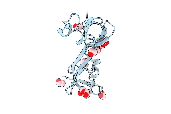 |
Jumonji Domain-Containing Protein 2B With Crystallization Epitope Mutations L916G:R917A:A918D
Organism: Homo sapiens
Method: X-RAY DIFFRACTION Resolution:1.80 Å Release Date: 2025-12-03 Classification: OXIDOREDUCTASE Ligands: EDO, PEG, NH4 |
Organism: Homo sapiens
Method: X-RAY DIFFRACTION
Release Date: 2025-12-03
Ligands: EDO, PEG, NH4
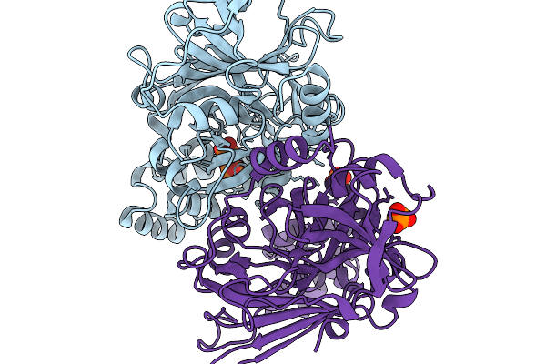 |
Structure-Guided Interface Engineering For Modifying Substrate Binding And Catalytic Activity Of 2-Keto-3-Deoxy-D-Xylonate Dehydratase
Organism: Caulobacter vibrioides na1000
Method: X-RAY DIFFRACTION Resolution:1.77 Å Release Date: 2025-11-19 Classification: CARBOHYDRATE Ligands: 2HP, NH4 |
Organism: Caulobacter vibrioides na1000
Method: X-RAY DIFFRACTION
Release Date: 2025-11-19
Ligands: 2HP, NH4
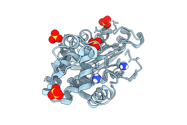 |
Structure Of P167S/D240G/D172A/S104G Blac From Mycobacterium Tuberculosis At Ph 5.5
Organism: Mycobacterium tuberculosis
Method: X-RAY DIFFRACTION Resolution:1.60 Å Release Date: 2025-10-22 Classification: HYDROLASE Ligands: NH4, SO4 |
Organism: Mycobacterium tuberculosis
Method: X-RAY DIFFRACTION
Release Date: 2025-10-22
Ligands: NH4, SO4
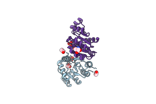 |
Crystal Structure Of De Novo Designed Atpase, Pl2X4_2
Organism: Synthetic construct
Method: X-RAY DIFFRACTION Resolution:2.29 Å Release Date: 2025-04-23 Classification: DE NOVO PROTEIN Ligands: NH4, SO4, ACT, TRS |
Organism: Synthetic construct
Method: X-RAY DIFFRACTION
Release Date: 2025-04-23
Ligands: NH4, SO4, ACT, TRS
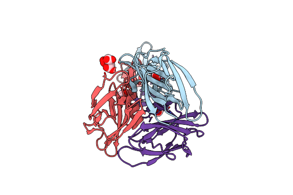 |
Structure Of Serratia Proteamaculans Antifeeding Prophage Fibre Foot Domain (Afpx13)
Organism: Serratia proteamaculans
Method: X-RAY DIFFRACTION Resolution:1.47 Å Release Date: 2025-03-19 Classification: STRUCTURAL PROTEIN Ligands: NH4, GOL, CIT |
Organism: Serratia proteamaculans
Method: X-RAY DIFFRACTION
Release Date: 2025-03-19
Ligands: NH4, GOL, CIT
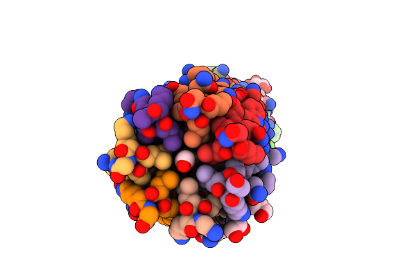 |
Cc-Hept-Hen2 Variant Peptide With Hendecad Repeat Substitution
Organism: Synthetic construct
Method: X-RAY DIFFRACTION Resolution:1.65 Å Release Date: 2025-01-22 Classification: DE NOVO PROTEIN Ligands: FMT, NA, GOL, NH4, P33 |
Organism: Synthetic construct
Method: X-RAY DIFFRACTION
Release Date: 2025-01-22
Ligands: FMT, NA, GOL, NH4, P33
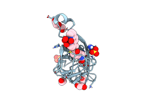 |
Artificial Metalloenzyme With A Nickel-Based 1,10-Phenanthroline Cofactor And Streptavidin N49M-S112V Mutant
Organism: Streptomyces avidinii
Method: X-RAY DIFFRACTION Resolution:1.27 Å Release Date: 2025-01-15 Classification: METAL BINDING PROTEIN Ligands: EDO, A1ICD, PEG, SO4, NH4 |
Organism: Streptomyces avidinii
Method: X-RAY DIFFRACTION
Release Date: 2025-01-15
Ligands: EDO, A1ICD, PEG, SO4, NH4