

Search Count: 40
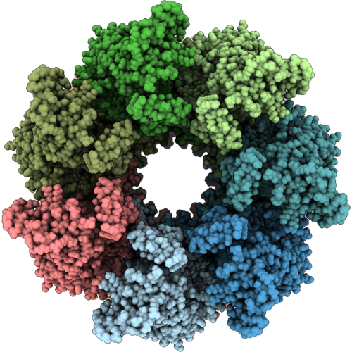 |
Mitochondrial Hsp60 From Acanthamoeba Castellanii
Organism: Acanthamoeba castellanii
Method: ELECTRON MICROSCOPY Resolution:3.76 Å Release Date: 2026-10-14 Classification: CHAPERONE |
Organism: Acanthamoeba castellanii
Method: ELECTRON MICROSCOPY
Release Date: 2026-10-14
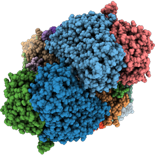 |
Cryoem Structure Of Respiratory Complex Iii Intermediate From Acanthamoeba Castellanii
Organism: Acanthamoeba castellanii
Method: ELECTRON MICROSCOPY Resolution:3.65 Å Release Date: 2026-10-14 Classification: OXIDOREDUCTASE Ligands: HEM |
Organism: Acanthamoeba castellanii
Method: ELECTRON MICROSCOPY
Release Date: 2026-10-14
Ligands: HEM
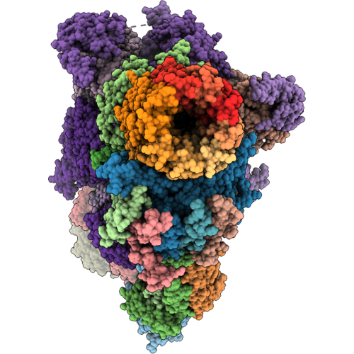 |
Mitochondrial Atp Synthase Monomer From Acanthamoeba Castellanii
Organism: Acanthamoeba castellanii
Method: ELECTRON MICROSCOPY Release Date: 2026-10-14 Classification: HYDROLASE Ligands: ATP, MG, ADP |
Organism: Acanthamoeba castellanii
Method: ELECTRON MICROSCOPY
Release Date: 2026-10-14
Ligands: ATP, MG, ADP
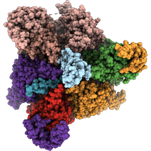 |
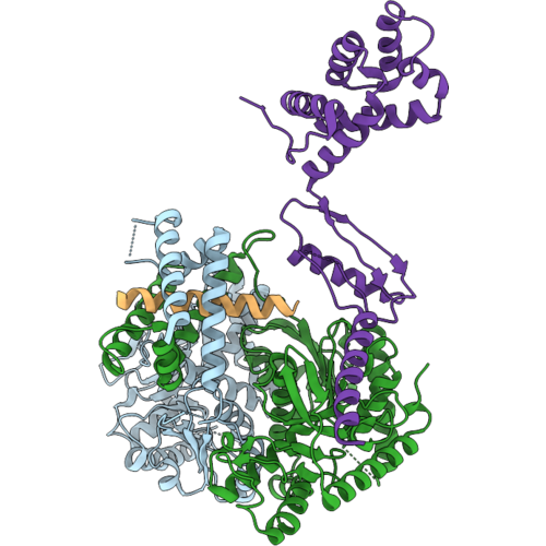
F1 Atpase From Acanthamoeba Castellanii
Organism: Acanthamoeba castellanii
Method: ELECTRON MICROSCOPY Release Date: 2026-10-14 Classification: HYDROLASE Ligands: ATP, MG, ADP |
Organism: Acanthamoeba castellanii
Method: ELECTRON MICROSCOPY
Release Date: 2026-10-14
Ligands: ATP, MG, ADP
 |
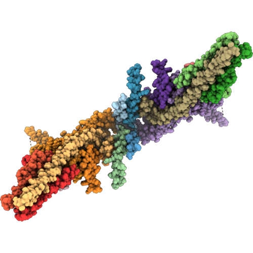
Mitochondrial Mdh And Oscp In The Peripheral Stalk Of Atp Synthase In Acanthamoeba Castellanii
Organism: Acanthamoeba castellanii
Method: ELECTRON MICROSCOPY Release Date: 2026-10-14 Classification: OXIDOREDUCTASE |
Organism: Acanthamoeba castellanii
Method: ELECTRON MICROSCOPY
Release Date: 2026-10-14
 |
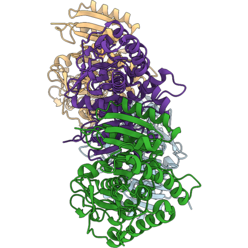
Membrane Region Of Acanthamoeba Castellanii Mitochondrial Atp Synthase Dimer
Organism: Acanthamoeba castellanii
Method: ELECTRON MICROSCOPY Resolution:4.23 Å Release Date: 2026-10-14 Classification: HYDROLASE |
Organism: Acanthamoeba castellanii
Method: ELECTRON MICROSCOPY
Release Date: 2026-10-14
 |
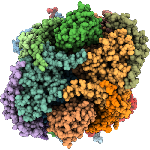
Mitochondrial Malate Dehydrogenase From Acanthamoeba Castellanii
Organism: Acanthamoeba castellanii
Method: X-RAY DIFFRACTION Resolution:2.33 Å Release Date: 2026-10-14 Classification: OXIDOREDUCTASE |
Organism: Acanthamoeba castellanii
Method: X-RAY DIFFRACTION
Release Date: 2026-10-14
 |
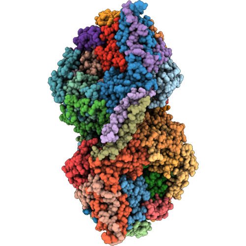
Cryo-Em Structure Of Leishmania Tarentolae Respiratory Complex Iii (Cytochrome Bc1 Complex)
Organism: Leishmania tarentolae
Method: ELECTRON MICROSCOPY Resolution:3.02 Å Release Date: 2026-10-07 Classification: ELECTRON TRANSPORT Ligands: HEM |
Organism: Leishmania tarentolae
Method: ELECTRON MICROSCOPY
Release Date: 2026-10-07
Ligands: HEM
 |
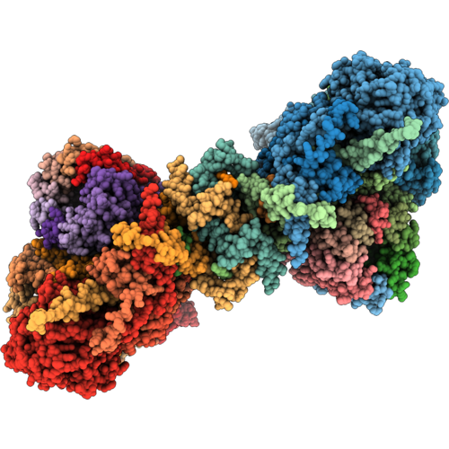
Cryo-Em Structure Of Leishmania Tarentolae Respiratory Complex Iv (Cytochrome C Oxidase Dimer)
Organism: Leishmania tarentolae
Method: ELECTRON MICROSCOPY Resolution:3.21 Å Release Date: 2026-10-07 Classification: ELECTRON TRANSPORT Ligands: CU, HEA |
Organism: Leishmania tarentolae
Method: ELECTRON MICROSCOPY
Release Date: 2026-10-07
Ligands: CU, HEA
 |
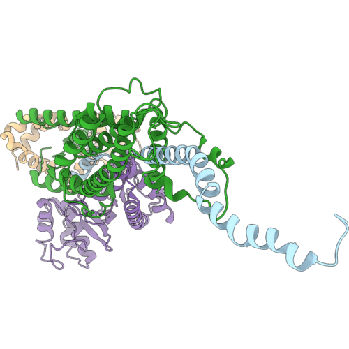
Cryo-Em Structure Of Leishmania Tarentolae Respiratory Complex V (Atp Synthase) Membrane Region
Organism: Leishmania tarentolae
Method: ELECTRON MICROSCOPY Resolution:3.73 Å Release Date: 2026-10-07 Classification: ELECTRON TRANSPORT |
Organism: Leishmania tarentolae
Method: ELECTRON MICROSCOPY
Release Date: 2026-10-07
 |
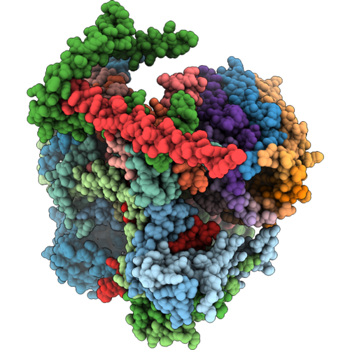
Cryo-Em Structure Of Leishmania Tarentolae Respiratory Complex V (Atp Synthase) Peripheral Stalk
Organism: Leishmania tarentolae
Method: ELECTRON MICROSCOPY Resolution:3.95 Å Release Date: 2026-10-07 Classification: ELECTRON TRANSPORT |
Organism: Leishmania tarentolae
Method: ELECTRON MICROSCOPY
Release Date: 2026-10-07
 |
Cryo-Em Structure Of Leishmania Tarentolae Respiratory Complex V (Atp Synthase) Fo Monomer Membrane Region
Organism: Leishmania tarentolae
Method: ELECTRON MICROSCOPY Resolution:3.62 Å Release Date: 2026-10-07 Classification: ELECTRON TRANSPORT |
Organism: Leishmania tarentolae
Method: ELECTRON MICROSCOPY
Release Date: 2026-10-07
 |
Small Subunit Of Yeast Mitochondrial Ribosome In Complex With Mettl17/Rsm22.
Organism: Saccharomyces cerevisiae
Method: ELECTRON MICROSCOPY Release Date: 2024-01-10 Classification: RIBOSOME Ligands: MG, K, LMT, ATP, SF4 Modified Residue(s): SAC |
Organism: Saccharomyces cerevisiae
Method: ELECTRON MICROSCOPY
Release Date: 2024-01-10
Ligands: MG, K, LMT, ATP, SF4
 |
Small Subunit Of Yeast Mitochondrial Ribosome In Complex With If3/Aim23.
Organism: Saccharomyces cerevisiae
Method: ELECTRON MICROSCOPY Release Date: 2024-01-10 Classification: RIBOSOME Ligands: MG, K, LMT, ATP |
Organism: Saccharomyces cerevisiae
Method: ELECTRON MICROSCOPY
Release Date: 2024-01-10
Ligands: MG, K, LMT, ATP
 |
Small Subunit Of Yeast Mitochondrial Ribosome.
Organism: Saccharomyces cerevisiae
Method: ELECTRON MICROSCOPY Release Date: 2024-01-10 Classification: RIBOSOME Ligands: MG, K, LMT, ATP |
Organism: Saccharomyces cerevisiae
Method: ELECTRON MICROSCOPY
Release Date: 2024-01-10
Ligands: MG, K, LMT, ATP
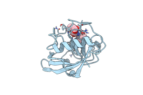 |
Structure Of Cyclophilin D Peptidyl-Prolyl Isomerase Domain Bound To Macrocyclic Inhibitor Jombt
Organism: Homo sapiens
Method: X-RAY DIFFRACTION Resolution:1.75 Å Release Date: 2022-08-24 Classification: ISOMERASE Ligands: I3Q |
Organism: Homo sapiens
Method: X-RAY DIFFRACTION
Release Date: 2022-08-24
Ligands: I3Q
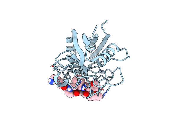 |
Structure Of Cyclophilin D Peptidyl-Prolyl Isomerase Domain Bound To Macrocyclic Inhibitor A26
Organism: Homo sapiens
Method: X-RAY DIFFRACTION Resolution:1.06 Å Release Date: 2022-08-24 Classification: ISOMERASE Ligands: I4F |
Organism: Homo sapiens
Method: X-RAY DIFFRACTION
Release Date: 2022-08-24
Ligands: I4F
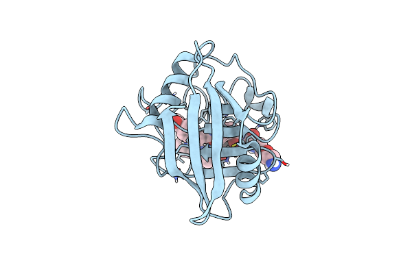 |
Structure Of Cyclophilin D Peptidyl-Prolyl Isomerase Domain Bound To Macrocyclic Inhibitor B1
Organism: Homo sapiens
Method: X-RAY DIFFRACTION Resolution:1.21 Å Release Date: 2022-08-24 Classification: ISOMERASE/ISOMERASE inhibitor Ligands: I44 |
Organism: Homo sapiens
Method: X-RAY DIFFRACTION
Release Date: 2022-08-24
Ligands: I44
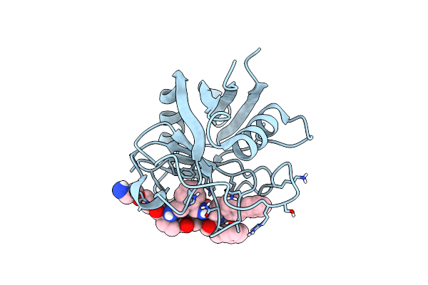 |
Structure Of Cyclophilin D Peptidyl-Prolyl Isomerase Domain Bound To Macrocyclic Inhibitor B2
Organism: Homo sapiens
Method: X-RAY DIFFRACTION Resolution:1.46 Å Release Date: 2022-08-24 Classification: ISOMERASE/ISOMERASE inhibitor Ligands: I3X |
Organism: Homo sapiens
Method: X-RAY DIFFRACTION
Release Date: 2022-08-24
Ligands: I3X
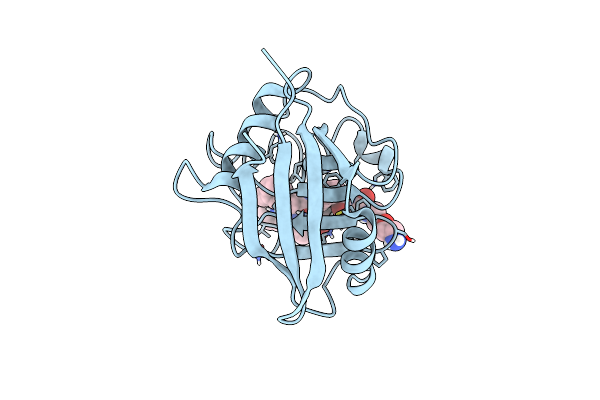 |
Structure Of Cyclophilin D Peptidyl-Prolyl Isomerase Domain Bound To Macrocyclic Inhibitor B3
Organism: Homo sapiens
Method: X-RAY DIFFRACTION Resolution:1.52 Å Release Date: 2022-08-24 Classification: ISOMERASE Ligands: I5J |
Organism: Homo sapiens
Method: X-RAY DIFFRACTION
Release Date: 2022-08-24
Ligands: I5J