

Search Count: 41
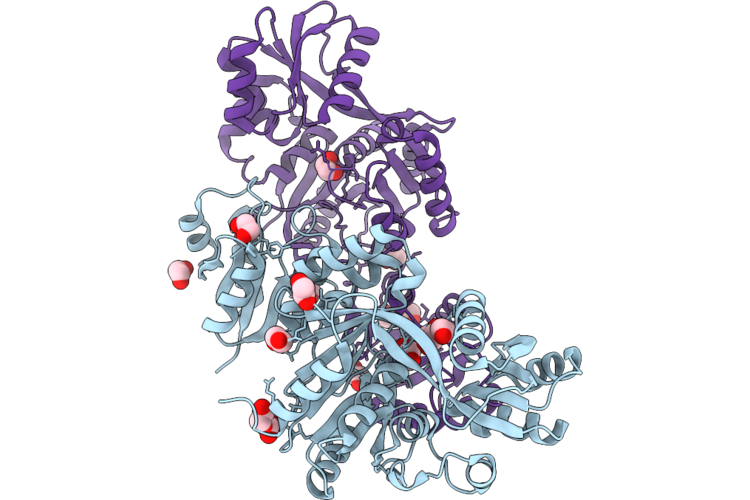 |
Thermococcus Sibiricus Cyclic 2,3-Diphosphoglycerate Syntethase
Organism: Thermococcus sibiricus
Method: X-RAY DIFFRACTION Resolution:2.28 Å Release Date: 2026-05-27 Classification: LYASE Ligands: EDO, GOL, PEG |
Organism: Thermococcus sibiricus
Method: X-RAY DIFFRACTION
Release Date: 2026-05-27
Ligands: EDO, GOL, PEG
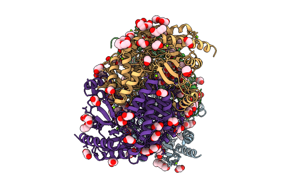 |
Bv333 Aminotransferase From Streptomyces Sp. Mutant W89A
Organism: Streptomyces sp. bv333
Method: X-RAY DIFFRACTION Resolution:1.24 Å Release Date: 2026-03-18 Classification: TRANSFERASE Ligands: PEG, PGE, EDO, GOL, 1PG, CL, MG, PG4 |
Organism: Streptomyces sp. bv333
Method: X-RAY DIFFRACTION
Release Date: 2026-03-18
Ligands: PEG, PGE, EDO, GOL, 1PG, CL, MG, PG4
 |
Bv333 Aminotransferase From Streptomyces Sp. Mutant F61C
Organism: Streptomyces sp. bv333
Method: X-RAY DIFFRACTION Resolution:1.31 Å Release Date: 2026-03-18 Classification: TRANSFERASE |
Organism: Streptomyces sp. bv333
Method: X-RAY DIFFRACTION
Release Date: 2026-03-18
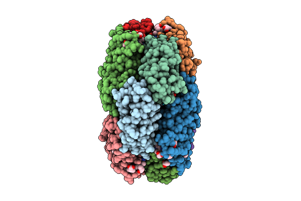 |
X-Ray Structure Of The Adduct Formed By Dirhodium Tetraacetate With A C-Phycocyanin
Organism: Galdieria phlegrea
Method: X-RAY DIFFRACTION Resolution:2.17 Å Release Date: 2025-12-17 Classification: PHOTOSYNTHESIS Ligands: CYC, A1JTN, ACT, DOD |
Organism: Galdieria phlegrea
Method: X-RAY DIFFRACTION
Release Date: 2025-12-17
Ligands: CYC, A1JTN, ACT, DOD
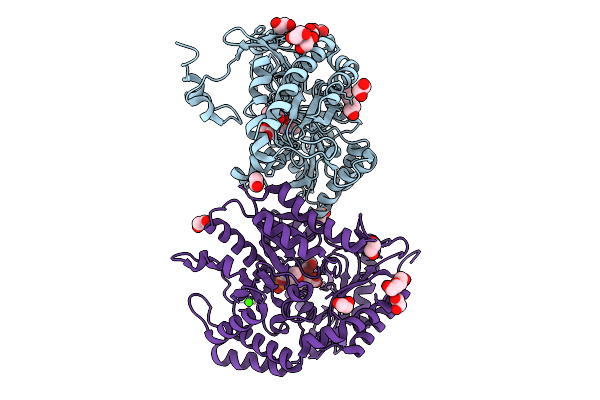 |
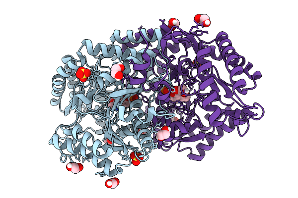
A Novel Aminotransferase From Streptomyces Sp. Gabaculine Complex
Organism: Streptomyces sp. bv333
Method: X-RAY DIFFRACTION Resolution:1.31 Å Release Date: 2025-09-10 Classification: TRANSFERASE Ligands: PXG, EDO, PEG, CA, CL |
Organism: Streptomyces sp. bv333
Method: X-RAY DIFFRACTION
Release Date: 2025-09-10
Ligands: PXG, EDO, PEG, CA, CL
 |
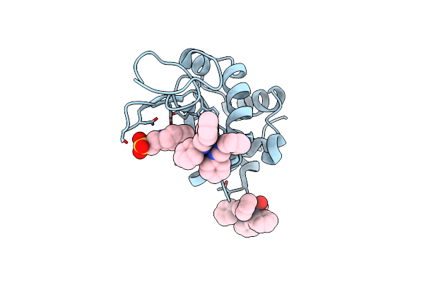
A Novel Aminotransferase From Streptomyces Sp.
Organism: Streptomyces sp. bv333
Method: X-RAY DIFFRACTION Resolution:1.49 Å Release Date: 2025-08-27 Classification: TRANSFERASE Ligands: EDO, PLP, SO4 |
Organism: Streptomyces sp. bv333
Method: X-RAY DIFFRACTION
Release Date: 2025-08-27
Ligands: EDO, PLP, SO4
 |
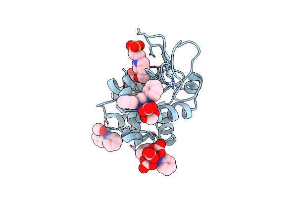
X-Ray Structure Of The Adduct Formed Upon Reaction Of Lysozyme With [Ru2Cl(Dphf)2(O2Cch3)2] In Condition B
Organism: Gallus gallus
Method: X-RAY DIFFRACTION Resolution:1.29 Å Release Date: 2023-09-13 Classification: PROTEIN BINDING Ligands: EPE, ZJK |
Organism: Gallus gallus
Method: X-RAY DIFFRACTION
Release Date: 2023-09-13
Ligands: EPE, ZJK
 |
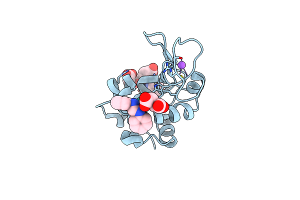
X-Ray Structure Of The Adduct Formed Upon Reaction Of Lysozyme With K2[Ru2(Dphf)(Co3)3] In Condition B
Organism: Gallus gallus
Method: X-RAY DIFFRACTION Resolution:1.07 Å Release Date: 2023-09-13 Classification: PROTEIN BINDING Ligands: EPE, ZWL, YWV, ZWO, ZXE |
Organism: Gallus gallus
Method: X-RAY DIFFRACTION
Release Date: 2023-09-13
Ligands: EPE, ZWL, YWV, ZWO, ZXE
 |
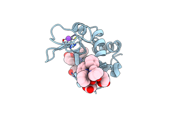
X-Ray Structure Of The Adduct Formed Upon Reaction Of Lysozyme With [Ru2Cl(Dphf)(O2Cch3)3] In Condition A
Organism: Gallus gallus
Method: X-RAY DIFFRACTION Resolution:1.41 Å Release Date: 2023-09-13 Classification: PROTEIN BINDING Ligands: ZQ2, SIN, ZWH, NA |
Organism: Gallus gallus
Method: X-RAY DIFFRACTION
Release Date: 2023-09-13
Ligands: ZQ2, SIN, ZWH, NA
 |
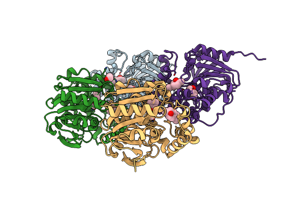
X-Ray Structure Of The Adduct Formed Upon Reaction Of Lysozyme With [Ru2Cl(Dphf)2(O2Cch3)2] In Condition A
Organism: Gallus gallus
Method: X-RAY DIFFRACTION Resolution:1.29 Å Release Date: 2023-09-13 Classification: PROTEIN BINDING |
Organism: Gallus gallus
Method: X-RAY DIFFRACTION
Release Date: 2023-09-13
 |
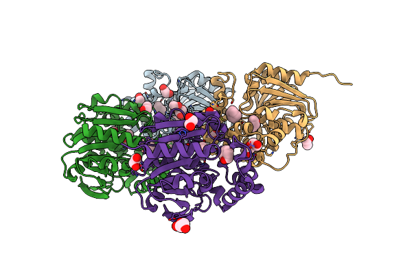
Limonene Epoxide Low Ph Soak Of Epoxide Hydrolase From Metagenomic Source Ch65
Organism: Metagenome
Method: X-RAY DIFFRACTION Resolution:1.97 Å Release Date: 2023-08-16 Classification: HYDROLASE Ligands: LEO, CL, EDO |
Organism: Metagenome
Method: X-RAY DIFFRACTION
Release Date: 2023-08-16
Ligands: LEO, CL, EDO
 |
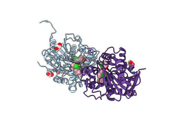
Cyclohexane Epoxide Low Ph Soak Of Epoxide Hydrolase From Metagenomic Source Ch65
Organism: Metagenome
Method: X-RAY DIFFRACTION Resolution:1.96 Å Release Date: 2023-08-16 Classification: HYDROLASE Ligands: 7EX, PEG, EDO |
Organism: Metagenome
Method: X-RAY DIFFRACTION
Release Date: 2023-08-16
Ligands: 7EX, PEG, EDO
 |
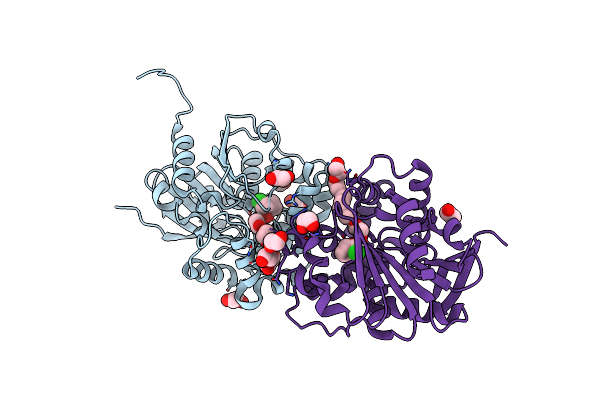
Halogenated Product Of Limonene Epoxide Turnover By Epoxide Hydrolase From Metagenomic Source Ch65
Organism: Metagenome
Method: X-RAY DIFFRACTION Resolution:1.49 Å Release Date: 2023-08-16 Classification: HYDROLASE Ligands: M1U, CL, EDO, NA |
Organism: Metagenome
Method: X-RAY DIFFRACTION
Release Date: 2023-08-16
Ligands: M1U, CL, EDO, NA
 |
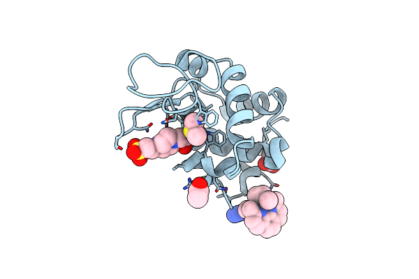
Cyclohexane Epoxide Soak Of Epoxide Hydrolase From Metagenomic Source Ch65 Resulting In Halogenated Compound In The Active Site
Organism: Metagenome
Method: X-RAY DIFFRACTION Resolution:1.61 Å Release Date: 2023-08-16 Classification: HYDROLASE Ligands: A1KDR, EDO, PGE, CL, P6G |
Organism: Metagenome
Method: X-RAY DIFFRACTION
Release Date: 2023-08-16
Ligands: A1KDR, EDO, PGE, CL, P6G
 |
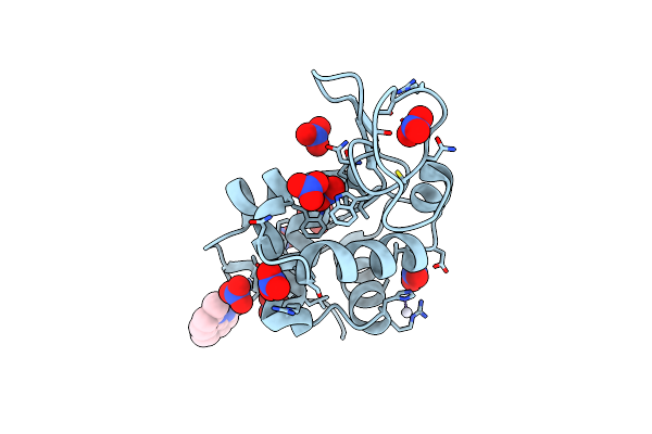
X-Ray Structure Of The Adduct Formed Upon Reaction Of The Five-Coordinate Pt(Ii) Complex, 1-Me,Me, With Hewl At Ph 7.5
Organism: Gallus gallus
Method: X-RAY DIFFRACTION Resolution:1.25 Å Release Date: 2023-02-22 Classification: HYDROLASE Ligands: EPE, ACT, R0I, DMS |
Organism: Gallus gallus
Method: X-RAY DIFFRACTION
Release Date: 2023-02-22
Ligands: EPE, ACT, R0I, DMS
 |
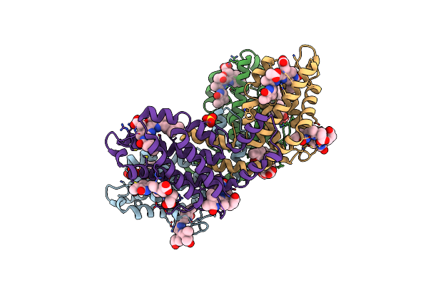
X-Ray Structure Of The Adduct Formed Upon Reaction Of The Five-Coordinate Pt(Ii) Complex, 1-Me,Me, With Hewl At Ph 4.0
Organism: Gallus gallus
Method: X-RAY DIFFRACTION Resolution:1.33 Å Release Date: 2023-02-22 Classification: HYDROLASE Ligands: NO3, R0I, PT |
Organism: Gallus gallus
Method: X-RAY DIFFRACTION
Release Date: 2023-02-22
Ligands: NO3, R0I, PT
 |
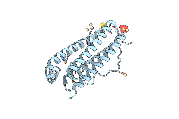
X-Ray Structure Of Phycoerythrin From Porphyridium Cruentum
Organism: Porphyridium purpureum
Method: X-RAY DIFFRACTION Resolution:1.60 Å Release Date: 2023-02-08 Classification: PHOTOSYNTHESIS Ligands: PEB, SO4 |
Organism: Porphyridium purpureum
Method: X-RAY DIFFRACTION
Release Date: 2023-02-08
Ligands: PEB, SO4
 |
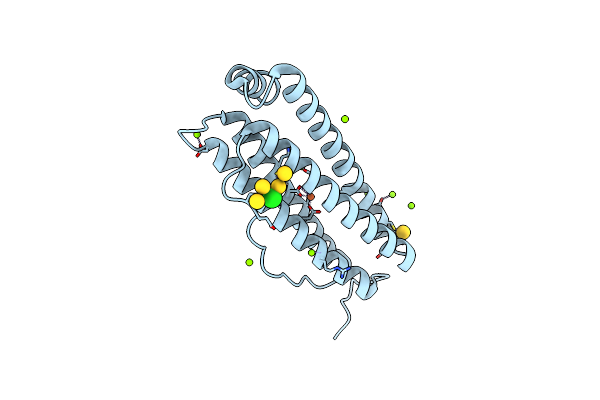
X-Ray Structure Of Auranofin-Horse Spleen Ferritin
Organism: Equus caballus
Method: X-RAY DIFFRACTION Resolution:1.24 Å Release Date: 2022-12-28 Classification: TRANSPORT PROTEIN Ligands: CD, SO4, AU, CL |
Organism: Equus caballus
Method: X-RAY DIFFRACTION
Release Date: 2022-12-28
Ligands: CD, SO4, AU, CL
 |
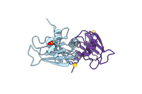
X-Ray Structure Of Auranofin-Human H-Chain Ferritin
Organism: Homo sapiens
Method: X-RAY DIFFRACTION Resolution:1.17 Å Release Date: 2022-12-28 Classification: TRANSPORT PROTEIN Ligands: CL, MG, FE, AU |
Organism: Homo sapiens
Method: X-RAY DIFFRACTION
Release Date: 2022-12-28
Ligands: CL, MG, FE, AU
 |
X-Ray Structure Of The Adduct Formed Upon Reaction Of The Gold(I) N-Heterocyclic Carbene Complex Au1 With Rnase A
Organism: Bos taurus
Method: X-RAY DIFFRACTION Resolution:1.42 Å Release Date: 2022-07-13 Classification: RNA BINDING PROTEIN Ligands: AU, SO4 |
Organism: Bos taurus
Method: X-RAY DIFFRACTION
Release Date: 2022-07-13
Ligands: AU, SO4