

Search Count: 30
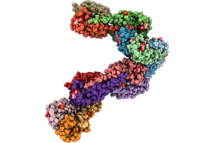 |
Xn-Il Lectin From Xenorhabdus Nematophila In Complex With Lactose
Organism: Xenorhabdus nematophila
Method: X-RAY DIFFRACTION Resolution:1.70 Å Release Date: 2026-06-24 Classification: SUGAR BINDING PROTEIN Ligands: EDO, CA, MG, SO4, PEG, BGC, PGE, CL, NA, 1PE |
Organism: Xenorhabdus nematophila
Method: X-RAY DIFFRACTION
Release Date: 2026-06-24
Ligands: EDO, CA, MG, SO4, PEG, BGC, PGE, CL, NA, 1PE
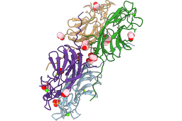 |
Apo Form Of Xn-Il Lectin From Xenorhabdus Nematophila
Organism: Xenorhabdus nematophila
Method: X-RAY DIFFRACTION Resolution:1.56 Å Release Date: 2026-06-24 Classification: SUGAR BINDING PROTEIN Ligands: CA, SO4, MG, EDO, PEG |
Organism: Xenorhabdus nematophila
Method: X-RAY DIFFRACTION
Release Date: 2026-06-24
Ligands: CA, SO4, MG, EDO, PEG
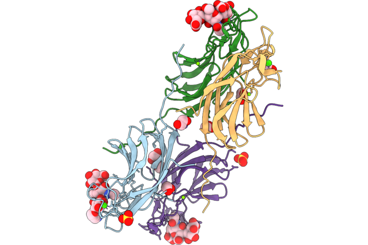 |
Xn-Il Lectin From Xenorhabdus Nematophila In Complex With Heparosan Trisaccharide
Organism: Xenorhabdus nematophila
Method: X-RAY DIFFRACTION Resolution:1.80 Å Release Date: 2026-06-24 Classification: SUGAR BINDING PROTEIN Ligands: EDO, CA, MG, SO4, NA |
Organism: Xenorhabdus nematophila
Method: X-RAY DIFFRACTION
Release Date: 2026-06-24
Ligands: EDO, CA, MG, SO4, NA
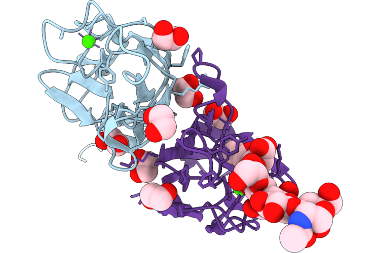 |
Xn-Il Lectin From Xenorhabdus Nematophila In Complex With Hyaluronan Tetrasaccharide
Organism: Xenorhabdus nematophila
Method: X-RAY DIFFRACTION Resolution:1.60 Å Release Date: 2026-06-24 Classification: SUGAR BINDING PROTEIN Ligands: EDO, CA |
Organism: Xenorhabdus nematophila
Method: X-RAY DIFFRACTION
Release Date: 2026-06-24
Ligands: EDO, CA
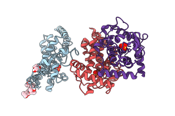 |
Crystal Structure Of Phoc Mutant Acid Phosphatase
Organism: Morganella morganii
Method: X-RAY DIFFRACTION Resolution:2.00 Å Release Date: 2026-01-14 Classification: BIOSYNTHETIC PROTEIN Ligands: MPD, PEG, SO4 |
Organism: Morganella morganii
Method: X-RAY DIFFRACTION
Release Date: 2026-01-14
Ligands: MPD, PEG, SO4
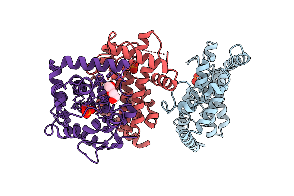 |
Crystal Structure Of Phoc Wild Type Acid Phosphatase
Organism: Morganella morganii
Method: X-RAY DIFFRACTION Resolution:2.24 Å Release Date: 2026-01-14 Classification: BIOSYNTHETIC PROTEIN Ligands: PO4, PEG |
Organism: Morganella morganii
Method: X-RAY DIFFRACTION
Release Date: 2026-01-14
Ligands: PO4, PEG
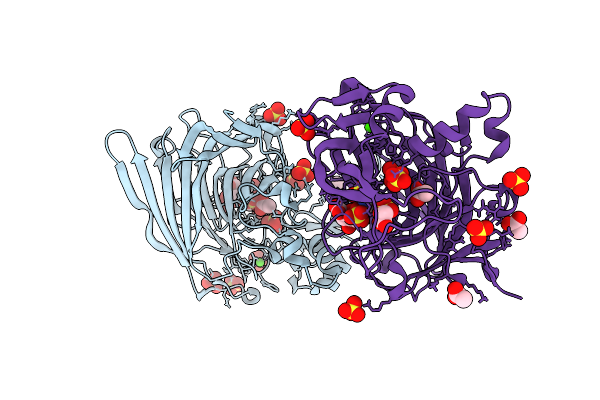 |
Structure Of Heparinase I From Bacteroides Eggerthii In Complex With Calcium Cofactor And Delta-Ua(1-4)Glc 3, 6, N-Sulphated Disaccharide
Organism: Bacteroides eggerthii
Method: X-RAY DIFFRACTION Resolution:1.72 Å Release Date: 2025-10-08 Classification: LYASE Ligands: ACT, SO4, CA |
Organism: Bacteroides eggerthii
Method: X-RAY DIFFRACTION
Release Date: 2025-10-08
Ligands: ACT, SO4, CA
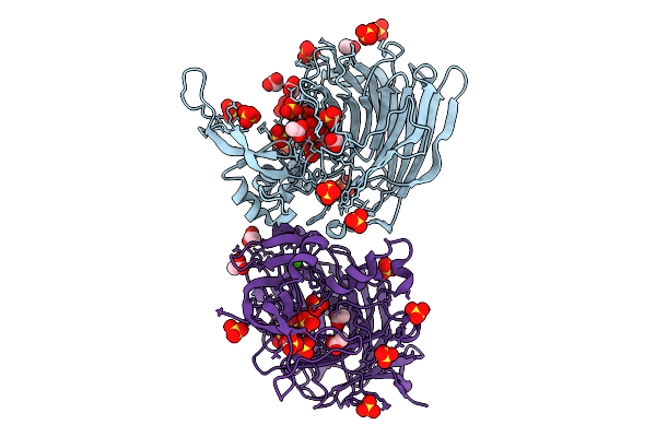 |
Structure Of Heparinase I From Bacteroides Eggerthii In Complex With Calcium Cofactor
Organism: Bacteroides eggerthii
Method: X-RAY DIFFRACTION Resolution:1.85 Å Release Date: 2025-09-24 Classification: LYASE Ligands: CA, ACT, SO4 |
Organism: Bacteroides eggerthii
Method: X-RAY DIFFRACTION
Release Date: 2025-09-24
Ligands: CA, ACT, SO4
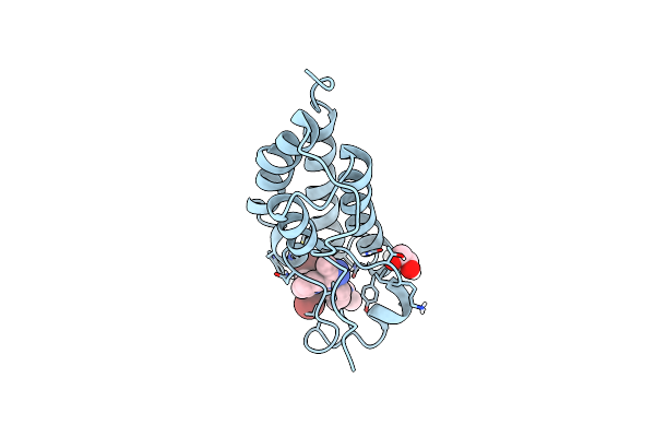 |
X-Ray Crystal Structure Of Human Brd4(D1) In Complex With 2-(4-{5-[6-(2,5-Dibromophenoxy)Pyridin-2-Yl]-4-Methyl-1H-1,2,3-Triazol-1-Yl}Piperidin-1-Yl)-N,N-Dimethylethan-1-Amine (Compound 26)
Organism: Homo sapiens
Method: X-RAY DIFFRACTION Resolution:1.32 Å Release Date: 2021-07-28 Classification: GENE REGULATION/INHIBITOR Ligands: EDO, ZHV, CL |
Organism: Homo sapiens
Method: X-RAY DIFFRACTION
Release Date: 2021-07-28
Ligands: EDO, ZHV, CL
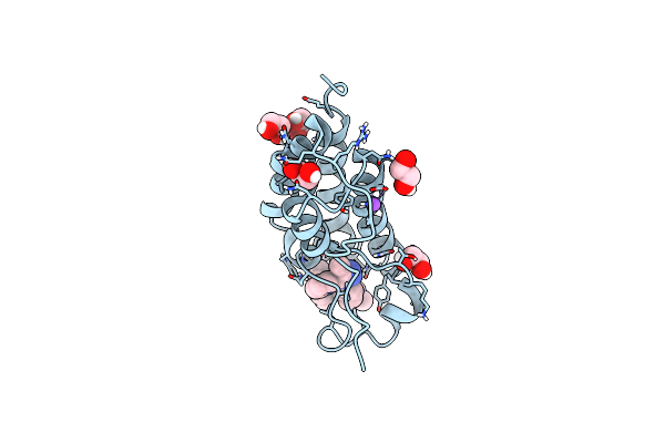 |
X-Ray Crystal Structure Of Human Brd4(D1) In Complex With 2-(4-{5-[6-(3,5-Dimethylphenoxy)Pyridin-2-Yl]-4-Methyl-1H-1,2,3-Triazol-1- Yl}Piperidin-1-Yl)-N,N-Dimethylethan-1-Amine (Dw34)
Organism: Homo sapiens
Method: X-RAY DIFFRACTION Resolution:1.20 Å Release Date: 2021-07-28 Classification: GENE REGULATION/INHIBITOR Ligands: EDO, ZHS, NA |
Organism: Homo sapiens
Method: X-RAY DIFFRACTION
Release Date: 2021-07-28
Ligands: EDO, ZHS, NA
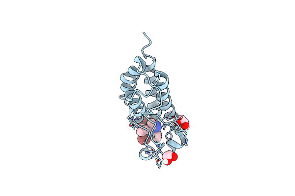 |
X-Ray Crystal Structure Of Human Brd4(D1) In Complex With 2-(2,5-Dibromophenoxy)-6-[4-Methyl-1-(Piperidin-4-Yl)-1H-1,2,3-Triazol-5-Yl]Pyridine (Compound 23)
Organism: Homo sapiens
Method: X-RAY DIFFRACTION Resolution:1.26 Å Release Date: 2021-07-28 Classification: GENE REGULATION/INHIBITOR Ligands: ZHM, EDO, CL |
Organism: Homo sapiens
Method: X-RAY DIFFRACTION
Release Date: 2021-07-28
Ligands: ZHM, EDO, CL
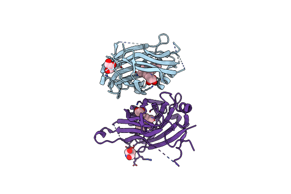 |
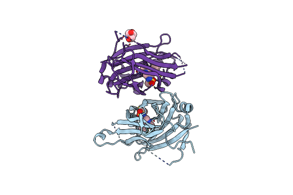
Crystal Structure Of Tead2 Bound To Compound 1
Organism: Homo sapiens
Method: X-RAY DIFFRACTION Resolution:1.54 Å Release Date: 2020-06-24 Classification: Transcription/Transcription Inhibitor Ligands: GOL, QSJ, TRS |
Organism: Homo sapiens
Method: X-RAY DIFFRACTION
Release Date: 2020-06-24
Ligands: GOL, QSJ, TRS
 |
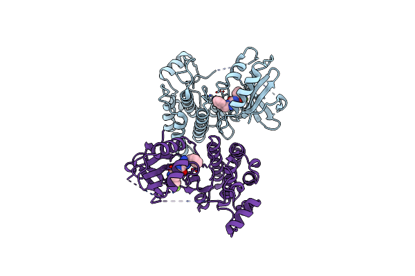
Crystal Structure Of Tead2 Bound To Compound 2
Organism: Homo sapiens
Method: X-RAY DIFFRACTION Resolution:1.66 Å Release Date: 2020-06-24 Classification: TRANSCRIPTION/INHIBITOR Ligands: QLV, TRS |
Organism: Homo sapiens
Method: X-RAY DIFFRACTION
Release Date: 2020-06-24
Ligands: QLV, TRS
 |
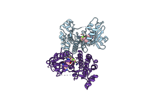
Crystal Structure Of Rip1 Kinase In Complex With Gsk3145095
Organism: Homo sapiens
Method: X-RAY DIFFRACTION Resolution:2.87 Å Release Date: 2019-07-03 Classification: TRANSFERASE Ligands: K8K |
Organism: Homo sapiens
Method: X-RAY DIFFRACTION
Release Date: 2019-07-03
Ligands: K8K
 |
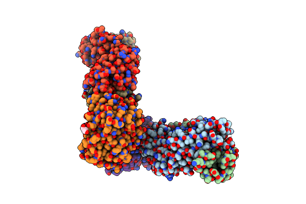
Crystal Structure Of Rip1 Kinase In Complex With Gsk547
Organism: Homo sapiens
Method: X-RAY DIFFRACTION Resolution:3.49 Å Release Date: 2018-12-12 Classification: TRANSFERASE Ligands: G4W |
Organism: Homo sapiens
Method: X-RAY DIFFRACTION
Release Date: 2018-12-12
Ligands: G4W
 |
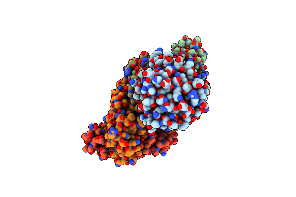
Structure Of Vaccinia Virus D8 Protein Bound To Human Fab Vv138
Organism: Vaccinia virus, Homo sapiens
Method: X-RAY DIFFRACTION Resolution:2.90 Å Release Date: 2017-11-22 Classification: VIRAL PROTEIN/immune system Ligands: GOL, NA |
Organism: Vaccinia virus, Homo sapiens
Method: X-RAY DIFFRACTION
Release Date: 2017-11-22
Ligands: GOL, NA
 |
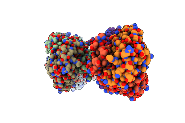
Structure Of Vaccinia Virus D8 Protein Bound To Human Fab Vv66
Organism: Vaccinia virus, Homo sapiens
Method: X-RAY DIFFRACTION Resolution:2.30 Å Release Date: 2017-09-20 Classification: VIRAL PROTEIN/Immune system |
Organism: Vaccinia virus, Homo sapiens
Method: X-RAY DIFFRACTION
Release Date: 2017-09-20
 |
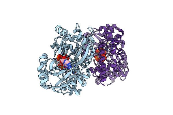
Structure Of Vaccinia Virus D8 Protein Bound To Human Fab Vv304
Organism: Vaccinia virus, Homo sapiens
Method: X-RAY DIFFRACTION Resolution:2.90 Å Release Date: 2017-09-20 Classification: VIRAL PROTEIN/Immune System |
Organism: Vaccinia virus, Homo sapiens
Method: X-RAY DIFFRACTION
Release Date: 2017-09-20
 |
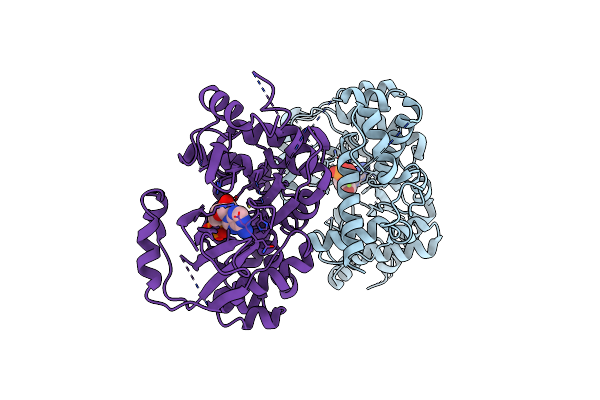
Crystal Structure Of Inositol 1,3,4,5,6-Pentakisphosphate 2-Kinase E82C/S142C
Organism: Arabidopsis thaliana
Method: X-RAY DIFFRACTION Resolution:2.60 Å Release Date: 2013-11-06 Classification: TRANSFERASE Ligands: IHP, ADP, MG, ZN |
Organism: Arabidopsis thaliana
Method: X-RAY DIFFRACTION
Release Date: 2013-11-06
Ligands: IHP, ADP, MG, ZN
 |
Inositol 1,3,4,5,6-Pentakisphosphate 2-Kinase From A. Thaliana In Complex With Adp.
Organism: Arabidopsis thaliana
Method: X-RAY DIFFRACTION Resolution:3.10 Å Release Date: 2012-03-14 Classification: TRANSFERASE Ligands: ADP, MG |
Organism: Arabidopsis thaliana
Method: X-RAY DIFFRACTION
Release Date: 2012-03-14
Ligands: ADP, MG