

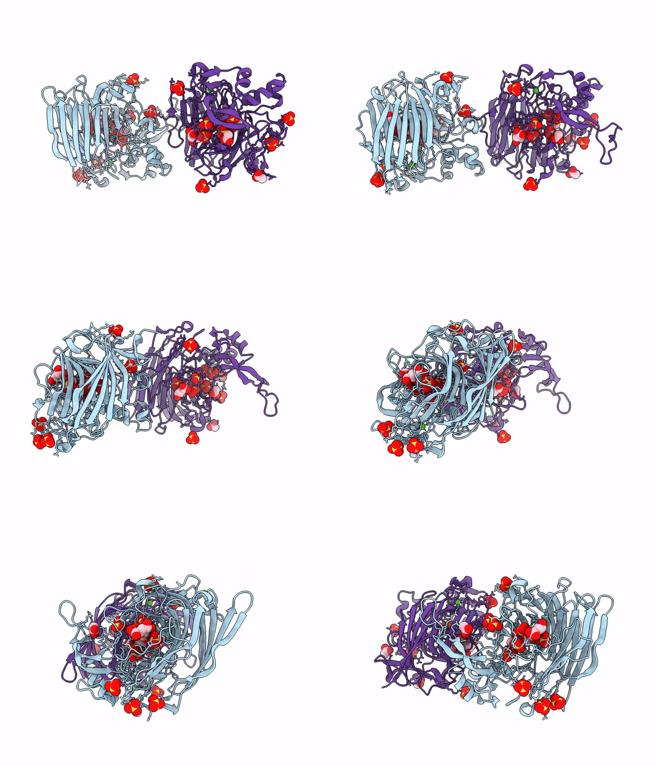
9GV9 image
Deposition Date 2024-09-23
Release Date 2025-10-08
Last Version Date 2026-10-21
Entry Detail
PDB ID:
9GV9
Keywords:
Title:
Structure of Heparinase I from Bacteroides eggerthii in complex with calcium cofactor and delta-UA(1-4)Glc 3, 6, N-Sulphated disaccharide
Biological Source:
Source Organism(s):
Expression System(s):
Method Details:
Experimental Method:
Resolution:
1.72 Å
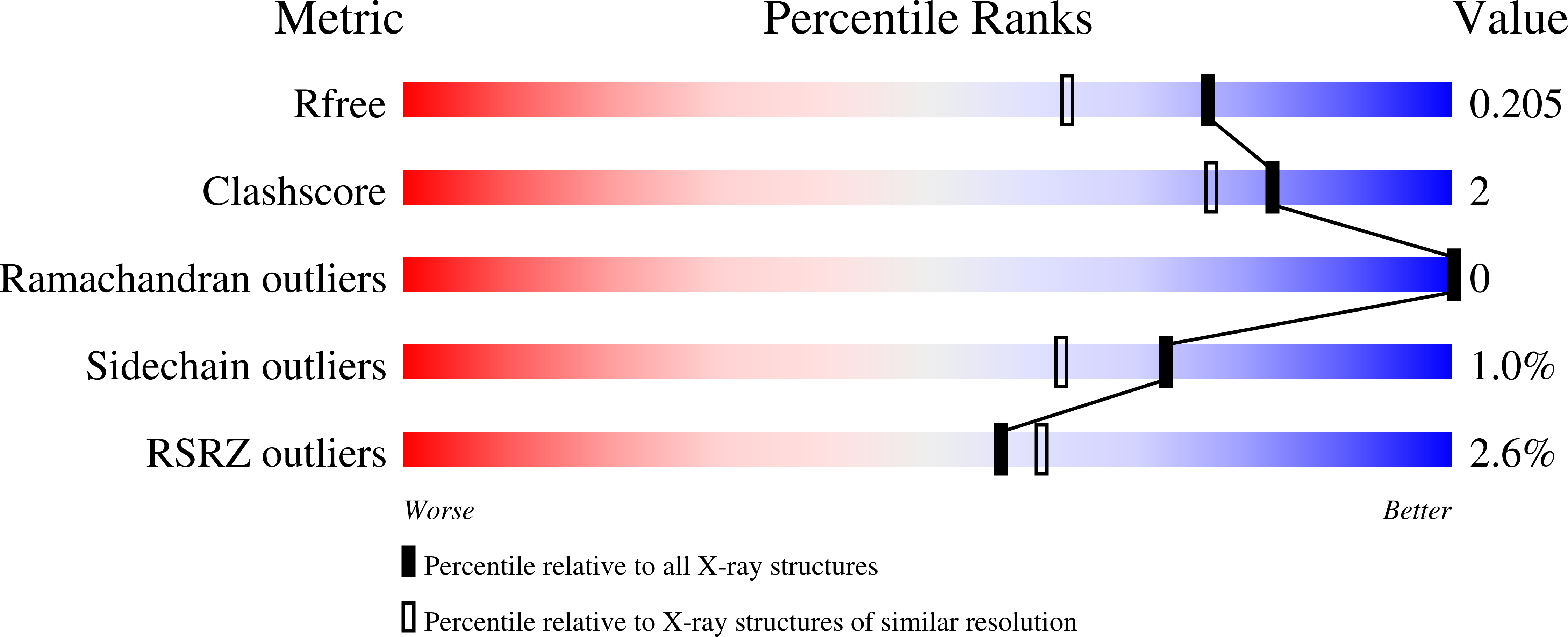
R-Value Free:
0.24
R-Value Work:
0.20
Space Group:
C 1 2 1
Macromolecular Entities
Polymer Type:polypeptide(L)
Molecule:Heparin lyase I
Gene (Uniprot):HMPREF1016_01920
Chain IDs:A (auth: B), B (auth: A)
Chain Length:382
Number of Molecules:2
Biological Source:Bacteroides eggerthii
Primary Citation
Functional characterization of a 3-O-sulfate tolerant heparin lyase from Bacteroides eggerthii.
Glycobiology ? ? ? (2026)
PMID: 42867289 DOI: 10.1093/glycob/cwag097

Abstact

Heparin is a sulfated glycosaminoglycan, and the most used pharmaceutical anticoagulant worldwide. Heparin is isolated primarily from porcine sources, and possesses a complex variable structure, wherein anticoagulant activity is principally derived from the pentasaccharide GlcNS6S-GlcA-GlcNS3S6S-IdoA2S-GlcNS6S (AGA*IA) motif, containing an infrequent 3-O-sulfated glucosamine that enhances antithrombin stabilization. Pedobacter heparinus heparin lyases are currently widely employed for characterization of heparin and its derivatives, but have limited activity near regions of 3-O-sulfation, hindering analyses of anticoagulation relevant sequences. Here, using a panel of biophysical and structural techniques, we show that Bacteroides eggerthii lyase I liberates DeltaUA-GlcNS3S6S disaccharides from the defined pentasaccharide, fondaparinux. This activity provides improved access to 3-O-sulfated heparin standards, with potential to aid heparin quality control.

Legend

Protein

Chemical

Disease

Primary Citation of related structures
Feedback Form
Name
Email
Institute
Feedback