
Search Count: 95
 |
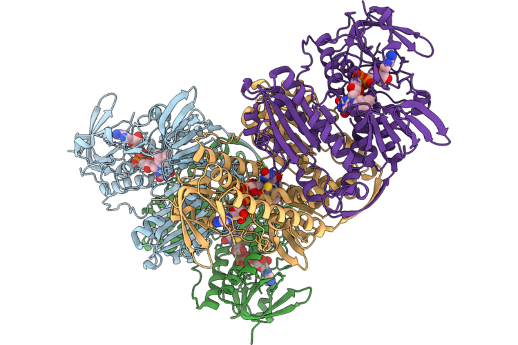
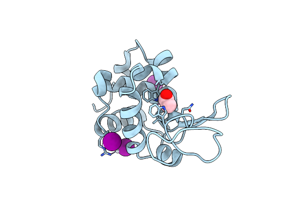
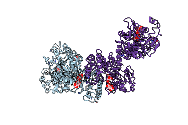
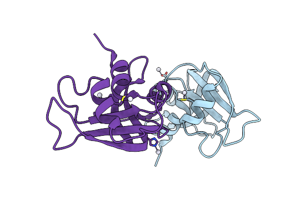
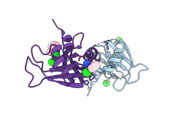
Crystal Structure Of Cryptosporidium Parvum Thioredoxin Reductase In Complex With The Au(Iii) Dithiocarbamato Complex Aul12
Organism: Cryptosporidium parvum
Method: X-RAY DIFFRACTION Resolution:2.15 Å Release Date: 2026-06-03 Classification: FLAVOPROTEIN Ligands: FAD, AU, PEG |
 |
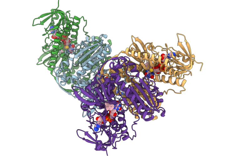
Crystal Structure Of Cryptosporidium Parvum Thioredoxin Reductase In Complex With The Auranofin Analogue Aup(Och3)3Cl
Organism: Cryptosporidium parvum
Method: X-RAY DIFFRACTION Resolution:2.20 Å Release Date: 2026-06-03 Classification: FLAVOPROTEIN Ligands: FAD, AU |
 |
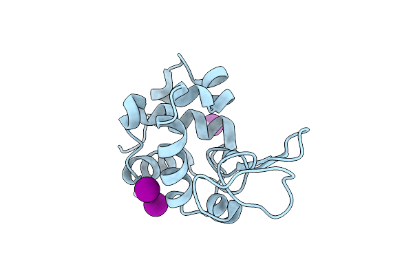
Crystal Structure Of Cryptosporidium Parvum Thioredoxin Reductase In Complex With Aurothiomalate
Organism: Cryptosporidium parvum
Method: X-RAY DIFFRACTION Resolution:2.30 Å Release Date: 2026-06-03 Classification: FLAVOPROTEIN Ligands: FAD, AU |
 |
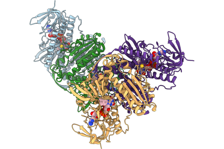
Crystal Structure Of Cryptosporidium Parvum Thioredoxin Reductase In Complex With The Dicarbene Au(I) Complex Au(Nhc)2Pf6
Organism: Cryptosporidium parvum
Method: X-RAY DIFFRACTION Resolution:2.20 Å Release Date: 2026-06-03 Classification: FLAVOPROTEIN Ligands: FAD, AU |
 |
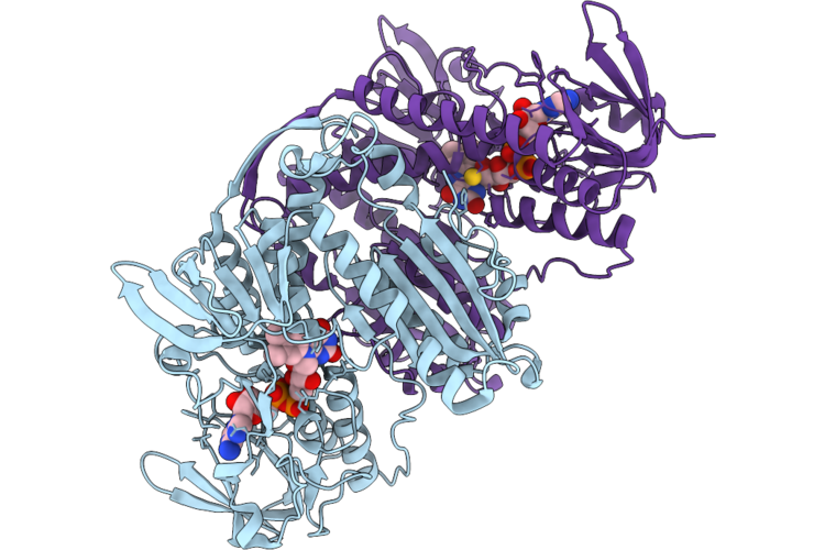
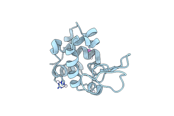
Crystal Structure Of Cryptosporidium Parvum Thioredoxin Reductase In Complex With Mono-Carbene Au(I) Complex Au(Nhc)Cl
Organism: Cryptosporidium parvum
Method: X-RAY DIFFRACTION Resolution:2.20 Å Release Date: 2026-06-03 Classification: FLAVOPROTEIN Ligands: FAD, AU |
 |
Crystal Structure Of Cryptosporidium Parvum Thioredoxin Reductase In Complex With Auranofin
Organism: Cryptosporidium parvum
Method: X-RAY DIFFRACTION Resolution:3.28 Å Release Date: 2026-06-03 Classification: FLAVOPROTEIN Ligands: FAD, AU |
 |
X-Ray Structure Of The Adduct Formed Upon Reaction Of The Diiodido Analogue Of Picoplatin With Lysozyme (Structure A)
Organism: Gallus gallus
Method: X-RAY DIFFRACTION Resolution:1.96 Å Release Date: 2025-05-14 Classification: HYDROLASE Ligands: NH3, ACT, CL, PT, IOD |
 |
X-Ray Structure Of The Adduct Formed Upon Reaction Of The Diiodido Analogue Of Picoplatin With Lysozyme (Structure B)
Organism: Gallus gallus
Method: X-RAY DIFFRACTION Resolution:1.48 Å Release Date: 2025-05-14 Classification: HYDROLASE Ligands: PT, IOD |
 |
X-Structure Of The Adduct Formed Upon Reaction Of The Diiodido Analogue Of Picoplatin With Lysozyme (Structure C)
Organism: Gallus gallus
Method: X-RAY DIFFRACTION Resolution:2.25 Å Release Date: 2025-05-14 Classification: HYDROLASE Ligands: PT, NH3, IOD |
 |
X-Ray Structure Of The Adduct Formed Upon Reaction Of The Diiodido Analogue Of Picoplatin With Ribonuclease A
Organism: Bos taurus
Method: X-RAY DIFFRACTION Resolution:1.77 Å Release Date: 2025-05-14 Classification: HYDROLASE Ligands: NH3, PT, IOD, CL |
 |
X-Ray Structure Of The Adduct Formed Upon Reaction Of The Diiodido Analogue Of Picoplatin With Human Serum Albumin
Organism: Homo sapiens
Method: X-RAY DIFFRACTION Resolution:3.90 Å Release Date: 2025-05-14 Classification: HYDROLASE Ligands: PT |
 |
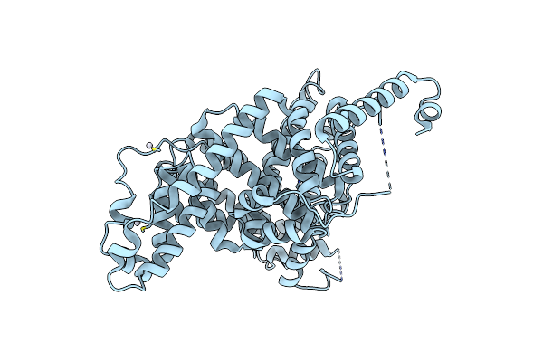
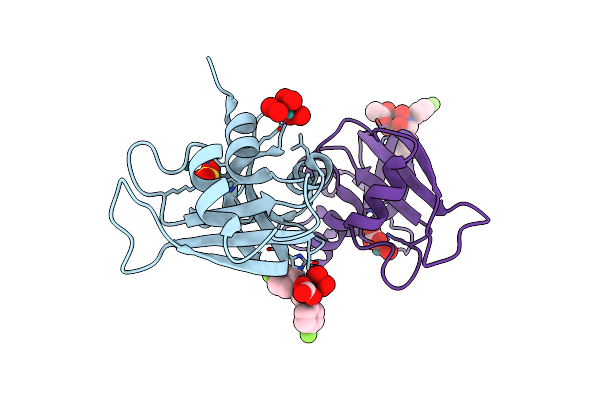
Structural Insights In The Huhf@Gold-Monocarbene Adduct: Aurophilicity Revealed In A Biological Context
Organism: Homo sapiens
Method: ELECTRON MICROSCOPY Resolution:1.51 Å Release Date: 2025-05-07 Classification: METAL BINDING PROTEIN Ligands: BM0, AU |
 |
Crystal Structure Of The Adduct Formed Upon Reaction Of Aurothiomalate With Human Serum Transferrin (Apo-Form)
Organism: Homo sapiens
Method: X-RAY DIFFRACTION Resolution:3.02 Å Release Date: 2025-02-12 Classification: METAL TRANSPORT Ligands: CIT, AU, NAG |
 |
X-Ray Structure Of The Adduct Formed Upon Reaction Of Rnase A With [Ru2(D-P-Fphf)(O2Cch3)2(O2Co)] Complex
Organism: Bos taurus
Method: X-RAY DIFFRACTION Resolution:1.74 Å Release Date: 2024-12-11 Classification: STRUCTURAL PROTEIN Ligands: SO4, A1IQW |
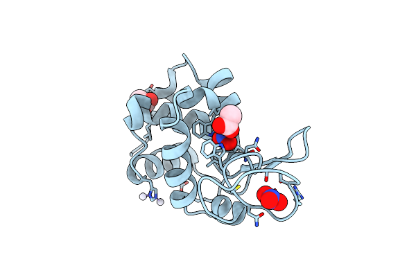 |
X-Ray Structure Of The Adduct Formed Upon Reaction Of Picoplatin With Lysozyme (Structure A)
Organism: Gallus gallus
Method: X-RAY DIFFRACTION Resolution:1.60 Å Release Date: 2024-05-22 Classification: HYDROLASE Ligands: NO3, ACT, NH3, PT |
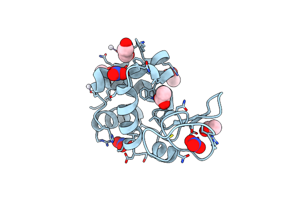 |
X-Ray Structure Of The Adduct Formed Upon Reaction Of Picoplatin With Lysozyme (Structure B)
Organism: Gallus gallus
Method: X-RAY DIFFRACTION Resolution:1.36 Å Release Date: 2024-05-22 Classification: HYDROLASE Ligands: NO3, GOL, ACT, PT |
 |
X-Ray Structure Of The Adduct Formed Upon Reaction Of Picoplatin With Bovine Pancreatic Ribonuclease (Structure C)
Organism: Bos taurus
Method: X-RAY DIFFRACTION Resolution:1.99 Å Release Date: 2024-05-22 Classification: RNA BINDING PROTEIN Ligands: NH3, PT |
 |
X-Ray Structure Of The Adduct Formed Upon Reaction Of Picoplatin With Bovine Pancreatic Ribonuclease (Structure D)
Organism: Gallus gallus
Method: X-RAY DIFFRACTION Resolution:1.76 Å Release Date: 2024-05-22 Classification: RNA BINDING PROTEIN Ligands: NH3, PT, CL, A1H58 |
 |
Crystal Structure Of Transplatin/B-Dna Adduct Obtained Upon 7 Days Of Soaking
Organism: Synthetic construct
Method: X-RAY DIFFRACTION Resolution:1.40 Å Release Date: 2024-02-28 Classification: DNA Ligands: MG, PT, NH3, CL |
 |
Crystal Structure Of Transplatin/B-Dna Adduct Obtained Upon 48 H Of Soaking
Organism: Dna molecule
Method: X-RAY DIFFRACTION Resolution:1.42 Å Release Date: 2024-02-07 Classification: DNA Ligands: MG, NH3, PT |