

Search Count: 24
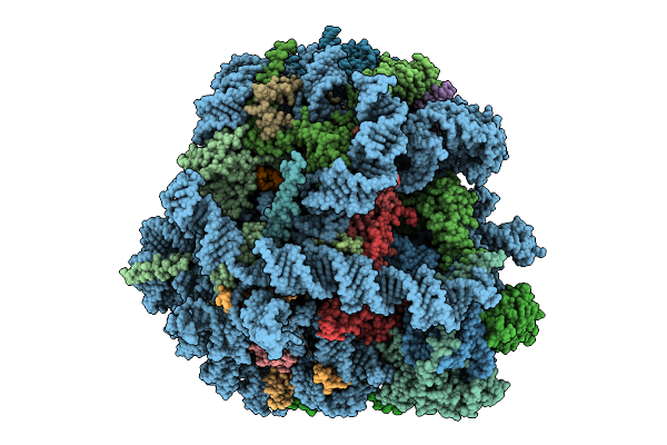 |
Structure Of Fluc-Xbp1U+ Stalled Human 60S Ribosome Nascent Chain Complex
Organism: Photinus pyralis, Homo sapiens
Method: ELECTRON MICROSCOPY Release Date: 2025-09-24 Classification: TRANSLATION Ligands: MG, ZN |
Organism: Photinus pyralis, Homo sapiens
Method: ELECTRON MICROSCOPY
Release Date: 2025-09-24
Ligands: MG, ZN
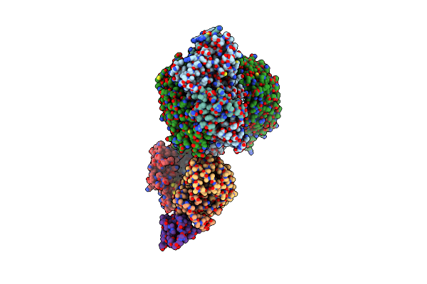 |
Cand1-Cul1-Rbx1-Skp1-Skp2-Cks1-Cdk2
Organism: Homo sapiens
Method: ELECTRON MICROSCOPY Release Date: 2023-06-28 Classification: LIGASE Ligands: ZN |
Organism: Homo sapiens
Method: ELECTRON MICROSCOPY
Release Date: 2023-06-28
Ligands: ZN
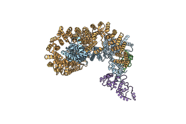 |
Cand1-Cul1-Rbx1-Dcnl1
Organism: Homo sapiens
Method: ELECTRON MICROSCOPY Release Date: 2023-06-28 Classification: LIGASE Ligands: ZN |
Organism: Homo sapiens
Method: ELECTRON MICROSCOPY
Release Date: 2023-06-28
Ligands: ZN
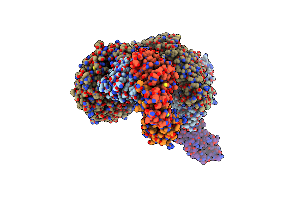 |
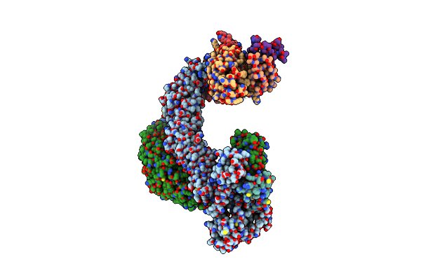
Cand1-Cul1-Rbx1-Skp1-Skp2-Dcnl1
Organism: Homo sapiens
Method: ELECTRON MICROSCOPY Release Date: 2023-06-28 Classification: LIGASE Ligands: ZN |
Organism: Homo sapiens
Method: ELECTRON MICROSCOPY
Release Date: 2023-06-28
Ligands: ZN
 |
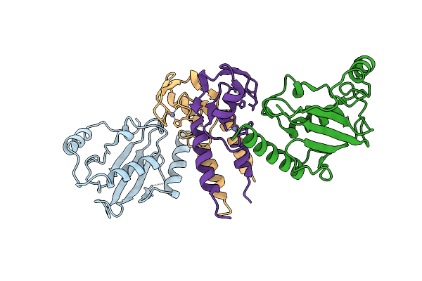
Partially Dissociated Cand1-Cul1-Rbx1-Skp1-Skp2-Cks1-Cdk2
|
Organism: Homo sapiens
Method: ELECTRON MICROSCOPY
Release Date: 2023-06-28
 |
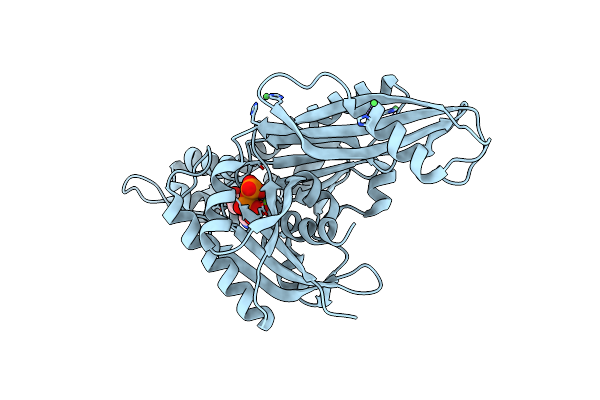
X-Ray Structure Of Trim21 Ring E3 Ligase In Complex With E2 Enzyme Ube2W
Organism: Homo sapiens
Method: X-RAY DIFFRACTION Resolution:2.25 Å Release Date: 2023-04-26 Classification: LIGASE Ligands: ZN |
Organism: Homo sapiens
Method: X-RAY DIFFRACTION
Release Date: 2023-04-26
Ligands: ZN
 |
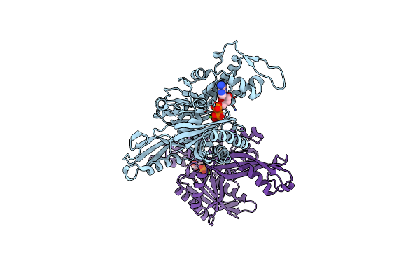
E. Coli Ftsa 1-405 Atp 3 Ni
Organism: Escherichia coli (strain k12)
Method: X-RAY DIFFRACTION Resolution:2.80 Å Release Date: 2022-09-21 Classification: CELL CYCLE Ligands: NI, MG, ATP |
Organism: Escherichia coli (strain k12)
Method: X-RAY DIFFRACTION
Release Date: 2022-09-21
Ligands: NI, MG, ATP
 |
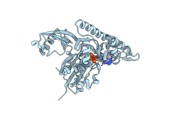
Vibrio Maritimus Ftsa 1-396 Atp, Double Filament
Organism: Vibrio maritimus
Method: X-RAY DIFFRACTION Resolution:3.31 Å Release Date: 2022-09-21 Classification: CELL CYCLE Ligands: ATP, MG |
Organism: Vibrio maritimus
Method: X-RAY DIFFRACTION
Release Date: 2022-09-21
Ligands: ATP, MG
 |
Xenorhabdus Poinarii Ftsa 1-396 Adp
Organism: Xenorhabdus poinarii g6
Method: X-RAY DIFFRACTION Resolution:2.80 Å Release Date: 2022-09-21 Classification: CELL CYCLE Ligands: ADP, MG |
Organism: Xenorhabdus poinarii g6
Method: X-RAY DIFFRACTION
Release Date: 2022-09-21
Ligands: ADP, MG
 |
Vibrio Maritimus Ftsa 1-396 Atp And Ftsn 1-29, Bent Tetramers In Double Filament Arrangement
Organism: Vibrio maritimus
Method: X-RAY DIFFRACTION Resolution:3.60 Å Release Date: 2022-09-21 Classification: CELL CYCLE Ligands: ATP, MG |
Organism: Vibrio maritimus
Method: X-RAY DIFFRACTION
Release Date: 2022-09-21
Ligands: ATP, MG
 |
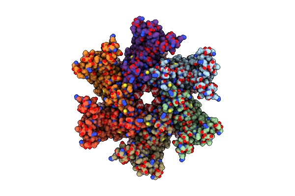
Human Complex Ii-Bats Bound To Membrane-Attached Rab5A-Gtp
Organism: Homo sapiens
Method: ELECTRON MICROSCOPY Resolution:9.80 Å Release Date: 2021-03-03 Classification: ENDOCYTOSIS Ligands: GTP, MG |
Organism: Homo sapiens
Method: ELECTRON MICROSCOPY
Release Date: 2021-03-03
Ligands: GTP, MG
 |
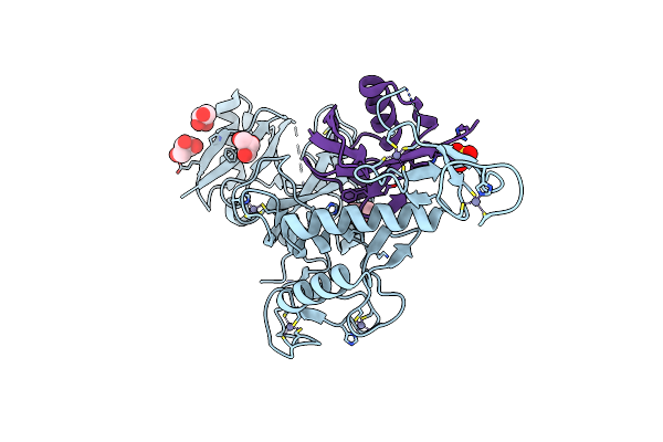
Cryoem Structure Of Serinc From Drosophila Melanogaster
Organism: Drosophila melanogaster
Method: ELECTRON MICROSCOPY Release Date: 2020-01-01 Classification: MEMBRANE PROTEIN Ligands: LMN, P5S, CDL |
Organism: Drosophila melanogaster
Method: ELECTRON MICROSCOPY
Release Date: 2020-01-01
Ligands: LMN, P5S, CDL
 |
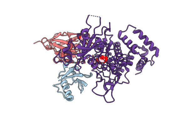
Structure Of Phospho-Parkin Bound To Phospho-Ubiquitin
Organism: Homo sapiens
Method: X-RAY DIFFRACTION Resolution:1.80 Å Release Date: 2018-06-13 Classification: LIGASE Ligands: ZN, GOL, MPD, SO4 |
Organism: Homo sapiens
Method: X-RAY DIFFRACTION
Release Date: 2018-06-13
Ligands: ZN, GOL, MPD, SO4
 |
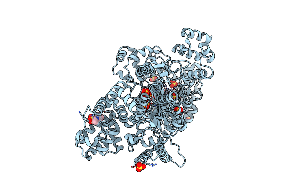
Structure Of Pink1 Bound To Ubiquitin
Organism: Homo sapiens, Lama glama, Pediculus humanus corporis
Method: X-RAY DIFFRACTION Resolution:3.10 Å Release Date: 2017-11-08 Classification: TRANSFERASE Ligands: GOL |
Organism: Homo sapiens, Lama glama, Pediculus humanus corporis
Method: X-RAY DIFFRACTION
Release Date: 2017-11-08
Ligands: GOL
 |
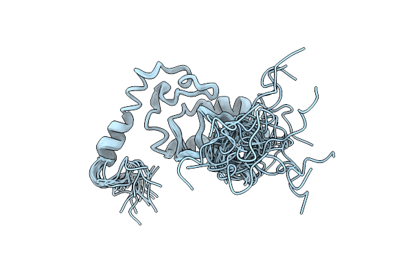
Dna Polymerase
Organism: Mycobacterium tuberculosis (strain atcc 25618 / h37rv)
Method: X-RAY DIFFRACTION Resolution:2.80 Å Release Date: 2017-10-25 Classification: TRANSFERASE Ligands: ZN, SO4, EPE |
Organism: Mycobacterium tuberculosis (strain atcc 25618 / h37rv)
Method: X-RAY DIFFRACTION
Release Date: 2017-10-25
Ligands: ZN, SO4, EPE
 |
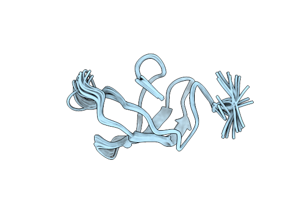
Structure Of The N-Terminal Domain Of The Escherichia Coli Proq Rna Binding Protein
Organism: Escherichia coli o45:k1 (strain s88 / expec)
Method: SOLUTION NMR Release Date: 2017-05-03 Classification: RNA |
Organism: Escherichia coli o45:k1 (strain s88 / expec)
Method: SOLUTION NMR
Release Date: 2017-05-03
 |
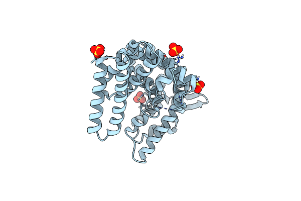
Structure Of The C-Terminal Domain Of The Escherichia Coli Proq Rna Binding Protein
Organism: Escherichia coli (strain k12)
Method: SOLUTION NMR Release Date: 2017-05-03 Classification: CHAPERONE |
Organism: Escherichia coli (strain k12)
Method: SOLUTION NMR
Release Date: 2017-05-03
 |
Crystal Structure Of The N-Terminal Domain Of Human Timeless
Organism: Homo sapiens
Method: X-RAY DIFFRACTION Resolution:1.85 Å Release Date: 2017-03-08 Classification: REPLICATION Ligands: SO4 |
Organism: Homo sapiens
Method: X-RAY DIFFRACTION
Release Date: 2017-03-08
Ligands: SO4
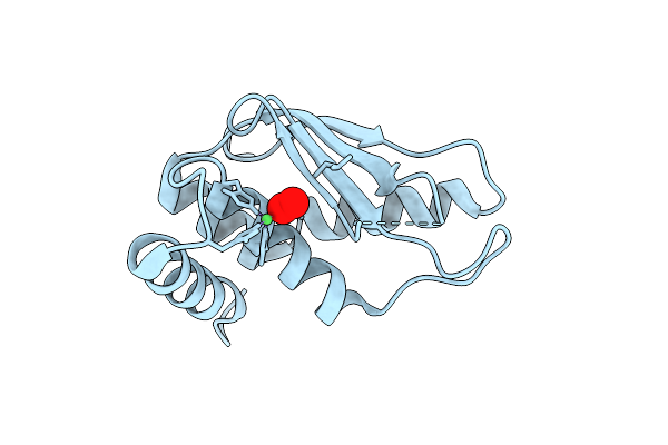 |
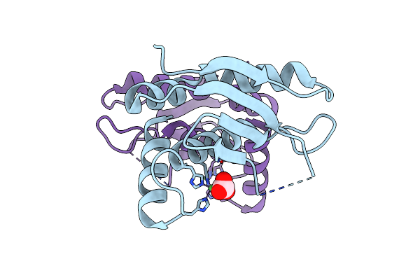
Crytsal Structure Of Wss1 From S. Pombe
Organism: Schizosaccharomyces pombe (strain 972 / atcc 24843)
Method: X-RAY DIFFRACTION Resolution:1.00 Å Release Date: 2016-11-09 Classification: HYDROLASE Ligands: NI, OXY |
Organism: Schizosaccharomyces pombe (strain 972 / atcc 24843)
Method: X-RAY DIFFRACTION
Release Date: 2016-11-09
Ligands: NI, OXY
 |
Crystal Structure Of The Wss1 E203Q Mutant From S. Pombe
Organism: Schizosaccharomyces pombe
Method: X-RAY DIFFRACTION Resolution:1.75 Å Release Date: 2016-11-09 Classification: HYDROLASE Ligands: NI, CO3 |
Organism: Schizosaccharomyces pombe
Method: X-RAY DIFFRACTION
Release Date: 2016-11-09
Ligands: NI, CO3